. 1997 Jan 27;136(2):345-54.
doi: 10.1083/jcb.136.2.345.

Identification of a mid-anaphase checkpoint in budding yeast

Affiliations

Identification of a mid-anaphase checkpoint in budding yeast

S S Yang et al. J Cell Biol. .

Abstract

Activation of a facultative, dicentric chromosome provides a unique opportunity to introduce a double strand DNA break into a chromosome at mitosis. Time lapse video enhanced-differential interference contrast analysis of the cellular response upon dicentric activation reveals that the majority of cells initiates anaphase B, characterized by pole-pole separation, and pauses in mid-anaphase for 30-120 min with spindles spanning the neck of the bud before completing spindle elongation and cytokinesis. The length of the spindle at the delay point (3-4 microm) is not dependent on the physical distance between the two centromeres, indicating that the arrest represents surveillance of a dicentric induced aberration. No mid-anaphase delay is observed in the absence of the RAD9 checkpoint gene, which prevents cell cycle progression in the presence of damaged DNA. These observations reveal RAD9-dependent events well past the G2/M boundary and have considerable implications in understanding how chromosome integrity and the position and state of the mitotic spindle are monitored before cytokinesis.


Figures

Figure 1
Figure 1
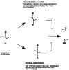
Dicentric chromosome breakage resulting from centromere movements during prometaphase or anaphase. A schematic diagram representing possible centromere attachments of a dicentric chromosome to the mitotic spindle during prometaphase and anaphase. During prometaphase, the two centromeres will independently form bipolar attachments to the mitotic spindle. A dicentric chromosome in which the two centromeres have formed initial monopolar attachments to opposite spindle poles is shown in the top figure. Centromere movements toward opposite spindle poles, as indicated by the arrows, may result in chromosome breakage. During anaphase B, when the two centromeres of a dicentric chromosome have successfully formed bipolar attachments, centromere movements at anaphase may result in chromosome breakage. If the two centromeres of each chromosome are oriented toward the same spindle pole (top), chromosome segregation is normal. If the centromeres are oriented toward opposite spindle poles (bottom), anaphase bridges are formed, and chromosome breakage can occur (Brock and Bloom, 1994).
Figure 2
Figure 2
High resolution DE-DIC images of mitosis upon dicentric chromosome activation. A pair of arrows on each frame demarcates the nucleus. The time of each frame is indicated in the upper right corner. Bottom cell: (A) Pre-anaphase B/metaphase spindle (1.62 μm) with the nucleus positioned at the neck; (B) spindle elongation into the bud (2.99 μm); (C–F) the nucleus persists in the neck at a length ranging from 2.57–3.45 μm for 38 min; (G) spindle elongation ensues (5.76 μm spindle); (H–K) the nucleus continues to elongate to the distal ends (7.85 μm) and septum formation occurs (J and K); (L) cytokinesis and new bud formation. Top cell: (I) Spindle elongation; (J–K) nucleus in the neck. (L) The appearance of septum formation through the nucleus.
Figure 3
Figure 3
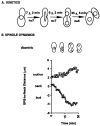
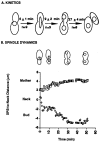
No mid-anaphase pause. (A) Kinetic analysis of changes in nuclear morphology and spindle dynamics in dicentric cells displaying wild-type kinetics. The time between spindle elongation and cytokinesis, seen as cell separation, was determined from the time lapse videos. 7 out of 29 cells filmed displayed these kinetics. (B) Spindle movements relative to the bud neck. The distance (μm) between the neck and the leading and lagging edge of the nuclear envelope was measured and plotted as a function of time (min). A schematic representation of the nuclear morphology and position at the different stages is shown above the plot. The neck is designated as 0 μm. The length from the leading edge to the neck (proximal to bud, diamonds) is designated with negative numbers. The length from the lagging edge to the neck (proximal to mother, squares) is designated with positive numbers. Preanaphase spindle is at T = 0.
Figure 4
Figure 4
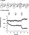
Mid-anaphase pause. (A) Kinetic analysis of changes in nuclear morphology and spindle dynamics in dicentric cells displaying mitotic delay. 22 out of 29 cells displayed these kinetics. (B) Spindle movements relative to the bud neck were determined as described in the legend to Fig. 3.
Figure 5
Figure 5
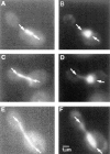
Progression of anaphase in cells containing a dicentric chromosome. Cells were fixed and processed for immunofluorescence after two hours of growth in glucose-containing medium. The panels on the left are cells stained with anti-tubulin antibodies; the panels on the right are the same cells stained with DAPI. The arrows mark the ends of the spindle or nucleus, respectively. (A and B) Cells in the G2 stage, medium budded with the DNA near the neck. (C and D) Cells in the early stages of anaphase, having a slightly elongated spindle with DNA just visible in the bud. (E and F) Typical of cells paused in mid-anaphase with a 4 μm spindle and DNA through the neck of large budded cells. Bar, 1 μm.
Figure 6
Figure 6
Quantitation of sister chromatid separation by FISH. Cells were exposed to glucose for 1 h and subjected to FISH analysis, as described in the Materials and Methods. (A) Micrographs of nuclei are shown. Chromosomal DNA, stained with propidium iodide, appears gray. Centromere-proximal chromosomes I are detected by FISH and appear as white dots. (B) The percentage of nuclei with one, two, or three signals was determined. Mean separation between two dots was determined.
Figure 6
Figure 6
Quantitation of sister chromatid separation by FISH. Cells were exposed to glucose for 1 h and subjected to FISH analysis, as described in the Materials and Methods. (A) Micrographs of nuclei are shown. Chromosomal DNA, stained with propidium iodide, appears gray. Centromere-proximal chromosomes I are detected by FISH and appear as white dots. (B) The percentage of nuclei with one, two, or three signals was determined. Mean separation between two dots was determined.
Figure 7
Figure 7
Nature of the mid-anaphase pause. The arrest in midanaphase B may be due to physical constraints on the chromosome during spindle elongation. This would predict a dependent relationship between spindle length and the distance between the two centromeres. Alternatively, activation of the dicentric chromosome and subsequent chromosomal alterations may signal a surveillance system resulting in cell cycle arrest. Spindle length at the mid-anaphase transition would be independent of the distance between the two centromeres.
Figure 8
Figure 8
Kinetics and spindle dynamics upon activation of a dicentric minichromosome. The distance between the two centromeres is 3 kb. As shown in Figs. 3 and 4, two cellular responses are observed upon dicentric activation. (A) No mid-anaphase pause: Approximately 50% of the cells (9/19) exhibit wild type kinetics. Mid-anaphase pause: The other 50% (11/19) are delayed in the 3–4 μm stage of spindle elongation (finger projection, gourd-shaped nucleus). The average time spent in mid-anaphase was 59 ± 10 min. (B) The distance (μm) between the neck and the leading edge of the nucleus was determined as a function of time (min). A schematic representation of the nuclear morphology and position at the different stages is shown above each plot. 0, neck; positive values, mother; negative values, bud.
Figure 9
Figure 9
The dicentric chromosome–induced mid-anaphase transition is dependent upon the RAD9 gene. A single cellular response was observed upon dicentric chromosome activation in the rad9 deletion strain. The behavior of the spindle and the timing of nuclear transitions were indistinguishable from wild type cells. (A) Kinetics of morphological transitions. (B) The distance between the neck and each spindle pole body was determined as a function of time (min). A schematic representation of the nuclear morphology and position at the different stages is shown above each plot. 0, neck; positive values, mother; negative values, bud. Spindle elongation is complete after 30 min with virtually no delay in spindle elongation at the 3–4 μm stage.
Figure 10
Figure 10
Anaphase spindle morphogenesis in yeast. The major transitions in spindle morphogenesis. Formation and maintenance of a 2 μm spindle are dependent upon members of the kinesin family (CIN8, KIP1 and KAR3; Saunders and Hoyt, 1992; Saunders et al., 1995), and checkpoint genes that limit anaphase onset (AO)(BUB1, 2, and 3, Hoyt et al., 1991; MAD1, 2 and 3, Li and Murray, 1991; RAD9, Weinert and Hartwell, 1988). Separation of the spindle poles (spindle elongation) and translocation of the spindle through the bud neck are simultaneous in wild type cells, characterized by fast spindle elongation (Yeh et al., 1995). VE-DIC microscopy of live cells and of serial reconstruction of yeast spindles indicates a transition between the fast (1 μm/min) to slow (0.3 μm/min) phases of spindle morphogenesis in wild type cells. This transition in mid-anaphase is prolonged upon activation of the dicentric chromosome and is dependent upon RAD9 function. Chromosome separation (noted morphologically by the bilobed structure) is delayed upon activation of the dicentric chromosome. The final stages of spindle elongation, disassembly, nuclear division, and cytokinesis are discrete morphological events (Copeland and Snyder, 1993; Yeh et al., 1995). We propose that this mid-anaphase checkpoint is essential for coordinating spindle morphogenesis and position with chromosome separation and the onset of cytokinesis.

References

    1. Allen JB, Zhou Z, Siede W, Friedberg EC, Elledge SJ. The SAD1/RAD53 protein kinase controls multiple checkpoints and DNA damage-induced transcription in yeast. Genes Dev. 1994;8:2401–2415. - PubMed
    1. Brock JA, Bloom K. A chromosome breakage assay to monitor mitotic forces in budding yeast. J Cell Sci. 1994;107:891–902. - PubMed
    1. Copeland CS, Snyder M. Nuclear pore complex antigens delineate nuclear envelope dynamics in vegetative and conjugating Saccharomyces cerevisiae. . Yeast. 1993;9:235–249. - PubMed
    1. Eshel D, Urrestarazu LA, Vissers S, Jauniaux JC, van Vliet-Reedijk JC, Planta RJ, Gibbons IR. Cytoplasmic dynein is required for normal nuclear segregation in yeast. Proc Natl Acad Sci USA. 1993;90:11172–11176. - PMC - PubMed
    1. Girard F, Fernandez A, Lamb N. Delayed cyclin A and B1 degradation in non-transformed mammalian cells. J Cell Sci. 1995;108:2599–2608. - PubMed

Publication types

MeSH terms