Comparative Study
. 1997 Jan 27;136(2):375-88.
doi: 10.1083/jcb.136.2.375.

NGF and neurotrophin-3 both activate TrkA on sympathetic neurons but differentially regulate survival and neuritogenesis

Affiliations
Comparative Study

NGF and neurotrophin-3 both activate TrkA on sympathetic neurons but differentially regulate survival and neuritogenesis

D J Belliveau et al. J Cell Biol. .

Abstract

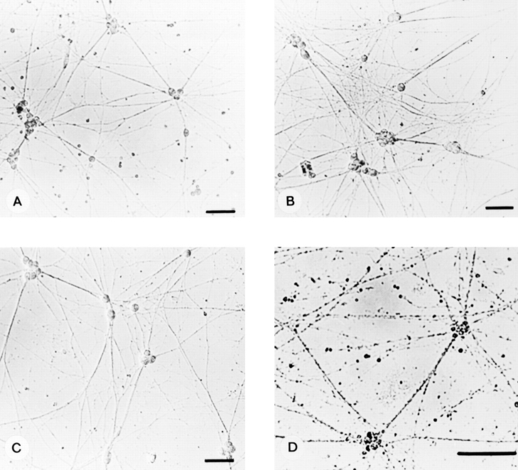
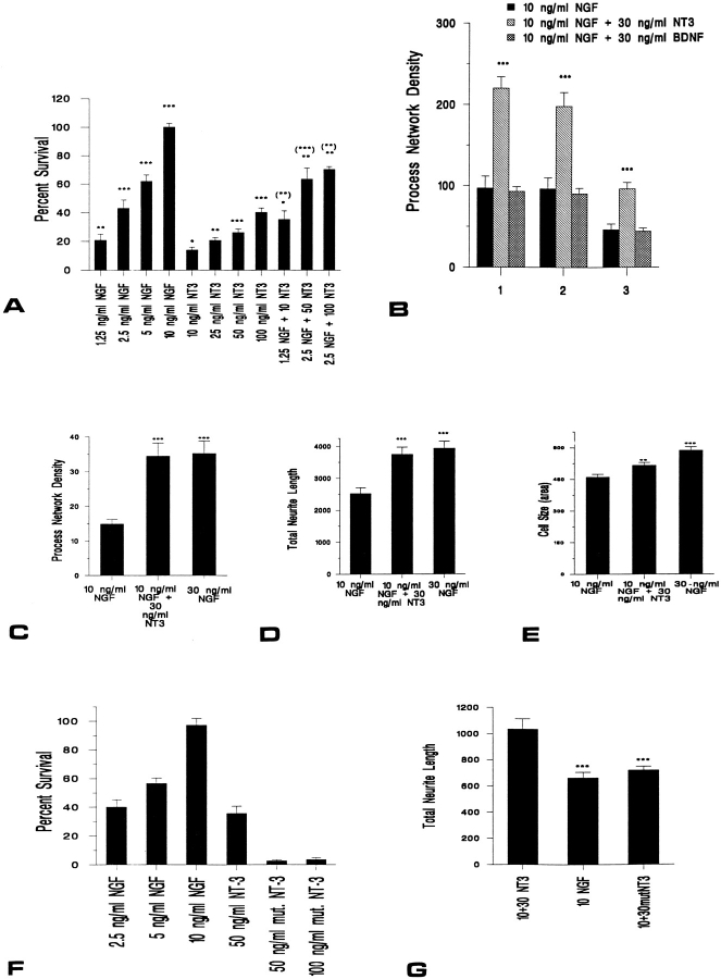
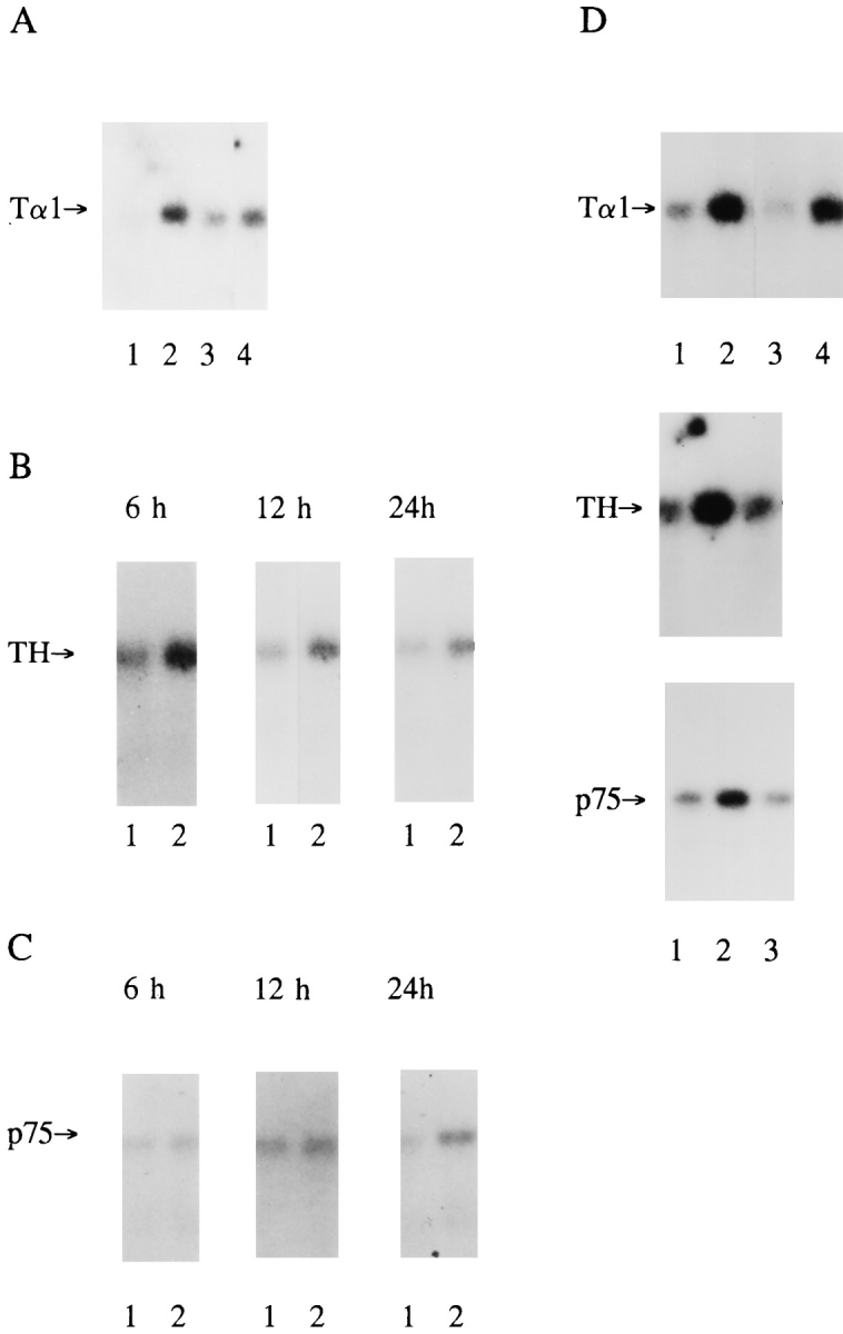
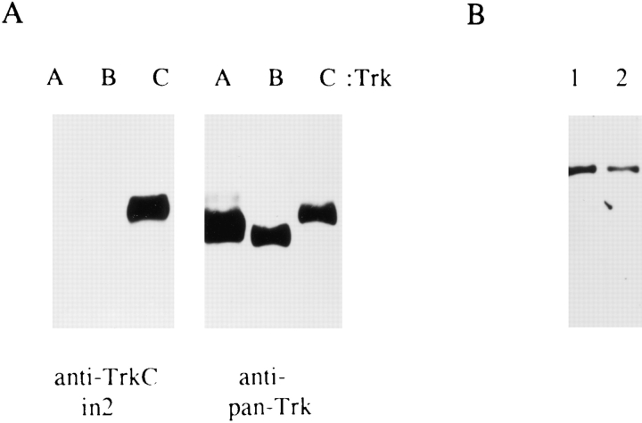
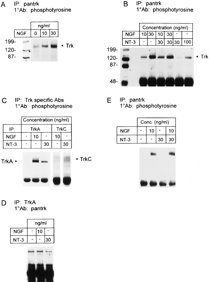
In this report we examine the biological and molecular basis of the control of sympathetic neuron differentiation and survival by NGF and neurotrophin-3 (NT-3). NT-3 is as efficient as NGF in mediating neuritogenesis and expression of growth-associated genes in NGF-dependent sympathetic neurons, but it is 20-40-fold less efficient in supporting their survival. Both NT-3 and NGF induce similar sustained, long-term activation of TrkA, while NGF is 10-fold more efficient than NT-3 in mediating acute, short-term TrkA activity. At similar acute levels of TrkA activation, NT-3 still mediates neuronal survival two- to threefold less well than NGF. However, a mutant NT-3 that activates TrkC, but not TrkA, is unable to support sympathetic neuron survival or neuritogenesis, indicating that NT-3-mediated TrkA activation is necessary for both of these responses. On the basis of these data, we suggest that NGF and NT-3 differentially regulate the TrkA receptor both with regard to activation time course and downstream targets, leading to selective regulation of neuritogenesis and survival. Such differential responsiveness to two ligands acting through the same Trk receptor has important implications for neurotrophin function throughout the nervous system.


Figures

Figure 1
Figure 1
NGF-dependent sympathetic neurons respond to NT-3 but not to BDNF. Phase-contrast micrographs of cultures of pure sympathetic neurons from the postnatal day 1 rat SCG maintained in 10 ng/ml NGF for 5 d (A) and then supplemented with 30 ng/ml NT-3 (B) or 30 ng/ml BDNF (C). NT-3 enhanced the number of neurites compared with BDNF when examined 2 d after addition. In similar cultures where the NGF was replaced with 30 ng/ml BDNF (D), obvious cell body and process deterioration was evident.
Figure 2
Figure 2
(A) NT-3 is 20–40-fold less efficient than NGF at supporting survival of NGF-dependent sympathetic neurons. Results of a colorimetric MTT assay to measure mitochondrial function and cell survival. Neonatal sympathetic neurons were cultured in 10 ng/ml NGF for 5 d in 48-well dishes, washed free of neurotrophin-containing medium, and then incubated for 2 d in various concentrations of NGF and/or NT-3, as indicated on the x axis. Each point represents the values pooled from three independent sets of survival assays, each of which was performed in quadruplicate. In these assays, absolute values are normalized so that the value obtained with 0 neurotrophin is 0% survival, while that obtained with 10 ng/ml NGF (in which the neurons were originally selected) is considered 100% survival. Error bars represent SEM; * = P < 0.05, ** = P < 0.01, and *** = P < 0.001. For NGF and NT-3 alone, comparisons were made using a t test with the 0 neurotrophin control. For the NGF/NT-3 combinations, comparisons were made against the survival supported by the same concentrations of NGF (no brackets) and NT-3 (brackets) alone. (B–D) NT-3 is equivalent to NGF in mediating neuritogenesis of NGF-dependent sympathetic neurons. (B) Three separate experiments were performed to determine the effect of NT-3 or BDNF on process outgrowth in sympathetic neurons. Sympathetic neurons were plated at moderate density on a collagen substrate, selected in 10 ng/ml NGF for 5 d, and then supplemented with either 30 ng/ml NT-3 or BDNF as indicated. The number of fields examined for each point ranged from 9–14. In all three experiments, significantly more neurite intersections were observed after exposure to 30 ng/ml NT-3 plus 10 ng/ml NGF (*** = P < 0.001) than 10 ng/ml NGF alone. (C and D) To confirm the results presented in B, and to directly compare NT-3 to NGF, neurons were plated at low density on a poly-d-lysine/laminin substrate, selected in 10 ng/ml NGF for 5 d, and then either maintained in 10 ng/ml NGF or switched to 30 ng/ml NGF or to 10 ng/ml NGF plus 30 ng/ml NT-3. Two independent experiments were performed, and 8–12 fields were quantitated per treatment in each experiment. The results of the two experiments were pooled. (C) 2–2.5-fold more neurite intersections were observed after exposure to either 30 ng/ml NGF or to 10 ng/ml NGF plus 30 ng/ ml NT-3 than after exposure to 10 ng/ml NGF alone (*** = P < 0.001). These NGF- and NT-3–induced increases were statistically similar to each other (P = 0.45). (D) Total neurite length was also significantly increased after exposure to 30 ng/ml NGF or to 10 ng/ml NGF plus 30 ng/ml NT-3, relative to 10 ng/ml NGF (*** = P < 0.001). Again, the increase was statistically similar for 30 ng/ml NGF vs 10 ng/ ml NGF plus 30 ng/ml NT-3 (P = 0.28). (E) NGF is more effective than NT-3 at mediating cell body hypertrophy. Cell body size of sympathetic neurons was quantitated in the experiments described in C and D. In this case, NT-3 elicited a small but statistically significant increase of ∼10%, whereas 30 ng/ml NGF led to an ∼25% hypertrophy of the sympathetic neuron cell bodies (** = P < 0.01, *** = P < 0.001). The hypertrophy observed in 10 ng/ml NGF plus 30 ng/ml NT-3 was significantly different from that seen in 30 ng/ml NGF (P = 0.0006). (F and G) Recombinant mutant NT-3 that activates TrkC but not TrkA is insufficient to support sympathetic neuron survival (F) or neuritogenesis (G). (F) Results of a colorimetric MTT assay to measure mitochondrial function and cell survival in response to NGF, NT-3, or a mutant NT-3 that binds only TrkC (mut. NT-3) (58, 59). Sympathetic neurons were cultured in 10 ng/ml NGF for 5 d, washed free of neurotrophin-containing media, and then incubated for 2 d in various concentrations of neurotrophins, as indicated on the x axis. Each point represents the values pooled from three independent sets of survival assays, each of which was performed in quadruplicate. Growth of sympathetic neurons in 50 or 100 ng/ml mutant NT-3 resulted in significantly less survival than 100 ng/ml NT-3 (*** = P < 0.001). In these assays, absolute values are normalized with regard to NGF-mediated survival, as described for A. (G) To compare sympathetic neuron process outgrowth in response to mutant NT-3 that activates only TrkC (mut. NT-3), neurons were cultured in 10 ng/ml NGF for 5 d, and then maintained for an additional 2 d in 10 ng/ml NGF plus or minus 30 ng/ml NT-3 or mutant NT-3. Two independent experiments were performed, and eight fields were quantitated per treatment. As shown in D, total neurite length was significantly increased after exposure to 10 ng/ml NGF plus 30 ng/ml NT-3, relative to 10 ng/ml NGF alone (*** = P < 0.0005). In contrast, no increase in neurite length was observed with mutant NT-3 addition relative to 10 ng/ml NGF alone (P = 0.14).
Figure 3
Figure 3
NT-3 selectively regulates expression of genes associated with morphological growth in a concentration-dependent fashion. (A) Expression of Tα1 α-tubulin mRNA (Tα1) in equal amounts of total RNA isolated from neonatal sympathetic neurons cultured for 5 d in the presence of 10 ng/ml NGF (lane 1) followed by addition of 30 ng/ml NT-3 for 6 h (lane 2), 24 h (lane 3), or 48 h (lane 4). (B and C) Expression of tyrosine hydroxylase (TH) (B) and p75 neurotrophin receptor (p75) (C) mRNAs in response to the addition of 30 ng/ml NT-3 for 6 , 12, or 24 h. In all panels, lane 1 includes total RNA from neurons maintained for the entire time in 10 ng/ml NGF, while lane 2 includes an equal amount of total RNA isolated from sister cultures treated with 10 ng/ml NGF plus 30 ng/ml NT-3. (D) Expression of Tα1 α-tubulin, tyrosine hydroxylase, and p75 neurotrophin receptor mRNAs after 24 h of treatment with 10 ng/ml NGF (lane 1), 10 ng/ml NGF plus 30 ng/ml NT-3 (lane 2), or 10 ng/ml NGF plus 10 ng/ml NT-3 (lane 3). Lane 4 for Tα1 mRNA contains an equal amount of total RNA isolated from neurons treated with 200 ng/ml NGF for 24 h.
Figure 6
Figure 6
(A–D) NT-3 induces a prolonged autophosphorylation of TrkA equivalent to that induced by NGF. Neonatal sympathetic neurons were cultured for 5 d in 10 ng/ml NGF, and then switched to neurotrophincontaining media for an additional 2 d. Cellular lysates were immunoprecipitated (IP) either with anti-pan Trk or anti-TrkA. Immunoprecipitates were then analyzed by Western blot analysis with anti-phosphotyrosine or antipan Trk (1°Ab). All samples were normalized for equal amounts of protein. (A) Trk autophosphorylation in response to 2 d of constant exposure to NGF and/or NT-3. Note that all three of these lanes are from the same autoradiograph of the same Western blot. (B) NGF and NT-3 do not alter Trk receptor levels in sympathetic neurons. In two separate experiments, total Trk levels in NGF-dependent sympathetic neurons were similar regardless of neurotrophintreatment, as evidenced when neurons are cultured in various concentrations of NGF, NT-3, and/or BDNF for 2 d. (C) TrkA autophosphorylation in response to 2 d of constant exposure to NGF and/or NT-3. (D) The same immunoprecipitates as in C were probed with antipan Trk on a separate Western blot to ensure that approximately equal amounts of TrkA were present in each sample. (E) NGF and NT-3 do not alter TrkA or TrkC levels. To determine whether TrkA or TrkC levels changed with increased levels of neurotrophins, NGF-dependent sympathetic neurons were cultured for 48 h in various concentrations of NGF and/or NT-3. Lysates were immunoprecipitated with the anti-pan Trk antibody, and then analyzed by Western blots with antibodies specific to TrkA or TrkC. The appropriately sized Trk bands are indicated by arrows. (F) NGF and NT-3 alter the ratio of Trk/p75 in NGF-dependent sympathetic neurons. After immunoprecipitation with anti-pan Trk, the lysates shown in B were immunoprecipitated for the p75 neurotrophin receptor using the mAb MC192. Immunoprecipitates were then probed on a Western blot with a polyclonal anti-p75 antibody.
Figure 4
Figure 4
Full-length TrkC is expressed in NGF-dependent sympathetic neurons. (A, left) Specificity of the anti-TrkCin2 antibody, which recognizes the intracellular domain of TrkC, was tested by probing lysates of insect cells expressing human TrkA, rat TrkB, or rat TrkC on Western blots. (A, right) Anti-pan Trk antibody 203 recognizes all three Trk family members on a similar Western blot. (B) Western blot analysis of full-length TrkC in NGF-dependent sympathetic neurons as detected by probing WGA precipitates with anti-TrkCin2. Lane 1 includes protein from sympathetic neurons cultured for 5 d in 10 ng/ml NGF, and lane 2 includes protein from sister cultures induced with 30 ng/ml NT-3 for 10 min. The immunoreactive TrkC band is detected at 145 kD, the size of full-length TrkC.
Figure 5
Figure 5
(A–D) NT-3 exposure leads to the autophosphorylation of both TrkA and TrkC on NGF-dependent sympathetic neurons. Neonatal sympathetic neurons were cultured for 5 d in 10 ng/ml NGF, washed for 3 h in neurotrophin-free media, and then induced with neurotrophins for 10 min. Cell lysates were immunoprecipitated (IP) with either anti-pan Trk 203 (pantrk), anti-TrkA, or anti-TrkC. Immunoprecipitates were analyzed by Western blot analysis with anti-phosphotyrosine or antipan Trk 203 as the primary antibodies (1°Ab). All samples were normalized for equal amounts of protein. (A) Trk autophosphorylation increased in a graded fashion in response to 10 ng/ml NGF, and in response to 30 ng/ml NGF, relative to base line levels of autophosphorylation with 0 NGF. (Numbers, left) Molecular weight markers; (Trk) size of the Trk band. (B) Trk autophosphorylation in sympathetic neurons in response to varying concentrations of neurotrophins. Note the presence of a single autophosphorylated Trk band in the samples treated with NT-3. (C) TrkA and TrkC autophosphorylation in response to NGF vs NT-3. Approximately 300,000 neurons were lysed and analyzed for TrkA-specific autophosphorylation in response to no neurotrophin addition, 10 ng/ml NGF, or 30 ng/ml NT-3. The same lysates were then immunoprecipitated for TrkC-specific autophosphorylation in response to 10 ng/ml NGF or 30 ng/ml NT-3. Note that the right two lanes containing the TrkC immunoprecipitates are both from the same autoradiograph of the same gel. (D) The same immunoprecipitates as in C were probed with anti-pan Trk on a separate Western blot to ensure that approximately equal amounts of TrkA were present in each sample. (E) TrkA in PC12 cells is not autophosphorylated in response to NT-3. PC12 cells were induced with NGF and/or NT-3 for 10 min, and cellular lysates were immunoprecipitated with anti-pan Trk 203, followed by a Western blot with anti-phosphotyrosine.

References

    1. Acheson A, Barker PA, Alderson RF, Miller FD, Murphy RA. Detection of brain-derived neurotrophic factor-like activity in fibroblasts and Schwann cells: inhibition by antibodies to NGF. Neuron. 1991;7:265–275. - PubMed
    1. Barbacid M. The trk family of neurotrophin receptors. J Neurobiol. 1994;25:1386–1403. - PubMed
    1. Barker PA, Shooter EM. Disruption of NGF binding to the low affinity neurotrophin receptor p75LNTR reduces NGF binding to TrkA on PC12 cells. Neuron. 1994;13:203–215. - PubMed
    1. Barker PA, Lomen-Hoerth C, Gensch EM, Meakin SO, Glass DJ, Shooter EM. Tissue-specific alternative splicing generates two isoforms of the trkA receptor. J Biol Chem. 1993;268:15150–15157. - PubMed
    1. Barrett GL, Bartlett PF. The p75 nerve growth factor receptor mediates survival or death depending on the stage of sensory neuron development. Proc Natl Acad Sci USA. 1994;91:6501–6505. - PMC - PubMed

Publication types

MeSH terms