. 1997 Jan 20;185(2):189-95.
doi: 10.1084/jem.185.2.189.

The common cytokine receptor gamma chain plays an essential role in regulating lymphoid homeostasis

Affiliations

The common cytokine receptor gamma chain plays an essential role in regulating lymphoid homeostasis

H Nakajima et al. J Exp Med. .

Abstract

In the immune system, there is a careful regulation not only of lymphoid development and proliferation, but also of the fate of activated and proliferating cells. Although the manner in which these diverse events are coordinated is incompletely understood, cytokines are known to play major roles. Whereas IL-7 is essential for lymphoid development, IL-2 and IL-4 are vital for lymphocyte proliferation. The receptors for each of these cytokines contain the common cytokine receptor gamma chain (gammac), and it was previously shown that gammac-deficient mice exhibit severely compromised development and responsiveness to IL-2, IL-4, and IL-7. Nevertheless, these mice exhibit an age-dependent accumulation of splenic CD4+ T cells, the majority of which have a phenotype typical of memory/activated cells. When gammac-deficient mice were mated to DO11.10 T cell receptor (TCR) transgenic mice, only the T cells bearing endogenous TCRs had this phenotype, suggesting that its acquisition was TCR dependent. Not only do the CD4+ T cells from gammac-deficient mice exhibit an activated phenotype and greatly enhanced incorporation of bromodeoxyuridine but, consistent with the lack of gammac-dependent survival signals, they also exhibit an augmented rate of apoptosis. However, because the CD4+ T cells accumulate, it is clear that the rate of proliferation exceeds the rate of cell death. Thus, surprisingly, although gammac-independent signals are sufficient to mediate expansion of CD4+ T cells in these mice, gammac-dependent signals are required to regulate the fate of activated CD4+ T cells, underscoring the importance of gammac-dependent signals in controlling lymphoid homeo-stasis.


Figures

Figure 1
Figure 1
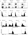
Activated CD4+ T cells accumulate in an age-dependent fashion in γc-deficient mice and exhibit increased BrdU uptake. (A) The number of splenic CD4+ T cells in γc-deficient (closed diamonds) and wildtype (open squares) mice was calculated as the product of the number of splenocytes and the percent of CD4+ T cells, based on staining with antiCD4 Cy-Chrome and analysis on a FACSort® (Becton Dickinson) using Lysis II software. Shown are mean ± SEM (n = 4–9 at each timepoint). (B) Size of CD4+ T cells. Splenocytes from 6-wk-old mice were stained with anti-CD4 Cy-Chrome and PI, and cell size of viable cells was assessed by forward light scatter (FSC); data are shown on a linear scale. (C) Activation markers expressed on CD4+ T cells. Splenocytes were stained with anti-CD4 Cy-Chrome and either PE conjugated anti-CD62L (L-selectin), anti-CD69, or anti-CD25. Data were gated on CD4+ cells and displayed on a log scale. The data shown are representative of six experiments. (D) BrdU uptake of thymocytes and splenic CD4+ T cells. Mice were injected twice with BrdU (see Material and Methods). 16 h after the second injection, purified splenic CD4+ T cells and thymocytes were stained with anti-BrdU FITC according to the Becton Dickinson protocol and analyzed on a FACSort®. Histograms show BrdU staining (log scale); the numbers above the gate represent the mean percentage of BrdU-positive cells from four experiments. When mice were injected with PBS instead of BrdU, less than 0.1% of thymocytes and splenic CD4+ T cells were stained with anti-BrdU FITC (data not shown).
Figure 2
Figure 2
Augmented cell death and diminished Bcl-2 expression in γc-deficient CD4+ T cells. (A) Spontaneous cell death in vitro. Purified splenic CD4+ T cells from 6-wk-old mice were cultured in RPMI medium containing 10% charcoal-treated fetal bovine serum (Cocalico Biological, Inc.) for 8–48 h, harvested, and cell survival determined by staining with 5 μg/ml of PI, followed by analysis using a FACSort®. Data for each mouse were analyzed in duplicate. Shown are the means ± SD of three mice in each group. At time 0, approximately 98% of cells from wild-type and 93% of cells from γc-deficient mice were PI negative. (B) Annexin V binding to CD4+ T cells. Splenocytes from 6-wk-old mice were stained with anti-CD4 Cy-Chrome and then with annexin V FITC. Annexin V binding (log scale) is shown for CD4+ PI cells. (C) Bcl-2 expression in splenic CD4+ T cells was analyzed approximately as previously described (14). The histograms show profiles for staining with anti-Bcl-2 (closed histograms) and control IgG (open histograms) in wild-type and γc-deficient mice (log scale).
Figure 3
Figure 3
Activation of splenic CD4+ T cells in γc-deficient mice is TCR dependent. γc-deficient mice were back-crossed to BALB/c mice (H-2d/d, Jackson Laboratory) for three generations. Since the γc gene is located on the X chromosome, DO10 TCR transgenic male mice (H-2d/d) were mated with BALB/c γc +/− heterozygous female mice (H-2d/d). These matings yielded four genotypes (assessed by PCR of tail DNA) of male mice (DO10γc +/Y, DO10+γc +/Y, DO10γc −/Y, and DO10+γc −/Y) (A). In (A), also shown are the total number and CD4/CD8 staining of splenocytes from 5-wk-old mice. As previously reported (32), DO10+ male mice expressing wild-type γc (DO10+γc +/Y mice) exhibit an increased CD4/CD8 ratio as compared with DO10γc +/Y mice (first two panels), but consistent with the increased CD4/CD8 ratios previously observed in γc-deficient mice (6, 7), DO10+γc −/Y mice exhibited an even greater CD4/CD8 ratio (fourth panel). Results are representative of three separate experiments. (B) Histograms of KJ1-26 mAb (anti-idiotype TCR) staining (gated on CD4+ T cells). (C) Splenocytes were stained by three color flow cytometric analysis for expression of KJ1-26, CD62L, and CD4. Data were gated on KJ1-26 CD4+ (endogenous TCR) (top histogram) or KJ1-26+CD4+ (transgenic TCR) (bottom histogram) T cells. The histograms show profiles for CD62L expression (log scale); the percent of CD62Lhigh cells is indicated.

References

    1. Noguchi M, Yi H, Rosenblatt HM, Filipovich AH, Adelstein S, Modi WS, McBride OW, Leonard WJ. Interleukin-2 receptor γ chain mutation results in X-linked severe combined immunodeficiency in human. Cell. 1993;73:147–157. - PubMed
    1. Leonard WJ, Noguchi M, Russell RM, McBride OW. The molecular basis of X-linked severe combined immunodeficiency: the role of the interleukin-2 receptor γ chain as a common γ chain, γc . Immunol Rev. 1994;138:61–86. - PubMed
    1. Sugamura K, Asao H, Kondo M, Tanaka N, Ishii N, Nakamura M, Takesha T. The common γ-chain for multiple cytokine receptors. Adv Immunol. 1995;59:225–277. - PubMed
    1. Leonard WJ. The molecular basis of X-linked severe combined immunodeficiency: defective cytokine receptor signaling. Annu Rev Med. 1996;47:229–239. - PubMed
    1. Leonard WJ, Shores EW, Love PE. Role of the common cytokine receptor γ chain in cytokine signaling and lymphoid development. Immunol Rev. 1995;148:97–114. - PubMed

Publication types