. 1997 Jan 20;185(2):207-18.
doi: 10.1084/jem.185.2.207.

Absence of respiratory burst in X-linked chronic granulomatous disease mice leads to abnormalities in both host defense and inflammatory response to Aspergillus fumigatus

Affiliations

Absence of respiratory burst in X-linked chronic granulomatous disease mice leads to abnormalities in both host defense and inflammatory response to Aspergillus fumigatus

D E Morgenstern et al. J Exp Med. .

Abstract

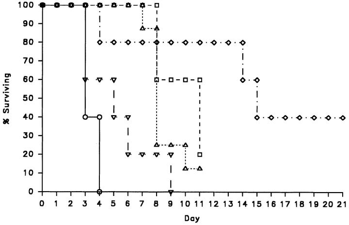
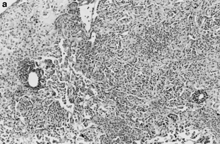
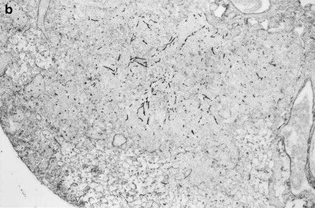
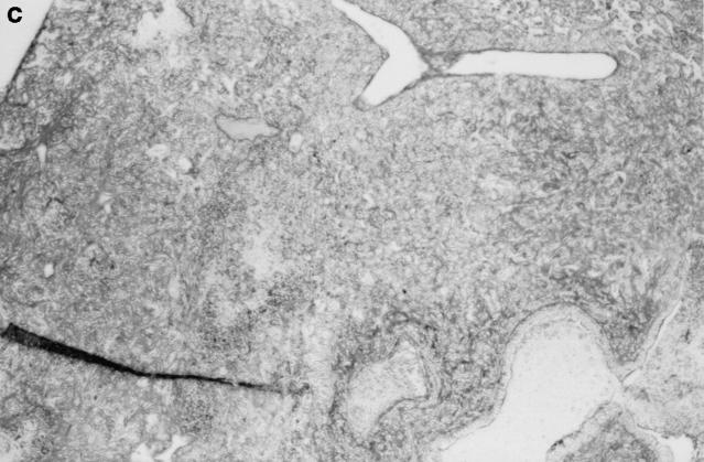
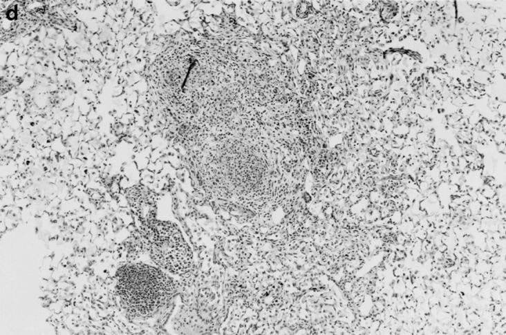
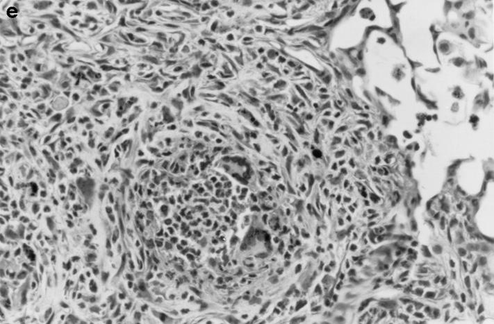
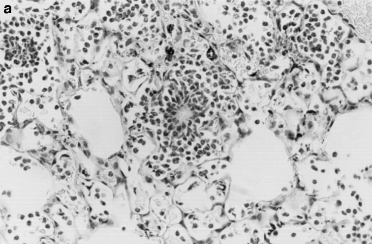
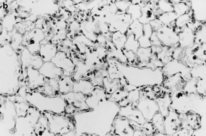
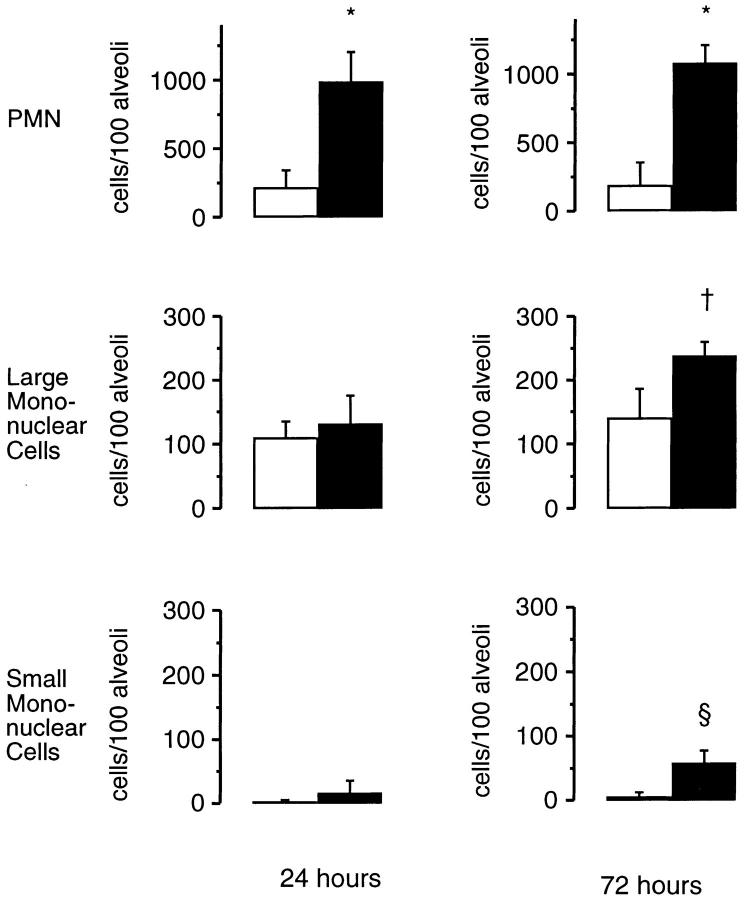
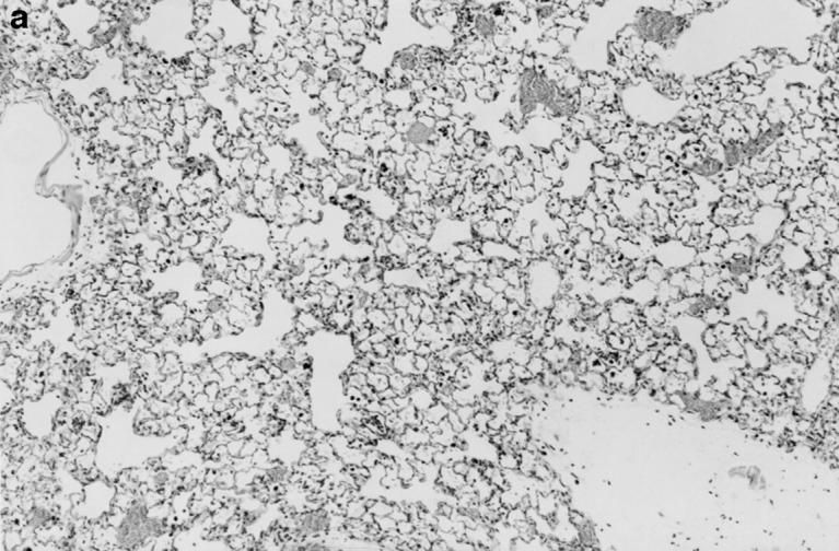
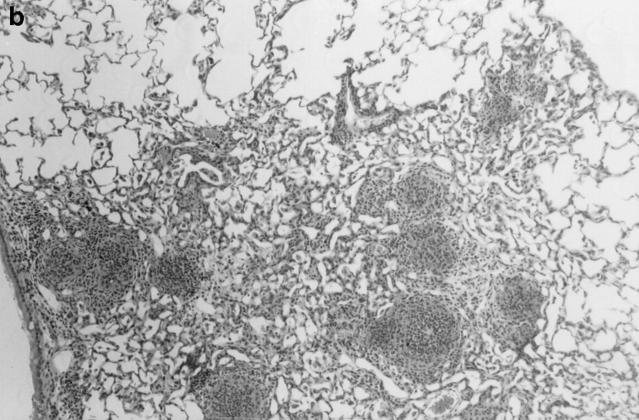
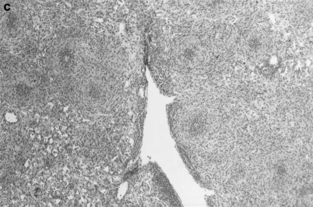
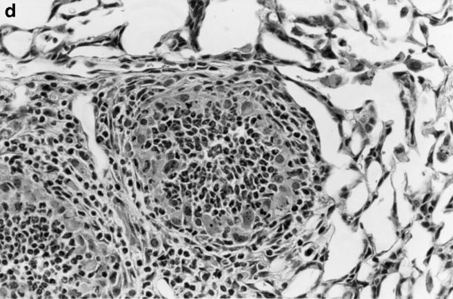
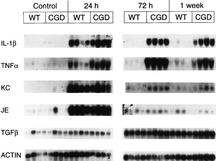
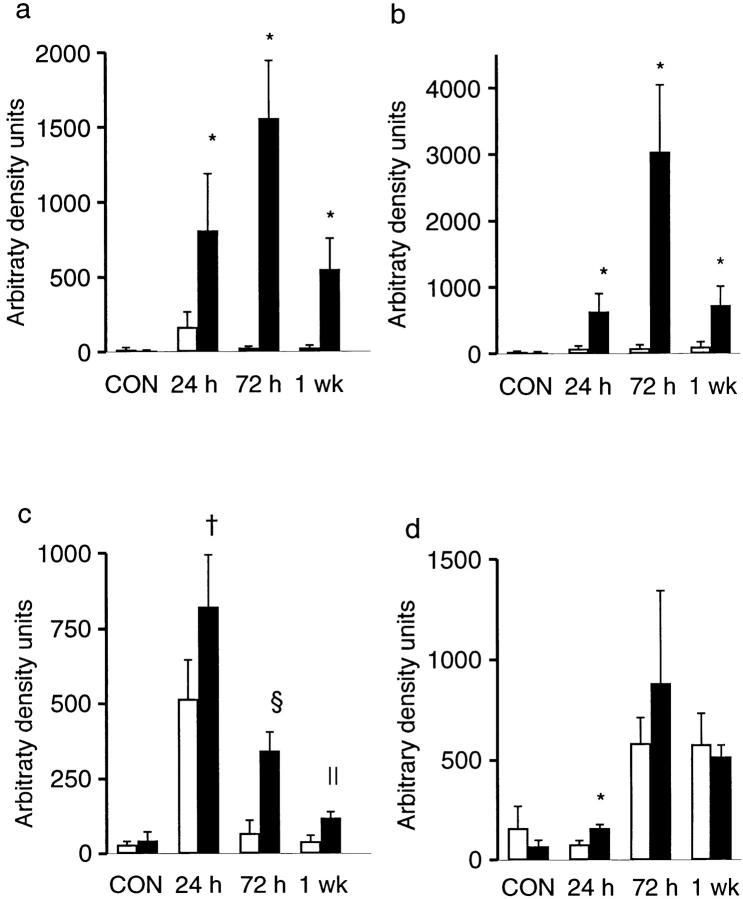
Mice with X-linked chronic granulomatous disease (CGD) generated by targeted disruption of the gp91phox subunit of the NADPH-oxidase complex (X-CGD mice) were examined for their response to respiratory challenge with Aspergillus fumigatus. This opportunistic fungal pathogen causes infection in CGD patients due to the deficient generation of neutrophil respiratory burst oxidants important for damaging A. fumigatus hyphae. Alveolar macrophages from X-CGD mice were found to kill A. fumigatus conidia in vitro as effectively as alveolar macrophages from wild-type mice. Pulmonary disease in X-CGD mice was observed after administration of doses ranging from 10(5) to 48 spores, none of which produced disease in wild-type mice. Higher doses produced a rapidly fatal bronchopneumonia in X-CGD mice, whereas progression of disease was slower at lower doses, with development of chronic inflammatory lesions. Marked differences were also observed in the response of X-CGD mice to the administration of sterilized Aspergillus hyphae into the lung. Within 24 hours of administration, X-CGD mice had significantly higher numbers of alveolar neutrophils and increased expression of the proinflammatory cytokines IL-1 beta and TNF-alpha relative to the responses seen in wild-type mice. By one week after administration, pulmonary inflammation was resolving in wild-type mice, whereas X-CGD mice developed chronic granulomatous lesions that persisted for at least six weeks. This is the first experimental evidence that chronic inflammation in CGD does not always result from persistent infection, and suggests that the clinical manifestations of this disorder reflect both impaired microbial killing as well as other abnormalities in the inflammatory response in the absence of a respiratory burst.


Figures

Figure 1
Figure 1
Survival curve of X-CGD mice receiving from 1 × 10⁵ to 48 A. fumigatus conidia. Mice were examined daily and the percent of mice surviving in each treatment group was plotted versus time from intratracheal administration of the indicated dose of conidia (day 0). ○, 1 × 10⁵ conidia/mouse (n = 5); ▿, 7 × 10³ conidia/mouse (n = 5); ▵, 1 × 10³ conidia/mouse (n = 8); □, 540 conidia/mouse (n = 5); ⋄, 48 conidia/mouse (n = 5).
Figure 2
Figure 2
Pulmonary disease in X-CGD mice after administration of A. fumigatus conidia. (a) Lung tissue obtained 11 d after challenge with 540 A. fumigatus conidia and stained with hematoxylin and eosin. Note areas of neutrophil inflammation surrounded by mononuclear cells (original magnification of 100). (b) Grocott methamine stain of lung from X-CGD mouse with neutrophil inflammation; note branching filamentous hyphae (original magnification of 100). (c) Grocott methamine silver staining of the same tissue shown in a with no visible hyphae (original magnification of 100). (d) Lung tissue obtained 21 d after challenge with 48 conidia and stained with hematoxylin and eosin. Low power view of a representative inflammatory lesion showing nodular region of inflammation and destruction of underlying lung tissue (original magnification of 100). (e) Higher power view of chronic inflammatory lesion consisting of neutrophils, epithelioid macrophages, and giant cells (original magnification of 400).
Figure 2
Figure 2
Pulmonary disease in X-CGD mice after administration of A. fumigatus conidia. (a) Lung tissue obtained 11 d after challenge with 540 A. fumigatus conidia and stained with hematoxylin and eosin. Note areas of neutrophil inflammation surrounded by mononuclear cells (original magnification of 100). (b) Grocott methamine stain of lung from X-CGD mouse with neutrophil inflammation; note branching filamentous hyphae (original magnification of 100). (c) Grocott methamine silver staining of the same tissue shown in a with no visible hyphae (original magnification of 100). (d) Lung tissue obtained 21 d after challenge with 48 conidia and stained with hematoxylin and eosin. Low power view of a representative inflammatory lesion showing nodular region of inflammation and destruction of underlying lung tissue (original magnification of 100). (e) Higher power view of chronic inflammatory lesion consisting of neutrophils, epithelioid macrophages, and giant cells (original magnification of 400).
Figure 3
Figure 3
Lung inflammation in wild-type mice and X-CGD mice 24 h after challenge with sterile A. fumigatus hyphae. Lung tissue was obtained 24 h after intratracheal administration of 5 μg of sterile hyphae and staining of representative sections with hematoxylin and eosin. (a) X-CGD lung with pneumonia and focal centers of neutrophil accumulation (original magnification of 400). (b) Lung from a wild-type mouse with comparatively mild, diffuse neutrophil infiltrate. (original magnification of 400).
Figure 3
Figure 3
Lung inflammation in wild-type mice and X-CGD mice 24 h after challenge with sterile A. fumigatus hyphae. Lung tissue was obtained 24 h after intratracheal administration of 5 μg of sterile hyphae and staining of representative sections with hematoxylin and eosin. (a) X-CGD lung with pneumonia and focal centers of neutrophil accumulation (original magnification of 400). (b) Lung from a wild-type mouse with comparatively mild, diffuse neutrophil infiltrate. (original magnification of 400).
Figure 4
Figure 4
Morphometric analysis of lung sections 24 and 72 h after challenge with sterile A. fumigatus hyphae. Lung tissue was obtained either 24 or 72 h after intratracheal administration of 5 μg of sterile hyphae. 100 randomly selected alveoli containing carbon were scored for numbers of neutrophils, large mononuclear cells, and small mononuclear cells. Data are expressed as mean ± SD. Open bars, wild-type mice; filled bars, X-CGD mice. *, significantly different from wild type (P <0.0005, t test); , significantly different from wild type (P <0.05, t test); §, significantly different from wild type (P <0.005, t test).
Figure 5
Figure 5
Inflammatory response in wild-type and X-CGD mice lungs 21 d after challenge with sterile A. fumigatus hyphae. Lung tissue was obtained 21 d after intratracheal administration of 5 μg of sterile hyphae and stained with hematoxylin and eosin. (a) Representative section of lung from a wild-type mouse. Inflammation has largely resolved, although some residual edema and peribronchiolar inflammation remain (original magnification of 100). (b) Lung from X-CGD mouse with scattered nodular inflammatory foci (original magnification of 100). (c) Lung from X-CGD mouse with almost complete consolidation and destruction of lung parenchyma (original magnification of 100). (d) Higher power of area in 6 b, showing central neutrophil microabscess and surrounding epithelioid cells (original magnification of 400).
Figure 5
Figure 5
Inflammatory response in wild-type and X-CGD mice lungs 21 d after challenge with sterile A. fumigatus hyphae. Lung tissue was obtained 21 d after intratracheal administration of 5 μg of sterile hyphae and stained with hematoxylin and eosin. (a) Representative section of lung from a wild-type mouse. Inflammation has largely resolved, although some residual edema and peribronchiolar inflammation remain (original magnification of 100). (b) Lung from X-CGD mouse with scattered nodular inflammatory foci (original magnification of 100). (c) Lung from X-CGD mouse with almost complete consolidation and destruction of lung parenchyma (original magnification of 100). (d) Higher power of area in 6 b, showing central neutrophil microabscess and surrounding epithelioid cells (original magnification of 400).
Figure 6
Figure 6
Northern analysis of total lung RNA in wild-type and X-CGD mice receiving sterile A. fumigatus hyphae. Lung tissue was obtained from wild-type (WT) and X-CGD (CGD) mice which were untreated (Control) or given 5 μg of sterile hyphal fragments by intratracheal administration. Mice given hyphal fragments were killed either at 24 h, 72 h, or 1 wk after instillation and the lungs removed and total RNA isolated as described in Materials and Methods. Blots were sequentially probed with the indicated cDNAs.
Figure 7
Figure 7
Densitometric analysis of cytokine expression in wild-type and X-CGD mice receiving A. fumigatus hyphae. Autoradiographs were scanned and optical density calculated after background subtraction. Optical density was expressed as arbitrary density units and then normalized to β-actin expression. Data expressed as mean ± SD. (a) IL1β; (b) TNF-α; (c) KC; (d) TGFβ1. Open bars, wild-type mice; filled bars, X-CGD mice. *, significantly different from wild type (P <0.05, Mann-Whitney U test); , significantly different from wild type (P <0.05, t test); §, significantly different from wild type (P <0.0005, t test); , significantly different from wild type (P <0.005, t test).

Similar articles

Cited by

References

    1. Dinauer MC. The respiratory burst oxidase and the molecular genetics of chronic granulomatous disease. Crit Rev Clin Lab Sci. 1993;30:329–369. - PubMed
    1. Carson MJ, Chadwick DL, Brubaker CA, Cleland RS, Landing BH. Thirteen boys with progressive septic granulomatosis. Pediatrics. 1965;35:405–412. - PubMed
    1. Johnston RB, Jr, McMurry J. Chronic familial granulomatosis. Am J Dis Child. 1967;114:370–378. - PubMed
    1. Gallin JI, Buescher ES, Seligmann BE, Nath J, Gaither T, Katz P. Recent advances in chronic granulomatous disease. Ann Intern Med. 1983;99:657–674. - PubMed
    1. Moskaluk CA, Pogrebniak HW, Pass HI, Gallin JI, Travis WD. Surgical pathology of the lung in chronic granulomatous disease. Am J Clin Pathol. 1994;102:684–691. - PubMed

Publication types

MeSH terms