. 1997 Jan 20;185(2):273-80.
doi: 10.1084/jem.185.2.273.

Estrogen protects lenses against cataract induced by transforming growth factor-beta (TGFbeta)

Affiliations

Estrogen protects lenses against cataract induced by transforming growth factor-beta (TGFbeta)

A M Hales et al. J Exp Med. .

Abstract

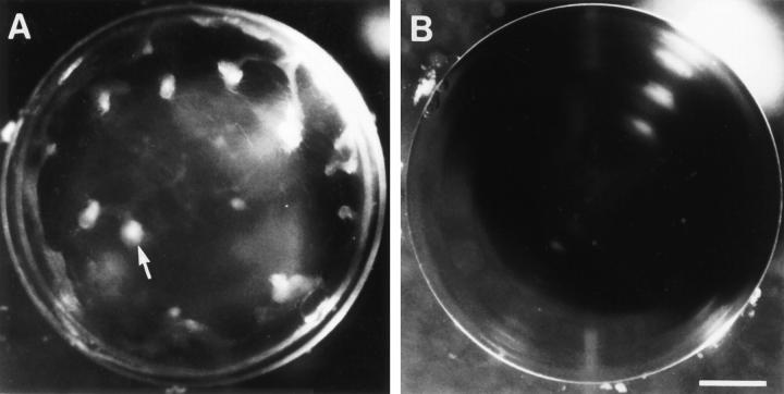
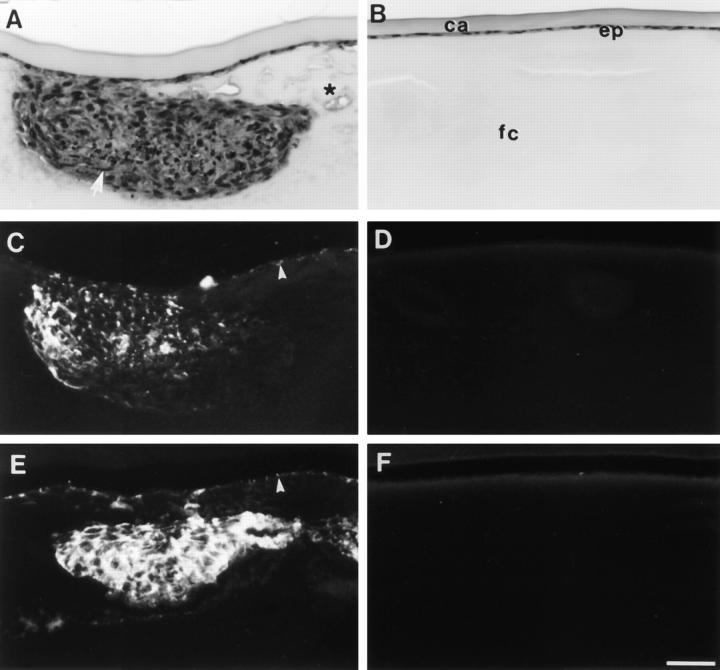
Cataract, already a major cause of visual impairment and blindness, is likely to become an increasing problem as the world population ages. In a previous study, we showed that transforming growth factor-beta (TGFP) induces rat lenses in culture to develop opacities and other changes that have many features of human subcapsular cataracts. Here we show that estrogen protects against cataract. Lenses from female rats are more resistant to TGFbeta-induced cataract than those from males. Furthermore, lenses from ovariectomized females show increased sensitivity to the damaging effects of TGFbeta and estrogen replacement in vivo, or exposure to estrogen in vitro, restores resistance. Sex-dependent and estrogen-related differences in susceptibility to cataract formation, consistent with a protective role for estrogen, have been noted in some epidemiological studies. The present study in the rat indicates that estrogen provides protection against cataract by countering the damaging effects of TGFbeP. It also adds to an increasing body of evidence that hormone replacement therapy protects postmenopausal women against various diseases.


Figures

Figure 1
Figure 1
Comparison of response of lenses from male and female rats to TGFβ. Lenses were cultured with 0.15 ng/ml TGFβ2 and photographed after 7 d. Lenses from male rats (A) developed distinct anterior opacities (arrow). Lenses from females remained transparent (B). Some flaring of the light source is evident in the upper righthand quadrant of each lens. Bar, 400 μm.
Figure 2
Figure 2
Influence of ovarian hormones on induction of cataract by TGFβ. Ovariectomized rats received vehicle alone (A), estrogen replacement (B), or progesterone replacement (C). Lenses were cultured with 0.15 ng/ml TGFβ2 and photographed after 7 d. Lenses from rats that received vehicle alone or progesterone developed distinct opacities (A and C), whereas lenses from rats that received estrogen remained transparent (B). Bar, 400 μm.
Figure 3
Figure 3
Histology and immunolocalization of cataract markers. Lenses from ovariectomized rats that received vehicle alone (A, C, and E) or estrogen replacement (B, D, and F) were cultured with 0.15 ng/ml TGFβ2 and fixed at the end of a 7 d culture period. Serial sections were stained with haematoxylin and eosin (A and B), or used for localization of α-smooth muscle actin (C and D) and type I collagen (E and F). Lenses from rats that received vehicle alone developed large anterior subcapsular plaques (A), which contained spindle-shaped cells (arrow) and many condensed nuclei. In addition, the fiber cells around the plaques appeared swollen and vacuoles were commonly present (asterisk). α-Smooth muscle actin (C) and type I collagen (E) were localized within the TGFβinduced subcapsular plaques and also in some of the cells that remained attached to the capsule (arrowheads). Lenses from rats that received estrogen retained normal lens morphology (B) with a monolayer of epithelial cells (ep) adjacent to the lens capsule (ca) and overlying the fibre cells (fc). These lenses showed no reactivity for either α-smooth muscle actin (D) or type I collagen (F). Bar, 40 μm.
Figure 4
Figure 4
Posterior migration of cells associated with induction of cataract by TGFβ. Lenses from ovariectomized rats that received vehicle alone (A and C) or estrogen replacement (B and D) were cultured with 0.15 ng/ml TGFβ2 and fixed at the end of the 7 d culture period. Serial sections were stained for routine histology with haematoxylin and eosin (A and B) or used for immunofluorescent localization of type I collagen (C and D). The lens equator is positioned at the top of each micrograph. In lenses from rats that received vehicle alone (A), nucleated cells were observed migrating along the lens capsule toward the posterior pole (A, arrowheads). Strong reactivity for type I collagen was associated with these posteriorly migrating cells (C, arrowheads). In contrast, lenses from rats that received estrogen replacement maintained a normal lens morphology with no abnormal migration of cells below the lens equator (B). No reactivity for type I collagen was observed (D). Bar, 40 μm.

Similar articles

Cited by

References

    1. Harding, J.J. 1991. Cataract: Biochemistry, Epidemiology and Pharmacology. Chapman and Hall, London. 83–124.
    1. Schwab IR, Armstrong MA, Frienman GD, Wong IG, Carpantieri AC, Dawson CR. Caratact extraction. Risk factors in a health maintenance organization population under 60 years of age. Arch Ophthalmol. 1988;106:1062–1065. - PubMed
    1. Klein BEK, Klein R, Linton KLP. Prevalence of age-related lens opacities in a population. The Beaver Dam eye study. Ophthalmology. 1992;99:546–552. - PubMed
    1. Thompson JR, Deane JS, Hall AB, Rosenthal AR. Oestrogen and lens opacities in the Melton eye study. Invest Ophthalmol Visual Sci. 1996;37:S585.
    1. Klein BEK. Lens opacities in women in Beaver Dam, Wisconsin: is there evidence of an effect of sex hormones? . Trans Am Ophthalmol Soc. 1993;91:517–544. - PMC - PubMed

Publication types