. 1997 Jan 20;185(2):317-28.
doi: 10.1084/jem.185.2.317.

Maturation stages of mouse dendritic cells in growth factor-dependent long-term cultures

Affiliations

Maturation stages of mouse dendritic cells in growth factor-dependent long-term cultures

C Winzler et al. J Exp Med. .

Abstract

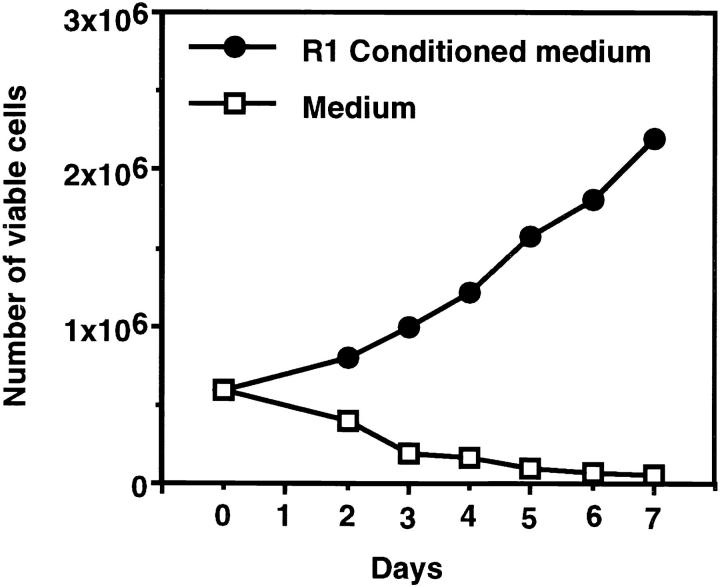
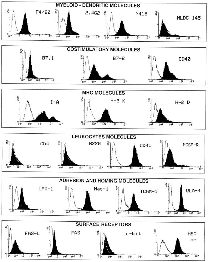
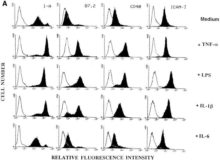
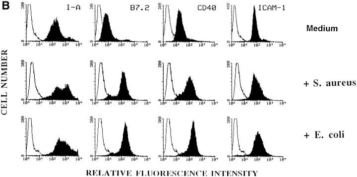
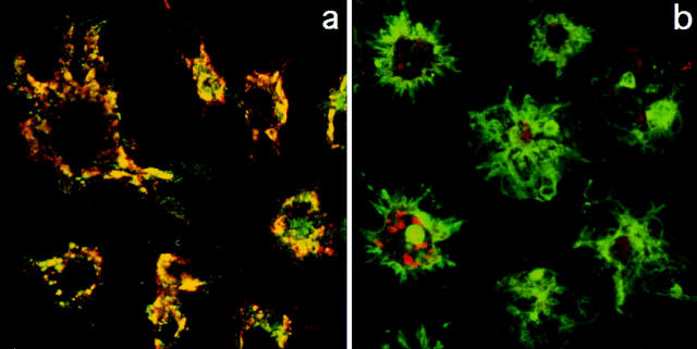
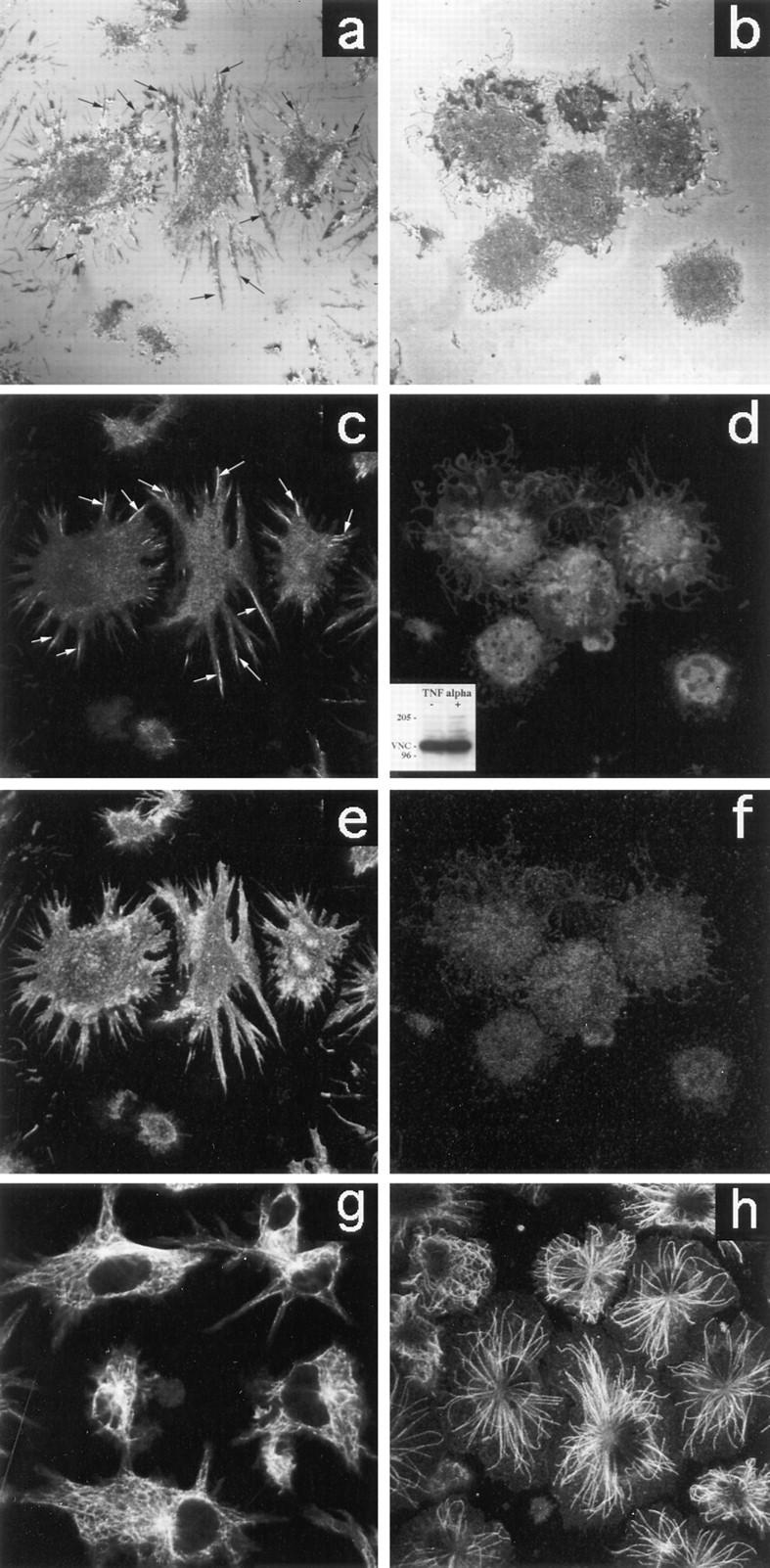
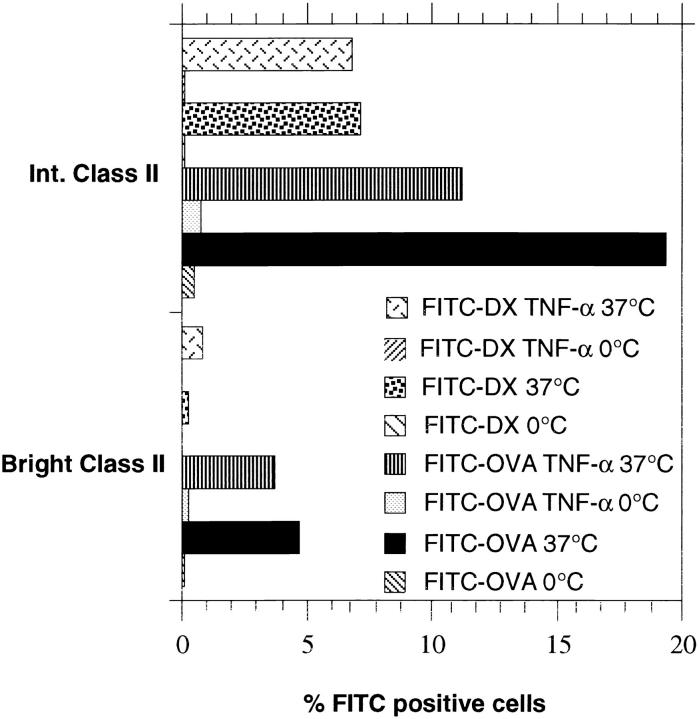
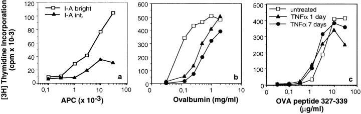
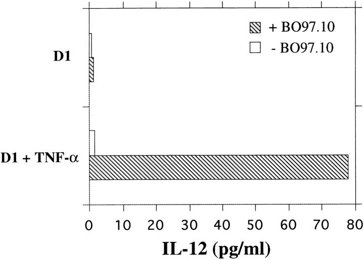
The signals controlling the checkpoints of dendritic cells (DC) maturation and the correlation between phenotypical and functional maturational stages were investigated in a defined model system of growth factor-dependent immature mouse DC. Three sequential stages of DC maturation (immature, mature, and apoptotic) were defined and characterized. Immature DC (stage 1) had low expression of costimulatory molecules, highly organized cytoskeleton, focal adhesion plaques, and slow motility; accordingly, they were very efficient in antigen uptake and processing of soluble proteins. Further, at this stage most of major histocompatibility complex class II molecules were within cytoplasmic compartments consistent with a poor allostimulatory capacity. Bacteria or cytokines were very efficient in inducing progression from stage 1 towards stage 2 (mature). Morphological changes were observed by confocal analysis including depolymerization of F-actin and loss of vinculin containing adhesive structures which correlates with acquisition of high motility. Antigen uptake and presentation of native protein antigen was reduced. In contrast, presentation of immunogenic peptides and allostimulatory activity became very efficient and secretion of IL-12 p75 was detectable after antigen presentation. This functional DC maturation ended by apoptotic cell death, and no reversion to the immature phenotype was observed.


Figures

Figure 1
Figure 1
Growth curve of  D1 cells in the presence of conditioned medium (R1) or growth factors deprived (medium alone).
Figure 2
Figure 2
Surface markers of D1 bulk culture by FACS® analysis. Filled histograms are showing binding of specific antibodies, whereas isotype matched control antibodies or secondary reagents are represented by open histograms. Cell surface phenotype was assessed with antibodies as indicated in Materials and Methods. Before all labeling experiments, FcR blocking was performed by incubating cells with 5% normal mouse serum or anti FcR (2.4G2) mAb.
Figure 3
Figure 3
Dot plot double-color FACS® analysis of MHC class II and B7.2 molecules of D1 bulk culture.
Figure 4
Figure 4
Phenotypical maturation of D1 cells. Surface markers by FACS® analysis after cytokine or living bacteria treatment. MHC class II (I-A), B7.2, CD40, and ICAM-1 molecules of D1 bulk culture growing in R1 medium or in medium supplemented with TNFα, LPS, IL-1β, or IL-6 (A) or after treatment with living E. coli or S. aureus (B).
Figure 4
Figure 4
Phenotypical maturation of D1 cells. Surface markers by FACS® analysis after cytokine or living bacteria treatment. MHC class II (I-A), B7.2, CD40, and ICAM-1 molecules of D1 bulk culture growing in R1 medium or in medium supplemented with TNFα, LPS, IL-1β, or IL-6 (A) or after treatment with living E. coli or S. aureus (B).
Figure 5
Figure 5
Redistribution of MHC class II molecules during maturation. Double-color confocal laser scanning microscopy analysis of class II–containing compartments in immature (a) and mature (b) D1 cells. Class II– positive (green) and H2-Mβ-positive (red) vesicles are mostly colocalized (yellow) in untreated D1 cells (a). Treatment of the cells with TNFα induces the redistribution of class II molecules from the cytoplasm to the cell surface (b) and no colocalization between class II (green) and H2Mβ (red) containing vesicles can be observed.
Figure 6
Figure 6
Cytoskeleton modifications in D1 cells after TNFα treatment. Immature (a) and mature (b) D1 cells morphology was analyzed by confocal microscopy using reflection interference contrast which gives the best visualization of the cell interface zone when attached to a glass support (37). Confocal laser scanning microscopy of D1 cells stained with anti-vinculin (c and d), phalloidin (e and f), and anti-tubulin (g and h) was also performed. Immature D1 cells (a, c, e, and g) appear to be adherent (a) and characterized by: vinculin containing adhesive structures (a and c, arrows), subcortical actin aggregates (e), and highly organized tubulin (g). D1 treatment with TNFα (b, d, f, and h) clearly induces morphological modification (b). Mature D1 cells lose adherence (b), vinculin (d), and subcortical actin organization (   f   ). No differences in the amount of vinculin protein are detectable by Western blot analysis (d, inset). Microtubules are only partially affected by TNFα treatment (h).
Figure 7
Figure 7
Antigen uptake (FITC-OVA and FITC-DX) by D1 bulk population in the presence or absence of TNFα was analyzed by doublecolor FACS® analysis. The D1 cells that better internalize soluble antigens at 37°C expressed comparatively low levels of MHC class II molecules, whereas class IIbright were less efficient in both FITC-OVA and FITC-DX uptake. Incubation of the cells with TNFα reduced the protein uptake of the immature D1 subpopulation and had no effects on the mature D1 subpopulation.
Figure 8
Figure 8
Allostimulatory and processing capacities of D1 cells. (a) Mixed lymphocyte reaction (MLR) by sorted I-Abright and I-Aint D1 cells shows that the mature I-Abright cells are the ones that have the highest allostimulatory capacity. Presentation of the OVA protein is downregulated by TNFα treatment. Antigenspecific presentation of exogenous OVA protein (b) or OVApeptide327-339 (c) by D1 cells treated or not with TNFα for 1 or 7 d: presentation of the OVA peptide is not affected by maturation but presentation of the OVA protein is downregulated by TNFα.
Figure 9
Figure 9
IL-12 p75 production by D1 cells pretreated or not with TNFα upon presentation of OVA protein to the specific hybridoma BO97.10 (striped bars or in the absence of the hybridoma open bars). IL-12 p75 was measured using an ELISA as described in Material and Methods. Il-12 p75 was quantified from two to three titration points using standard curves generated by purified recombinant mouse IL-12 and results expressed as cytokine concentration in pg/ml. Detection limit was 10 pg/ml.

References

    1. Steinman RM. The dendritic cell system and its role in immunogenicity. Annu Rev Immunol. 1991;9:271–296. - PubMed
    1. Schuler G, Steinman RM. Murine epidermal Langerhans cells mature into potent immunostimulatory dendritic cells in vitro. J Exp Med. 1985;161:526–546. - PMC - PubMed
    1. Inaba K, Witmer-Pack M, Inaba M, Hathcock KS, Sakuta H, Azuma M, Yagita H, Okumura K, Linsley PS, Ikehara S, et al. The tissue distribution of the B7-2 costimulator in mice: abundant expression on dendritic cells in situ and during maturation in vitro. J Exp Med. 1994;180:1849–1860. - PMC - PubMed
    1. Roake JA, Rao AS, Morris PJ, Larsen CP, Hankins DF, Austyn JM. Dendritic cell loss from nonlymphoid tissues after systemic administration of lipopolysaccharide, tumor necrosis factor, and interleukin 1. J Exp Med. 1995;181:2237–2247. - PMC - PubMed
    1. Witmer-Pack MD, Olivier W, Valinsky J, Schuler G, Steinman RM. Granulocyte/macrophage colonystimulating factor is essential for the viability and function of cultured murine epidermal Langerhans cells. J Exp Med. 1987;166:1484–1498. - PMC - PubMed

Publication types