. 1997 Feb 4;94(3):849-54.
doi: 10.1073/pnas.94.3.849.

Demonstration of mechanical connections between integrins, cytoskeletal filaments, and nucleoplasm that stabilize nuclear structure

Collaborators, Affiliations

Demonstration of mechanical connections between integrins, cytoskeletal filaments, and nucleoplasm that stabilize nuclear structure

A J Maniotis et al. Proc Natl Acad Sci U S A. .

Abstract

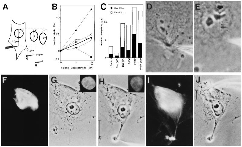
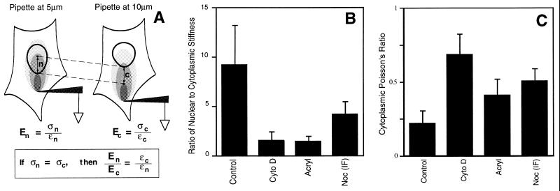
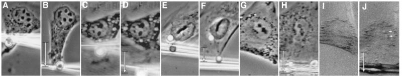
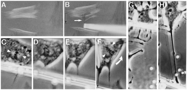
We report here that living cells and nuclei are hard-wired such that a mechanical tug on cell surface receptors can immediately change the organization of molecular assemblies in the cytoplasm and nucleus. When integrins were pulled by micromanipulating bound microbeads or micropipettes, cytoskeletal filaments reoriented, nuclei distorted, and nucleoli redistributed along the axis of the applied tension field. These effects were specific for integrins, independent of cortical membrane distortion, and were mediated by direct linkages between the cytoskeleton and nucleus. Actin microfilaments mediated force transfer to the nucleus at low strain; however, tearing of the actin gel resulted with greater distortion. In contrast, intermediate filaments effectively mediated force transfer to the nucleus under both conditions. These filament systems also acted as molecular guy wires to mechanically stiffen the nucleus and anchor it in place, whereas microtubules acted to hold open the intermediate filament lattice and to stabilize the nucleus against lateral compression. Molecular connections between integrins, cytoskeletal filaments, and nuclear scaffolds may therefore provide a discrete path for mechanical signal transfer through cells as well as a mechanism for producing integrated changes in cell and nuclear structure in response to changes in extracellular matrix adhesivity or mechanics.


Figures

Figure 3
Figure 3
Analysis of the molecular basis of stress transfer between the CSK and the nucleus. (A) Diagram of the method used to determine changes in nuclear strain and movement (see text for details). (B and C) Effects of CSK-modifying drugs on nuclear strain (B) and movement in the direction of pull (C); standard error was consistently less than 10% of the mean. •/Control, absence of drugs; ▪/Noc (MF), cells plated in 10 μg/ml Noc for 5 hr and harpooned in the pole of the cell containing only microfilaments; □/Noc (IF), the same Noc-treated cells that were harpooned in the opposite pole containing intermediate filaments; ⋄/Acryl, cells treated with 5 mM Acryl for 24 hr; ▴/CytoD, cells treated with 0.1 μg/ml CytoD for 2 hr; ▿/Noc+CytoD, cells in Noc for 4 hr and then in CytoD for 1 hr. (D) Control cell harpooned in the cytoplasm 2–4 μm from the nuclear border; arrow indicates a local tongue-like protrusion of the nuclear envelope. (E) Invagination of the nuclear envelope (large arrow) in response to harpooning the nucleoplasm. Four small arrows indicate the stressed nucleoplasmic thread stretching to the pipette tip. (F–J) Parallel immunofluorescence (F, I, Insets in G and H) and phase-contrast (G, H, and J) views of a cell that was plated in the presence of Noc, which induced formation of a vimentin-positive intermediate filament cap at one pole of the cell (F), although it did not prevent cell or nuclear spreading (G). (H) Harpooning and pulling the intermediate filament-free pole of the cell caused nuclear elongation in the direction of the pull; however, cytoplasmic tearing also resulted. (I) Rhodamine-phalloidin staining of cell depicted in H, showing tearing of the F actin-rich pole of the cell that lacked intermediate filaments. (J) Cell in H after pipette was removed and used to harpoon the cytoplasm on the opposite side of the same cell. Note extensive deformation of the nucleus and narrowing in the perpendicular direction. (Insets) Nuclei stained for DNA with 4′,6-diamidino-2-phenylindole (DAPI). (D, ×1500; E, ×2200; FJ, ×1000.)
Figure 4
Figure 4
Analysis of mechanical stiffness and connectivity (Poisson’s ratio) in the cytoplasm and nucleus. (A) Diagram of the method used to estimate the ratio of nuclear to cytoplasmic stiffness. (B) Ratio of nuclear to cytoplasmic stiffness in cells cultured in the absence (Control) or presence of CytoD, Acryl, or Noc (IF), using the conditions described for Fig. 3C. (C) Poisson’s ratio measured in the cytoplasm of control cells (Control) and in cells treated with CytoD, Acryl, or Noc (IF).
Figure 1
Figure 1
Phase-contrast (AH) and polarization optics (I and J) views of endothelial cells before (A, C, E, G, and I) and after (B, D, F, H, and J) mechanical stresses were applied to cell surface receptors. (A and B) Pulling on a single RGD-coated microbead (4.5-μm diameter) 15 min after binding to integrins using an uncoated glass micropipette; only 2 sec passed between A and B. (C and D) Similar displacement of a surface-bound AcLDL-coated microbead. (E and F) Mechanical displacement of RGD-coated beads bound to the surface of a cell permeabilized with 0.5% Triton X-100 prior to force application. (G and H) A spread cell before (G) and after (H) a fibronectin-coated micropipette was bound to cell surface integrins for 5 min and pulled laterally (downward in this view). (I and J) The same cell shown in G and H viewed under polarization optics; arrowheads indicate white birefringent spots in the region of nucleoli. The movement of the pipette is downward, and vertical black arrows indicate the extent of pipette displacement in all views. (×900.)
Figure 2
Figure 2
Polarization optics (A and B) and phase-contrast (C–H) views of interphase (A, B, G, and H) and mitotic (C–F) cells whose integrin receptors were mechanically stressed by using surface-bound glass micropipettes coated with fibronectin. (A) Cells exhibiting positively (white) and negatively (black) birefringent CSK bundles aligned horizontally and vertically, respectively. (B) White arrow indicates birefringent CSK bundles that originally appeared white in A and immediately changed black as they turned 90° and realigned vertically along the axis of the applied tension field when integrins were pulled. (C–F) Series of micrographs showing a living mitotic cell. Pulling on a fibronectin-coated micropipette bound to the cell surface resulted in counterclockwise rotation of the spindle axis. Partial separation of chromosomes also can be seen in D. Arrowheads point to the main axis of the spindle in C and F; curved arrow indicates the direction of spindle rotation. (G) An interphase cell treated with 0.1 μg/ml CytoD for 1 hr. (H) The same cell as in G after tension was applied to integrins by pulling on a surface-bound, matrix-coated micropipette (uncoated 4.5-μm diameter beads were included only for size reference). (A and B, ×400; C–G, ×870; and G and H, ×520.)

References

    1. Ingber D E. Curr Opin Cell Biol. 1991;3:841–848. - PubMed
    1. Ingber D E, Folkman J. J Cell Biol. 1989;109:317–330. - PMC - PubMed
    1. Singhvi R, Kumar A, Lopez G, Stephanopoulos G N, Wang D I C, Whitesides G M. Science. 1994;264:696–698. - PubMed
    1. Maniotis A J, Schliwa M. Cell. 1991;67:495–504. - PubMed
    1. Li M L, Ageller J, Farson D A, Hatier C, Hassell J, Bissell M J. Proc Natl Acad Sci USA. 1987;84:136–140. - PMC - PubMed

Publication types

MeSH terms

LinkOut - more resources