. 1997 Feb 4;94(3):855-60.
doi: 10.1073/pnas.94.3.855.

Activation of the cell death program by inhibition of proteasome function

Affiliations

Activation of the cell death program by inhibition of proteasome function

H C Drexler. Proc Natl Acad Sci U S A. .

Abstract

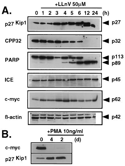
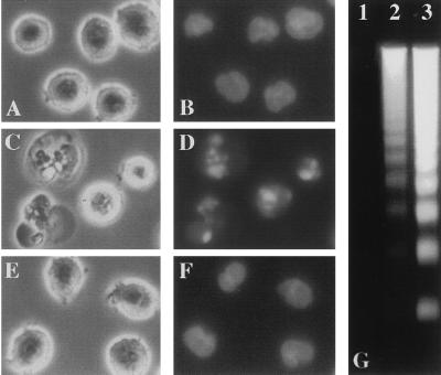
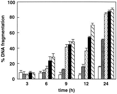
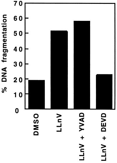
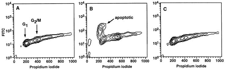
Activation of proteolytic enzymes, including cysteine proteases of the ced-3/ICE family, is a characteristic feature of the apoptotic program. In contrast, the role of the proteasome as the major nonlysosomal machinery to degrade or process proteins by ATP/ubiquitin-dependent proteolysis in this process is less clear. In human leukemic HL60 cells, inhibition of proteasome-mediated proteolysis by specific proteasomal inhibitors leads to the rapid induction of apoptosis as judged by morphological changes as well as by nuclear condensation and DNA fragmentation. HL60 apoptosis is due to activation of CPP32, a member of the ced-3/ICE family of cysteine proteases, and appears to occur independently from ICE activity. HL60 apoptosis is accompanied by an increase in the concentration of the cyclin-dependent kinase inhibitor p27Kip1. Labeling of the cells by the TUNEL technique demonstrates that HL60 cells undergoing apoptosis are primarily in the G1 phase of the cell cycle. Proteasomal activity therefore appears to be required in proliferating, but not in quiescent, HL60 cells for cell survival as well as normal progression through the cell cycle.


Figures

Figure 3
Figure 3
(A) Relative changes in protein levels after treatment with the proteasomal inhibitor LLnV. (B) Relative changes of c-Myc and p27Kip1 during PMA-induced differentiation. Lysates of cells incubated with 50 μM LLnV (A) or 10 ng/ml PMA (B) for the indicated times were subjected to SDS/PAGE followed by Western blotting.
Figure 1
Figure 1
(A–F) Phase-contrast photomicrographs (A, C, and E) of HL60 cells and photomicrographs of the same cells after nuclear staining with Hoechst 33324 (B, D, and F) viewed 6 h after the following treatments: 0.1% DMSO (A and B), 50 μM PSI (C and D), or 50 μM E64 (E and F). (G) Oligonucleosomal DNA fragmentation from PSI-treated cells: 0.1% DMSO (lane 1), 10 μM PSI (lane 2), or 50 μM PSI (lane 3). Soluble DNA fragments were extracted, separated on a 1.7% agarose gel, and stained with ethidium bromide. The DNA fragmentation patterns were similar in cells treated with TPCK, DCI, LLnL, or LLnV (not shown).
Figure 2
Figure 2
Dose–response relationship of LLnV-induced apoptosis. HL60 cells were maintained in medium containing 10% FBS, and treatments were as follows: 0.2% DMSO (empty bars); 10 μM LLnV (shaded bars); 20 μM LLnV (stippled bars); 50 μM LLnV (black bars); and 100 μM LLnV (hatched bars).
Figure 4
Figure 4
Effect of Ac-DEVD-cho or Ac-YVAD-cmk on LLnV-induced apoptosis in HL60 cells. The tetrapeptide protease inhibitors Ac-DEVD-cho and Ac-YVAD-cmk were loaded into HL60 cells by osmotic lysis of pinosomes as described. Cell death was then induced by incubation with LLnV for 6 h, and the extent of DNA fragmentation was determined. Data shown are a representative sample of four independent experiments with similar results.
Figure 5
Figure 5
LLnV-treated HL60 cells become apoptotic primarily in the G1 phase of the cell cycle. The cells were incubated for 5 h in RPMI 1640 medium containing 10% FBS supplemented with 0.1% DMSO (A), 50 μM LLnV (B), or 50 μM E64 (C). Apoptotic cells were labeled with fluorescein isothiocyanate (FITC) by the TUNEL technique, DNA was stained with propidium iodide, and the distribution of apoptotic cells within the cell cycle was analyzed by using a Becton Dickinson FACScan. Data shown are a representative example of three independent experiments with similar results.
Figure 6
Figure 6
Suggested model of proteasome function in the control of apoptotic cell death. See Discussion for further explanations.

References

    1. Steller H. Science. 1995;267:1445–1449. - PubMed
    1. Ellis R E, Yuan J, Horvitz H R. Annu Rev Cell Biol. 1991;7:663–698. - PubMed
    1. Yuan J Y, Shaham S, Ledoux S, Ellis H M, Horvitz H R. Cell. 1993;75:641–652. - PubMed
    1. Maki C G, Huibregtse J M, Howley P M. Cancer Res. 1996;56:2649–2654. - PubMed
    1. Sudakin V, Ganoth D, Dahan A, Heller H, Hershko J, Luca F C, Ruderman J V, Hershko A. Mol Biol Cell. 1995;6:185–197. - PMC - PubMed