. 1997 Feb 4;94(3):861-6.
doi: 10.1073/pnas.94.3.861.

Oncogenic H-ras stimulates tumor angiogenesis by two distinct pathways

Affiliations

Oncogenic H-ras stimulates tumor angiogenesis by two distinct pathways

J L Arbiser et al. Proc Natl Acad Sci U S A. .

Abstract

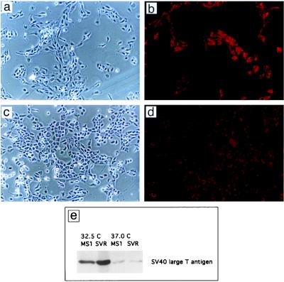
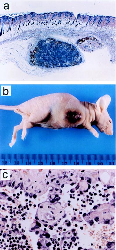
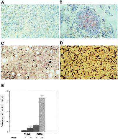
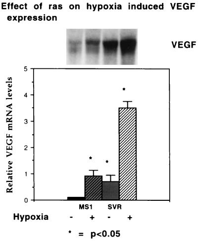
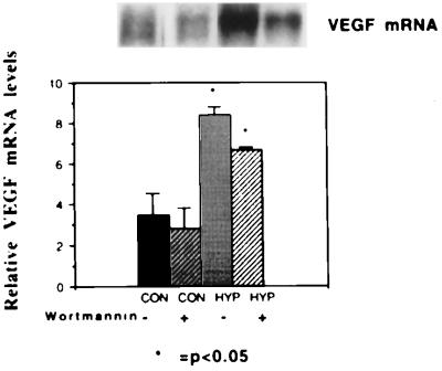
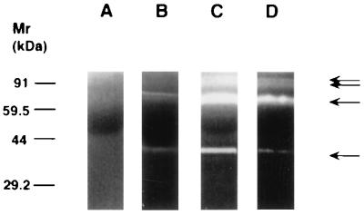
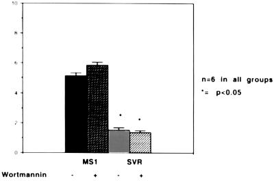
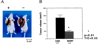
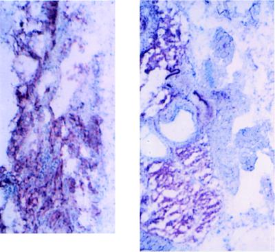
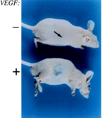
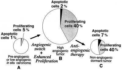
The switch from a quiescent tumor to an invasive tumor is accompanied by the acquisition of angiogenic properties. This phenotypic change likely requires a change in the balance of angiogenic stimulators and angiogenic inhibitors. The nature of the angiogenic switch is not known. Here, we show that introduction of activated H-ras into immortalized endothelial cells is capable of activating the angiogenic switch. Angiogenic switching is accompanied by up-regulation of vascular endothelial growth factor and matrix metalloproteinase (MMP) bioactivity and downregulation of tissue inhibitor of MMP. Furthermore, we show that inhibition of phosphatidylinositol-3-kinase leads to partial inhibition of tumor angiogenesis, thus demonstrating that activated H-ras activates tumor angiogenesis through two distinct pathways. Finally, we show evidence for two forms of tumor dormancy.


Figures

Figure 1
Figure 1
Morphology of endothelial cells containing SV40 large T antigen alone (MS1 cells) or both SV40 large T antigen and activated c-Ha-ras (SVR cells). (a) MS1 cells. (×37.) (b) MS1 cells. (×37; stained with diI-Ac-LDL.) (c) SVR cells. (×37.) (d) SVR cells. (×37; stained with diI-Ac-LDL.) (e) Western blot of MS1 and SVR cells for SV40 large T antigen at 32.5°C and 37°C.
Figure 2
Figure 2
Histology and appearance of tumors induced by MS1 cells and SVR cells in nude mice. (a) Subcutaneous MS1 tumor stained with hematoxylin and eosin. (×10.) (b) Tumor in nude mouse derived from SVR cells injected subcutaneously into 5- to 6-week-old male nude mice. (c) SVR tumors stained with hematoxylin and eosin. (×132.)
Figure 3
Figure 3
Bromodeoxyuridine and TUNEL staining of MS1 and SVR tumors. Tumors were fixed in formalin and UTP end-labeled with terminal transferase (TUNEL assay) (A and B) or stained with anti-bromodeoxyuridine antibody (C and D). (AD, ×40.) (A and C) MS1 tumors stained with terminal transferase (A) or bromodeoxyuridine (B). (B and D) SVR tumors labeled in the same manner as MS1. (E) Percentage of cells in MS1 and SVR tumors that are positive for uptake of bromodeoxyuridine or label with terminal transferase.
Figure 4
Figure 4
Effect of ras on hypoxic induction of VEGF. (Upper) mRNA from a representative Northern blot of MS1 and SVR cells hybridized with a murine VEGF probe. From left to right, the first lane represents MS1 cells under normoxic conditions, the second lane represents MS1 cells under hypoxic conditions, the third lane represents SVR cells under normoxic conditions, and the fourth lane represents SVR cells under hypoxic conditions. (Lower) Relative densitometric values normalized to β-actin mRNA. All experiments were performed in triplicate; the asterisk demonstrates significant differences, P < 0.05.
Figure 5
Figure 5
Effect of wortmannin on hypoxic induction of VEGF. (Upper) Northern blot. From left to right, the first lane represents mRNA from SVR cells under normoxic conditions in the absence of wortmannin. The second lane represents SVR cells under normoxic conditions in the presence of 1 μg/ml wortmannin. The third lane represents mRNA from SVR cells under hypoxic conditions in the absence of wortmannin, and the fourth lane represents mRNA from SVR cells under hypoxic conditions in the presence of wortmannin. (Lower) Normalized mRNA levels, labeled as in Fig. 4.
Figure 6
Figure 6
Effect of ras and wortmannin on MMP activity. Lane A represents control medium not conditioned by cells. The other lanes represent a gelatin substrate zymogram containing conditioned media from an equivalent number of MS1 (lane B), SVR (lane C), or SVR cells treated with wortmannin (lane D). A 30% decrease was seen in gelatinase activity after wortmannin treatment (n = 9).
Figure 7
Figure 7
Assay of TIMP bioactivity. Conditioned media from MS1 and SVR cells in the presence or absence of wortmannin were incubated in the presence of 14C-labeled collagen. 14C released into the medium was measured by scintillography. The y axis represents inhibitory units per 105 cells.
Figure 8
Figure 8
Effect of wortmannin on SVR growth in nude mice. (A) Effect of wortmannin on the growth of SVR tumors. The mouse on the right was treated with vehicle alone, while the mouse on the left was treated with wortmannin. (B) Effect of wortmannin on tumor volume. The asterisk indicates P < 0.05.
Figure 9
Figure 9
Hemangiomas and angiosarcomas are derived primarily from tumor tissue. (A) Frozen section of tumor derived from MS1 cells. (B) Frozen section of tumor derived from SVR cells. Both sections are stained with a primary biotinylated mouse monoclonal antibody directed against H-2Kb, coupled to avidin-alkaline phosphatase. (×67.5.)
Figure 10
Figure 10
Effect of overexpression of VEGF in immortalized endothelial cells. The upper mouse contains a hemangioma expressing puromycin resistance alone, while the lower mouse contains a hemangioma expressing VEGF.
Figure 11
Figure 11
Proposed model for tumor dormancy. (A) Dormant tumor with low proliferation and apoptotic indices. Introduction of ras activates the angiogenic switch and enhances the ability of the tumor to proliferate in vivo (B). Treatment of a proliferative tumor (B) with an angiogenesis inhibitor results in a dormant tumor (C), in which the proliferative index is elevated, as in B, but the apoptotic index is increased.

References

    1. Folkman J, Hanahan D. Cell. 1996;86:353–364. - PubMed
    1. Folkman J, Watson K, Ingber D, Hanahan D. Nature (London) 1989;339:58–61. - PubMed
    1. Kandel J, Bossy-Wetzel E, Radvanyi F, Klagsbrun M, Folkman J, Hanahan D. Cell. 1991;66:1095–1104. - PubMed
    1. Moses M A, Sudhalter J, Langer R. Science. 1990;248:1408–1410. - PubMed
    1. Liotta L A, Steeg P S, Stetler-Stevenson W G. Cell. 1991;64:327–336. - PubMed

Publication types

MeSH terms