. 1997 Feb 4;94(3):884-9.
doi: 10.1073/pnas.94.3.884.

Targeted disruption of the mouse alpha A-crystallin gene induces cataract and cytoplasmic inclusion bodies containing the small heat shock protein alpha B-crystallin

Affiliations

Targeted disruption of the mouse alpha A-crystallin gene induces cataract and cytoplasmic inclusion bodies containing the small heat shock protein alpha B-crystallin

J P Brady et al. Proc Natl Acad Sci U S A. .

Abstract

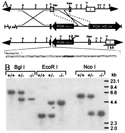
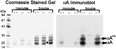
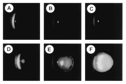
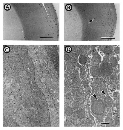
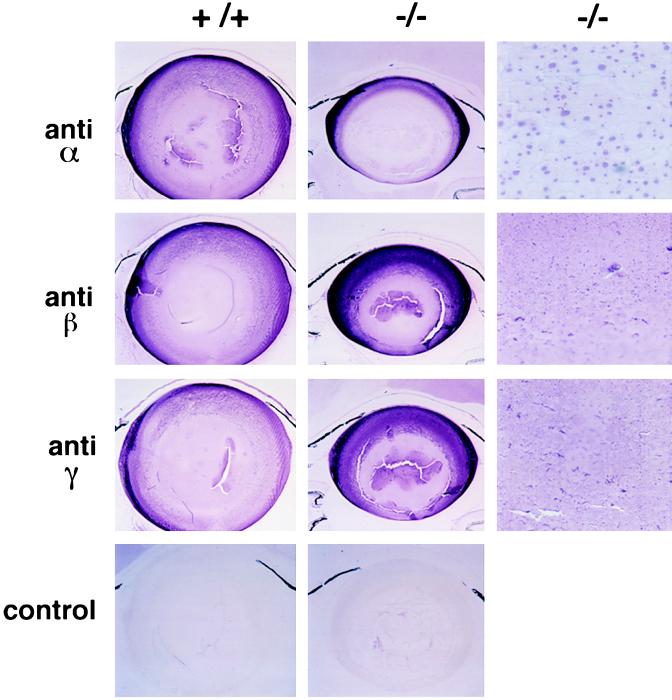
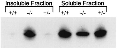
alpha A-crystallin (alpha A) and alpha B-crystallin (alpha B) are among the predominant proteins of the vertebrate eye lens. In vitro, the alpha-crystallins, which are isolated together as a high molecular mass aggregate, exhibit a number of properties, the most interesting of which is their ability to function as molecular chaperones for other proteins. Here we begin to examine the in vivo functions of alpha-crystallin by generating mice with a targeted disruption of the alpha A gene. Mice that are homozygous for the disrupted allele produce no detectable alpha A in their lenses, based on protein gel electrophoresis and immunoblot analysis. Initially, the alpha A-deficient lenses appear structurally normal, but they are smaller than the lenses of wild-type littermates. alpha A-/- lenses develop an opacification that starts in the nucleus and progresses to a general opacification with age. Light and transmission electron microscopy reveal the presence of dense inclusion bodies in the central lens fiber cells. The inclusions react strongly with antibodies to alpha B but not significantly with antibodies to beta- or gamma-crystallins. In addition, immunoblot analyses demonstrate that a significant portion of the alpha B in alpha A-/- lenses shifts into the insoluble fraction. These studies suggest that alpha A is essential for maintaining lens transparency, possibly by ensuring that alpha B or proteins closely associated with this small heat shock protein remain soluble.


Figures

Figure 1
Figure 1
Targeted disruption of the mouse αA gene. (A) Strategy used to disrupt the αA gene. (Top) Normal αA locus. (Middle) Targeting vector. (Bottom) Disrupted αA locus. The sequence of an oligonucleotide inserted between the 5′ αA sequences and PGK/neo containing multiple stop codons in all three reading frames and a polyadenylylation signal (enclosed in box) is shown beneath a diagram of the targeted allele. Depicted restriction sites include NcoI (N), BglI (B), EcoRI (E), AatII (A), XhoI, and XmnI. HSVtk, herpes simplex virus thymidine kinase. (B) Southern blot analysis of genomic DNA from wild-type (+/+), heterozygous (+/−), and homozygous (−/−) αA knockout mice. Liver DNA (15 μg) digested with BglI, EcoRI, and NcoI was hybridized with an XmnI/XhoI restriction fragment encompassing most of the first intron and the beginning of exon 2 (see A).
Figure 2
Figure 2
Gel electrophoresis and immunoblot analysis of lens proteins. Lens homogenates from αA+/+, αA−/−, and αA+/− mice were separated into soluble and insoluble fractions and subjected to SDS/PAGE. Each lane contains protein from the equivalent of 2.5 μg of lens wet weight. Coomassie staining of the gel (Left) shows the absence of a prominent αA band in the αA−/− lens (arrowhead). Immunoblot analysis of a duplicate gel with antiserum to recombinant human αA (Right) confirms the absence of αA in the αA−/− lenses. Bands corresponding to αA and αAinsert are identified. Weak cross-reactivity of this antiserum with a ≈30-kDa lens protein is observed in all three samples.
Figure 3
Figure 3
Lens opacity in an αA-deficient mouse. Eyes were dilated and examined by slit lamp. (AC) Wild-type mice ages 7, 10, and 20 weeks. (DF) αA−/− mice ages 7, 10, and 18 weeks. Normal reflection of the slit lamp from the surface of the cornea and lens are visible in all panels. Light scattering within the lens, as evinced by a white haze in the photograph, is significantly higher in the αA−/− mice than in wild-type mice at each age. Mild cataract is seen in the 7-week-old αA−/− lens and a dense opacity is evident in the 18-week-old lens.
Figure 4
Figure 4
Histological and transmission electron microscopic analysis of normal and αA-deficient lenses. Methacrylate-embedded eye sections from 11-week-old αA+/+ (A) and αA−/− (B) sibling mice were stained with hematoxylin and eosin. Weakly staining spherical bodies are observed in the nuclear and inner cortical areas of the αA−/− lens; the tip of the arrow in B marks the right boundary of the area containing the spherical bodies. (A and B, Bar = 100 μm.) Transmission electron micrographs of αA+/− (C) and αA−/− (D) lens inner cortex from 38-week-old sibling mice. Electron opaque spherical bodies (arrowheads) are evident in αA−/− lenses. (C and D, Bar = 1 μm.)
Figure 5
Figure 5
Immunohistochemistry of αA+/+ and αA−/− lenses with anti-crystallin antibodies. Sections of lenses cut parallel to the optic axis are shown with anteriors of the lenses toward the top of the figure. αA+/+ and αA−/− lenses were labeled with antibodies to bovine α-, β-, or γ-crystallin (anti-α, anti-β, or anti-γ) or with secondary antibody in the absence of a primary antibody (control). (×24 or ×300.)
Figure 6
Figure 6
Immunoblot analysis of soluble and insoluble lens proteins. Lens homogenates from αA+/+, αA−/−, and αA+/− mice were separated into soluble and insoluble fractions and subjected to immunoblot analysis with a polyclonal antibody to recombinant human αB. Soluble and insoluble proteins representing 2.5 μg of lens wet weight were analyzed.

References

    1. Benedek G B. Appl Opt. 1971;10:459–473. - PubMed
    1. Piatigorsky J. Differentiation (Berlin) 1981;19:134–153. - PubMed
    1. de Jong W W. In: Molecular and Cellular Biology of the Eye Lens. Bloemendal H, editor. New York: Wiley; 1981. pp. 221–278.
    1. Kato K, Shinohara H, Goto S, Inaguma Y, Morishita R, Asano T. J Biol Chem. 1992;267:7718–7725. - PubMed
    1. Srinivasan A N, Nagineni C N, Bhat S P. J Biol Chem. 1992;267:23337–23341. - PubMed