. 1997 Feb 4;94(3):961-6.
doi: 10.1073/pnas.94.3.961.

Foreign (M13) DNA ingested by mice reaches peripheral leukocytes, spleen, and liver via the intestinal wall mucosa and can be covalently linked to mouse DNA

Affiliations

Foreign (M13) DNA ingested by mice reaches peripheral leukocytes, spleen, and liver via the intestinal wall mucosa and can be covalently linked to mouse DNA

R Schubbert et al. Proc Natl Acad Sci U S A. .

Abstract

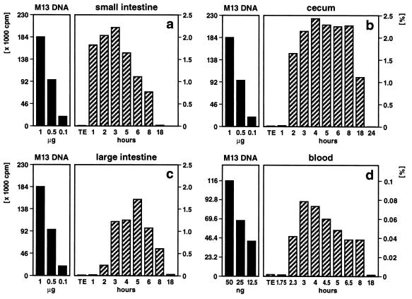
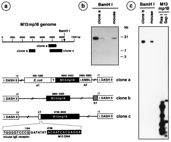
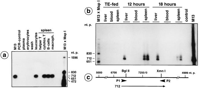
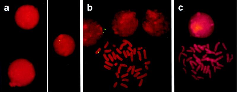
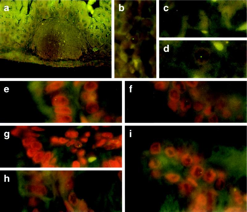
Food-ingested foreign DNA is not completely degraded in the gastrointestinal tract of mice. Phage M13mp18 DNA as a test molecule devoid of homology to mouse DNA was pipette-fed to or added to the food supply of mice. The fate of this foreign DNA in the animals was followed by several methods. In 84 animals, fragments of M13mp18 DNA were detected in the contents of the small intestine, the cecum (until 18 h), the large intestine, or the feces. In 254 animals, M13mp18 DNA fragments of up to 976 bp were found in blood 2-8 h after feeding. In buffer-fed control animals, M13mp18 DNA could not be detected. M13mp18 DNA fragments were traced by PCR in peripheral leukocytes and located by fluorescent in situ hybridization in about 1 of 1000 white cells between 2 and 8 h, and in spleen or liver cells up to 24 h after feeding, but not later. M13mp18 DNA could be traced by fluorescent in situ hybridization in the columnar epithelial cells, in the leukocytes in Peyer's patches of the cecum wall, in liver cells, and in B cells, T cells, and macrophages from spleen. These findings suggest transport of foreign DNA through the intestinal wall and Peyer's patches to peripheral blood leukocytes and into several organs. Upon extended feeding, M13mp18 DNA could be recloned from total spleen DNA into a lambda vector. Among about 2.5 x 10(7) lambda plaques, one plaque was isolated that contained a 1299 nucleotide pair fragment (nt 4736-6034) of sequence-identified M13mp18 DNA. This fragment was covalently linked to an 80 nt DNA segment with 70% homology to the mouse IgE receptor gene. The DNA from another lambda plaque also contained mouse DNA, bacterial DNA, and rearranged lambda DNA. Two additional plaques contained M13mp18 DNA fragments of at least 641 (nt 2660-3300) or 794 (nt 4640-5433) nucleotide pairs. The medical and evolutionary implications of these observations may be considerable.


Figures

Figure 1
Figure 1
Quantitation of M13mp18 foreign DNA detectable by dot blot hybridization in different segments of the intestine (ac) or in peripheral blood (d). Fifty micrograms of M13mp18 DNA (19) (7250 bp) was pipette-fed to 2–6 (12)-month-old mice. As indicated, at times after feeding, the DNA was extracted (1) from the contents of the small intestine (a), the cecum (b), the large intestine (c), or from blood (d). The persisting M13mp18 DNA was identified by dot blot hybridization (1). Aliquot portions of the extracted DNA were fixed on a positively charged Qiagen nylon membrane and hybridized to 32P-labeled (20) M13mp18 DNA followed by autoradiography. In reconstitution experiments, amounts of M13mp18 DNA ranging from 5 ng to 1 μg were also bound to the membranes. Subsequently, autoradiographically localized dots were excised, and the amounts of 32P label membrane bound in these spots were determined by Cerenkov counting. Considering the fraction of the total reextracted DNA from gut segments or blood applied to the dot blot matrix, percentages (scales on right) of the 50 μg of orally administered M13mp18 test DNA were calculated (hatched bars). The scales in cpm and the solid bars on the left referred to the actual 32P Cerenkov cpm for reference M13mp18 DNA.
Figure 5
Figure 5
Recloning of M13mp18 DNA from spleen DNA of mice fed with M13mp18 DNA. In brief, the segments of M13mp18 DNA present in the DNA of the M13mp18 DNA-positive recombinant λ clones were identified by hybridizing the 32P-labeled recombinant λ DNA to RsaI- or SspI-cleaved M13mp18 DNA (e.g., c, right lanes). On the basis of the known nucleotide sequence of M13mp18 DNA (19), primers were selected and the exact M13mp18 DNA represented was determined. By using primers from the known M13mp18 DNA (solid boxes in a), the adjacent nucleotide sequences were determined and identified by computer-aided National Center for Biotechnology Information searches. The λDASHII clone a was derived from feeding experiment ii, the clones b and c from the schedule iii experiment. (a) Map of the linearized M13mp18 genome. The locations of the nucleotide sequence-identified M13mp18 segments present in λDASHII clones a–c are indicated as solid blocks. In clone a, a1 represents the Escherichia coli DNA sequence of an ATP-dependent Clp-protease, the question mark a sequence of unknown, possibly mouse, origin. The segment a2 carries rearranged DNA from the left arm of λ DNA. In clone b, the DNA sequence shown as a hatched segment b1 exhibits homologies to mouse DNA (129/sv Clora cell 10 kDa, mCC10 gene). (b) Clone a DNA was 32P-labeled by nick-translation and hybridized to BamHI-restricted clone a DNA or genomic mouse DNA on a Southern blot under stringent hybridization (68°C, 2 × SSC) and wash conditions (68°C, 2 × 15 min with 2 × SSC, 0.1% SDS; 2 × 25 min with 0.2 × SSC, 0.1% SDS). (c) A similar experiment for clone b DNA, which is also shown to hybridize to m13mp18 DNA and to genomic mouse DNA. The 80 bp DNA segment in c1 was 70% homologous to the mouse IgE receptor gene.
Figure 2
Figure 2
Detection of M13mp18 DNA fragments in peripheral blood leukocytes, in liver, and in different spleen cells by PCR. (a) A male C57BL/6 mouse was pipette-fed 50 μg of M13mp18 DNA. At 4 h after feeding, blood was drawn by heart puncture and the plasma, erythrocyte, and leukocyte fractions were prepared by Ficoll gradient centrifugation. The leukocytes were washed twice in Tris-saline or PBS and incubated for 2 min in 0.15 M NaCl, 10 mM Tris·HCl (pH 7.5), 2 mM MgCl2, 1% Nonidet P-40. The thus liberated nuclei were sedimented, resuspended in 10 mM Tris·HCl (pH 7.5), 1 mM EDTA, 1% SDS, 1.3 mg/ml proteinase K, and incubated for 2 h at 37°C. Nucleic acids were then prepared by phenol/chloroform extraction. Similarly, the plasma, erythrocyte, and wash fractions were extracted for possibly present DNA. By using the synthetic oligodeoxyribonucleotide primers P1 and P2 designated on the M13mp18 DNA map in c, the indicated M13mp18 DNA segments were amplified as described earlier (1). (a) At 18 h after feeding a different animal, the different spleen cells were fractionated by the magnetic bead method as described. DNA from the designated cell fractions was prepared and PCR analyzed. (b) Detection of M13mp18 DNA in spleen and liver cells by PCR at 12 or 18 h after feeding. DNA samples from TE-fed animals or from blood of animals 12 or 18 h after feeding M13mp18 DNA did not contain M13mp18 DNA. The PCR products were blotted to positively charged membranes, and M13mp18 DNA was identified by hybridization to 32P-labeled M13mp18 DNA and by autoradiography.
Figure 3
Figure 3
Ingested foreign DNA detected in peripheral leukocytes and in spleen cells by FISH. (a) Peripheral blood leukocytes from M13mp18 DNA-fed mice were analyzed by the FISH technique using biotinylated M13mp18 DNA as hybridization probe and FITC-tagged avidin for the detection of biotinylated DNA (–15). (b) Spleen cells from M13mp18 DNA-fed mice were incubated at 37°C for 48 h in RPMI 1640 medium containing 10% fetal calf serum, 2.4 mM glutamine, 10 mM 2-mercaptoethanol, 30 μg/ml lipopolysaccharide, 300 μg/ml penicillin, 100 μg/ml streptomycin. Subsequently, ethidium bromide (10 μg/ml) and colcemid (50 μg/ml) were added, and incubation was continued for 2 h at 37°C. Finally, cells were incubated for 20 min at 37°C in 0.075 KCl, fixed at 4°C in methanol:acetic acid (3:1), and attached to precleaned glass slides. (c) Control with spleen cells from a TE-fed mouse.
Figure 4
Figure 4
Histological sections through the cecum wall (a, b, and ei) or through liver (c and d) from mice that had been fed 50 μg of M13mp18 DNA (a, b, d, and ei) or TE-buffer (c). Tissue samples were prepared as described. M13mp18 DNA was identified by FISH. Tissues were counterstained in propidium iodide solution (200 ng/ml of H2O). (a and b) Peyer’s patch inside the cecum wall 5 h after feeding; (ei) cecum epithelia from mice 3–5 h after feeding; (c) liver from a TE-fed control mouse; (d) liver from an animal 6 h after feeding M13mp18 DNA. (a, ×50; bi, ×1250.)

References

    1. Schubbert R, Lettmann C, Doerfler W. Mol Gen Genet. 1994;242:495–504. - PubMed
    1. Doerfler W. Proc Natl Acad Sci USA. 1968;60:636–643. - PMC - PubMed
    1. Groneberg J, Brown D T, Doerfler W. Virology. 1975;64:115–131. - PubMed
    1. Doerfler W. Adv Cancer Res. 1995;66:313–344. - PubMed
    1. Doerfler W. Biochim Biophys Acta Rev Cancer. 1996;1288(2):F79–F99. - PubMed

Publication types

MeSH terms