. 1997 Feb 4;94(3):967-72.
doi: 10.1073/pnas.94.3.967.

Transcriptional activation of mucin by Pseudomonas aeruginosa lipopolysaccharide in the pathogenesis of cystic fibrosis lung disease

Affiliations

Transcriptional activation of mucin by Pseudomonas aeruginosa lipopolysaccharide in the pathogenesis of cystic fibrosis lung disease

J D Li et al. Proc Natl Acad Sci U S A. .

Abstract

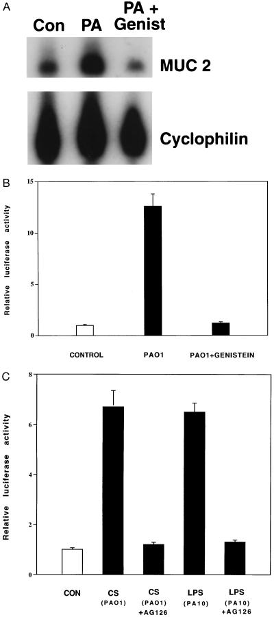
An unresolved question in cystic fibrosis (CF) research is how mutations of the CF transmembrane conductance regulator, a Cl ion channel, cause airway mucus obstruction leading to fatal lung disease. Recent evidence has linked the CF transmembrane conductance regulator mutation to the onset and persistence of Pseudomonas aeruginosa infection in the airways, and here we provide evidence directly linking P. aeruginosa infection to mucus overproduction. We show that P. aeruginosa lipopolysaccharide profoundly upregulates transcription of the mucin gene MUC 2 in epithelial cells via inducible enhancer elements and that this effect is blocked by the tyrosine kinase inhibitors genistein and tyr-phostin AG 126. These findings improve our understanding of CF pathogenesis and suggest that the attenuation of mucin production by lipopolysaccharide antagonists and tyrosine kinase inhibitors could reduce morbidity and mortality in this disease.


Figures

Figure 1
Figure 1
In situ hybridization analysis of MUC 2 mRNA expression in non-CF and CF human bronchial explants. In situ hybridization was performed with the human airway mucin 1 anti-sense probe recognizing human MUC 2 mRNA as described (7). MUC 2 mRNA expression is shown in vehicle control-treated and P. aeruginosa-treated bronchial explant surface epithelia from non-CF (A and B) and CF (C and D) individuals and in vehicle control-treated and P. aeruginosa-treated bronchial explant submucosal glands from non-CF (E and F) and CF (G and H) individuals. Arrowheads in A–D indicate the position of the epithelial basement membrane. In these experiments, both non-CF and CF bronchial explants were exposed for 6 h to P. aeruginosa culture supernatants. Similar results were observed in bronchial explants from four CF and four non-CF individuals treated with P. aeruginosa strain PAO1 or PA103. L, airway lumen; SG, submucosal glands.
Figure 2
Figure 2
RPA analysis of MUC 2 mRNA expression in human MUC 2-expressing epithelial cell lines. Up-regulation of MUC 2 mRNA by P. aeruginosa culture supernatants occurred in both HM3 (human colon epithelial) and NCIH292 (human airway epithelial) cells. Cells were treated with P. aeruginosa culture supernatants (PA) or vehicle (Con) for 6 h before cell lysis and RNA extraction. The results are typical of four separate experiments for each cell line. GAPDH, glyceraldehyde-3-phosphate dehydrogenase.
Figure 3
Figure 3
Up-regulation of MUC 2 transcriptional activity by P. aeruginosa. (A) A 2.8-kb DNA fragment of the 5′ flanking region of the human MUC 2 gene cloned into a luciferase reporter gene (p-2864luc) was transfected into NCIH292, HM3, and CFTE29O cells. Luciferase activity was then assessed in P. aeruginosa-treated and nontreated cells. Induction by P. aeruginosa was detected in all cell lines. (B) Comparison of the P. aeruginosa responsiveness of p-2864luc and p-73luc in NCIH292, HM3, and CFTE29O cells. Response elements reside between −2864 and −73 bp. (C) Comparison of the P. aeruginosa responsiveness of p-73luc, p-343luc, p-621luc, p-1308luc, and p-2864luc in HM3 cells. Transfected cells were treated with either P. aeruginosa culture supernatants or vehicle for 6 h before cell lysis. All transfections were carried out in triplicate. Values represent means ± SD (n = 5). Luciferase activity was normalized with respect to β-galactosidase activity.
Figure 4
Figure 4
Effect of P. aeruginosa LPS on MUC 2 transcriptional activity. HM3 cells were transfected with p-2864luc. (A) After 42 h, the cells were exposed to culture supernatants (CS) and LPS (5 μg/ml) purified from PAO1 and PA10 (serotype 10) (Sigma). (B) As in A, the cells were exposed to culture supernatants or purified LPS from PAO1 (wild-type) or PAO-pmm (algC) mutants. (C) The cells were exposed to LPS (5 μg/ml) (P. aeruginosa serotype 10) and E. coli lipid A (5 μg/ml) (Sigma). After 6 h, the cells were harvested for luciferase activity measurement. All transfections were carried out in triplicate. Values represent means ± SD (n = 4). Luciferase activity was normalized with respect to β-galactosidase.
Figure 5
Figure 5
Inhibition of P. aeruginosa-induced MUC 2 up-regulation by tyrosine kinase inhibitors. (A) HM3 cells were or were not pretreated with genistein (Genist) (100 μg/ml) (Sigma) for 2 h and then were exposed to P. aeruginosa PAO1 culture supernatants (PA) or vehicle (Con) for 6 h before RNA extraction and RPA analysis. The results are typical of four separate experiments. (B) HM3 cells were transfected with p-2864luc. After 40 h, the cells were pretreated with genistein (100 μg/ml) for 2 h and then were exposed to P. aeruginosa culture supernatants (PAO1) for 6 h before harvesting for luciferase analysis. (C) Forty hours after being transfected with p-2864luc, HM3 cells were pretreated with tyrphostin AG 126 (25 μM) (Calbiochem) for 3 h and then were exposed to PAO1 culture supernatant (CS) or LPS from PA10 (serotype 10) (5 μg/ml) for 6 h before harvesting. Luciferase activity was measured as described above. All transfections in B and C were carried out in triplicate. Values represent means ± SD (n = 4). Luciferase activity was normalized with respect to β-galactosidase.
Figure 6
Figure 6
Effect of P. aeruginosa culture supernatant on MUC 2 transcriptional activity in CFTE 29O ΔF508 cells cotransfected with p-2864luc ± the CFTR wild-type expression plasmid pREP4.7kbCFTR. Forty-two hours after transfection, PAO1 culture supernatant (CS) was added to cells; 6 h later, the cells were harvested for luciferase activity measurement. All transfections were carried out in triplicate. Values represent means ± SD (n = 4). Luciferase activity was normalized with respect to β-galactosidase.

References

    1. Collins F S, Wilson J M. Nature (London) 1992;358:708–709. - PubMed
    1. Saiman L, Prince A. J Clin Invest. 1993;92:1875–1880. - PMC - PubMed
    1. Pier G B, Grout M, Zaidi T S, Olsen J C, Johnson L G, Yankaskas J R, Goldberg J B. Science. 1996;271:64–67. - PMC - PubMed
    1. Smith J J, Travis S M, Greenberg E P, Welsh M J. Cell. 1996;85:229–236. - PubMed
    1. Matthews L. Am Rev Respir Dis. 1963;88:199–207. - PubMed

Publication types

MeSH terms

Associated data