. 1997 Feb 4;94(3):1019-22.
doi: 10.1073/pnas.94.3.1019.

Functional nonequality of the cardiac and skeletal ryanodine receptors

Affiliations

Functional nonequality of the cardiac and skeletal ryanodine receptors

J Nakai et al. Proc Natl Acad Sci U S A. .

Abstract

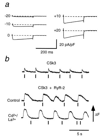
Dihydropyridine receptors (DHPRs), which are voltage-gated Ca2+ channels, and ryanodine receptors (RyRs), which are intracellular Ca2+ release channels, are expressed in diverse cell types, including skeletal and cardiac muscle. In skeletal muscle, there appears to be reciprocal signaling between the skeletal isoforms of both the DHPR and the RyR (RyR-1), such that Ca2+ release activity of RyR-1 is controlled by the DHPR and Ca2+ channel activity of the DHPR is controlled by RyR-1. Dyspedic skeletal muscle cells, which do not express RyR-1, lack excitation-contraction coupling and have an approximately 30-fold reduction in L-type Ca2+ current density. Here we have examined the ability of the predominant cardiac and brain RyR isoform, RyR-2, to substitute for RyR-1 in interacting with the skeletal DHPR. When RyR-2 is expressed in dyspedic muscle cells, it gives rise to spontaneous intracellular Ca2+ oscillations and supports Ca2+ entry-induced Ca2+ release. However, unlike RyR-1, the expressed RyR-2 does not increase the Ca2+ channel activity of the DHPR, nor is the gating of RyR-2 controlled by the skeletal DHPR. Thus, the ability to participate in skeletal-type reciprocal signaling appears to be a unique feature of RyR-1.


Figures

Figure 1
Figure 1
Intracellular Ca2+ transients in intact dyspedic myotubes expressing RyR-2. (a) Photomicrograph showing the vesiculated appearance of a dyspedic myotube in the region injected with RyR-2 cDNA (enlargement on right). (Bar = 100 μm.) (b and c) Pseudocolor images, separated in time by 67 ms (b) or 100 ms (c), of spontaneous Ca2+ transients in a normal myotube and an RyR-2-injected dyspedic myotube, respectively. Color scale from blue to red indicates increasing Ca2+ concentration in arbitrary units of Fluo-3 fluorescence. (d and e) Fluorescence intensity as a function of time is plotted for the positions indicated on the adjacent images. (d, bar = 500 μm, and applies to be.) (f) Spatially averaged Ca2+ responses in a RyR-2-expressing dyspedic myotube; the bars indicate exposure to 0.1 mM caffeine (C) or 0.1 mM ryanodine (R).
Figure 2
Figure 2
Voltage-clamp analysis of a RyR-2-expressing dyspedic myotube. (a) Simultaneous measurement of membrane current (upper trace) and intracellular Ca2+ during a 15-ms depolarization to +20 mV. Depolarization failed to elicit an intracellular Ca2+ transient. (b) Expression of RyR-2 failed to restore expression of a large-amplitude slow Ca2+ current. Depolarizations of 200 ms to the indicated potentials. The patch pipette contained 10 mM EGTA and 200 μM Fluo-3 as Ca2+ buffers.
Figure 3
Figure 3
Behavior of dyspedic cells expressing only CSk3 (a chimeric DHPR) or coexpressing CSk3 and RyR-2. (a) Presence of a large, rapidly activated calcium current in a voltage-clamped dyspedic myotube expressing CSk3. (b) Electrical stimulation (vertical bars) elicited only small, brief Ca2+ transients in an intact dyspedic myotube injected only with CSk3 cDNA (top trace). In an intact dyspedic myotube injected with both CSk3 and RyR-2 cDNA (middle trace), electrical stimulation elicited large, long-lasting Ca2+ transients (a spontaneous transient occurred between the second and third electrical stimuli). After addition of Cd2+ and La3+ to block Ca2+ current, electrical stimulation failed to evoke transients, although spontaneous transients still occurred (bottom trace, same cell as in middle trace). The cDNA for CSk3 (23) encodes a protein containing amino acids 1–787 and 923-2171 of the cardiac DHP receptor (3) joined by amino acids 666–792 of the skeletal DHP receptor (2).
Figure 4
Figure 4
Electron micrographs illustrating membrane junctions in RyR-2-expressing dyspedic myotubes. (a) A peripheral coupling between the SR (arrowhead) and plasmalemma (asterisks). A pair of feet (double arrows) spans the junctional gap. Extracellular debris is present in proximity of the cell (lower half of photograph). (b) A coupling which contains feet between the SR and a primitive transverse tubule. In addition to dyadic couplings (as shown here), there were also occasional triadic couplings (data not shown). The width of the junctional gaps containing feet was 13.2 ± 1.8 nm; 51 measurements). (c) A junction lacking feet between an SR vesicle and a plasmalemmal invagination in an RyR-2-injected myotube. Note that the junctional gap is smaller (9.0 ± 1.7 nm; 62 measurements) than in junctions with visible feet. Calibration = 0.1 μm (applies to a–c).

References

    1. Hofmann F, Biel M, Flockerzi V. Annu Rev Neurosci. 1994;17:399–418. - PubMed
    1. Tanabe T, Takeshima H, Mikami A, Flockerzi V, Takahashi H, Kangawa K, Kojima M, Matsuo H, Hirose T, Numa S. Nature (London) 1987;328:313–318. - PubMed
    1. Mikami A, Imoto K, Tanabe T, Niidome T, Mori Y, Takeshima H, Narumiya S, Numa S. Nature (London) 1989;340:230–233. - PubMed
    1. Williams M E, Feldman D H, McCue A F, Brenner R, Velicelebi G, Ellis S B, Harpold M M. Neuron. 1992;8:71–84. - PubMed
    1. McPherson P S, Campbell K P. J Biol Chem. 1993;268:13765–13768. - PubMed

Publication types

MeSH terms

LinkOut - more resources