. 1997 Feb 10;136(3):531-44.
doi: 10.1083/jcb.136.3.531.

Interphase nuclei of many mammalian cell types contain deep, dynamic, tubular membrane-bound invaginations of the nuclear envelope

Affiliations

Interphase nuclei of many mammalian cell types contain deep, dynamic, tubular membrane-bound invaginations of the nuclear envelope

M Fricker et al. J Cell Biol. .

Abstract

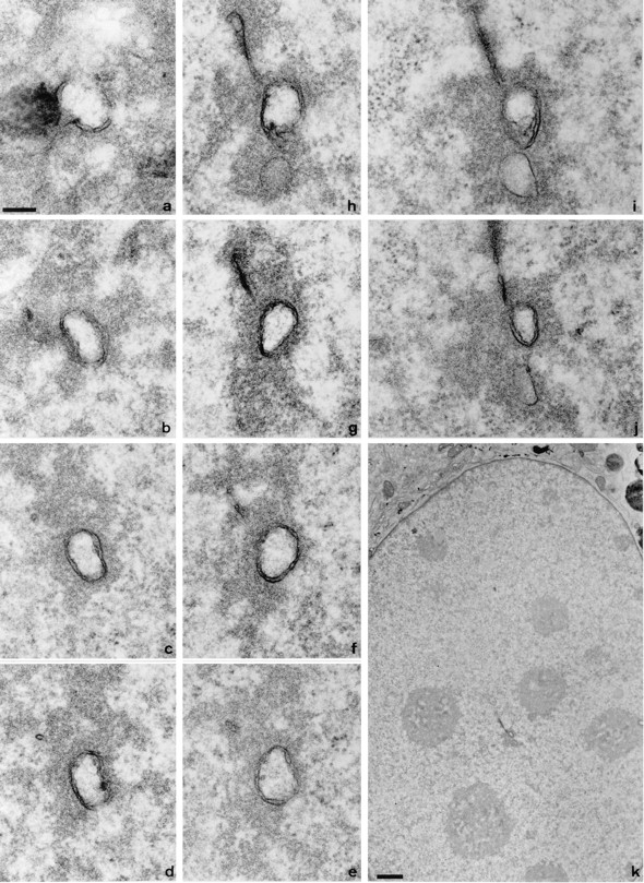
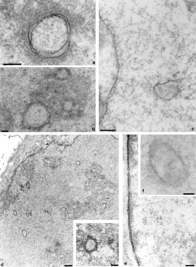
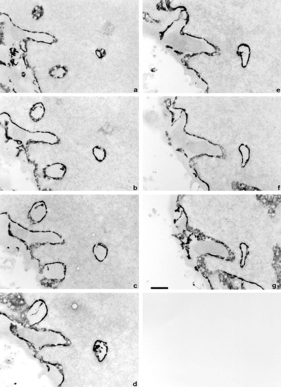
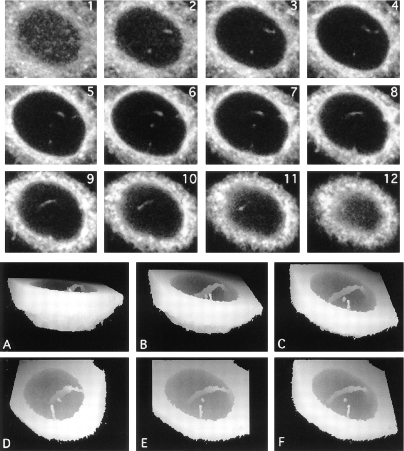
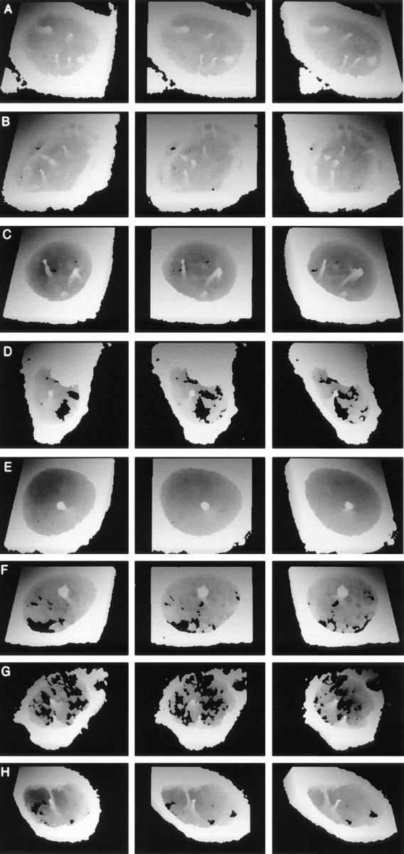
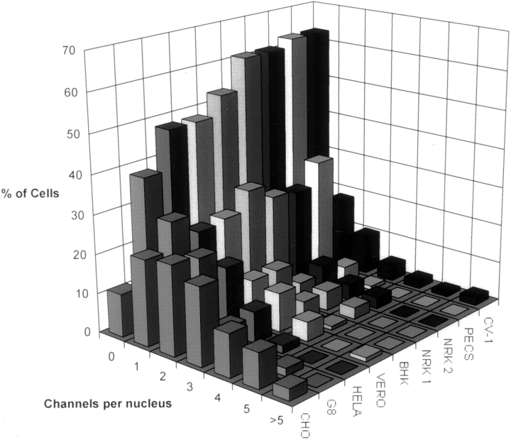
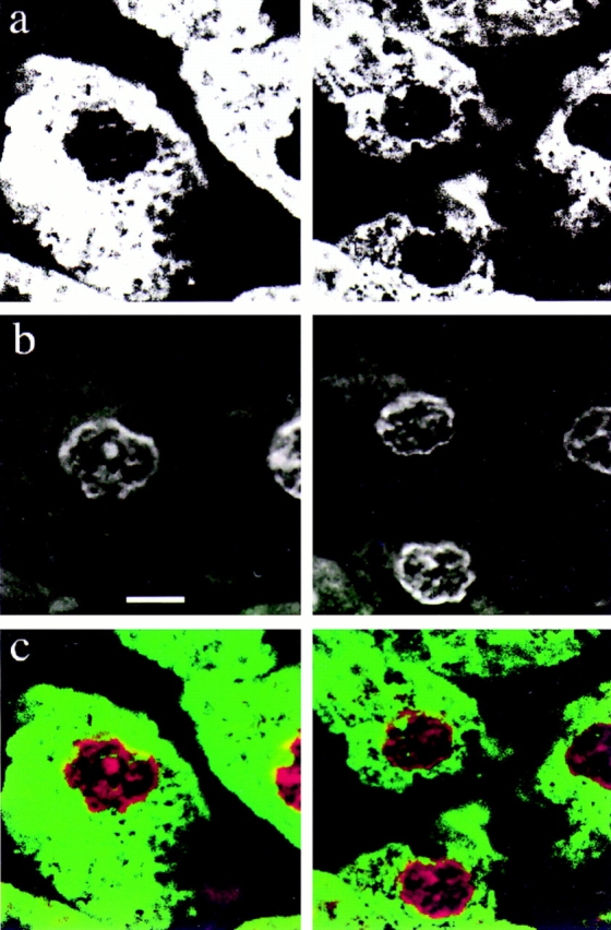
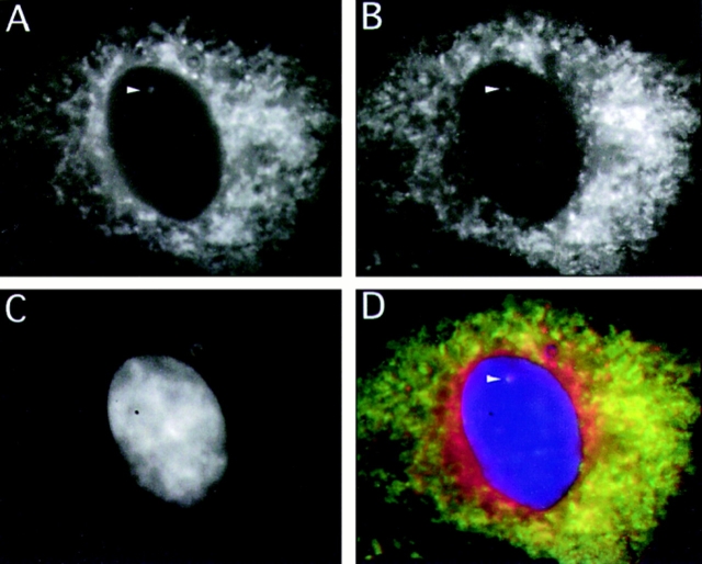
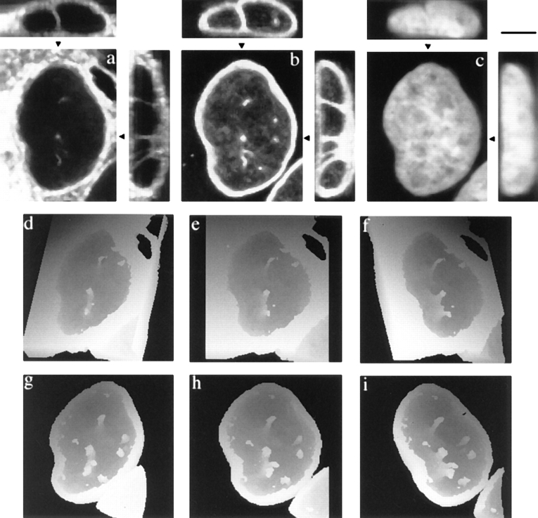
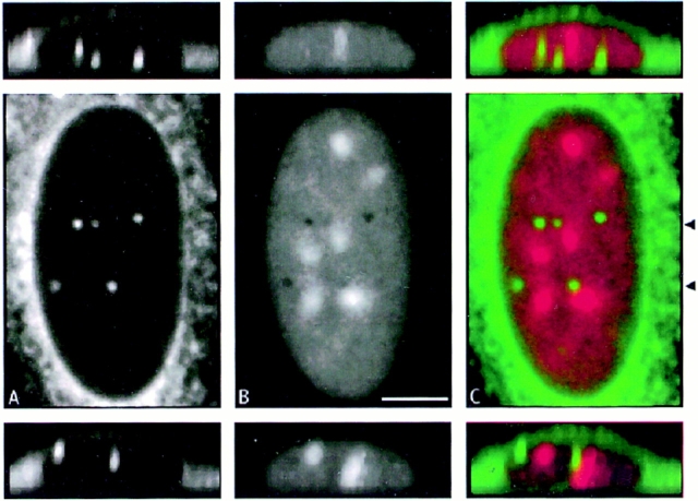
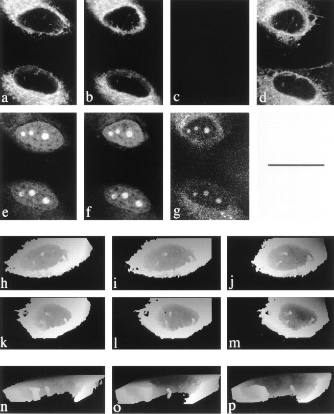
The nuclear envelope consists of a double-membraned extension of the rough endoplasmic reticulum. In this report we describe long, dynamic tubular channels, derived from the nuclear envelope, that extend deep into the nucleoplasm. These channels show cell-type specific morphologies ranging from single short stubs to multiple, complex, branched structures. Some channels transect the nucleus entirely, opening at two separate points on the nuclear surface, while others terminate at or close to nucleoli. These channels are distinct from other topological features of the nuclear envelope, such as lobes or folds. The channel wall consists of two membranes continuous with the nuclear envelope, studded with features indistinguishable from nuclear pore complexes, and decorated on the nucleoplasmic surface with lamins. The enclosed core is continuous with the cytoplasm, and the lumenal space between the membranes contains soluble ER-resident proteins (protein disulphide isomerase and glucose-6-phosphatase). Nuclear channels are also found in live cells labeled with the lipophilic dye DiOC6. Time-lapse imaging of DiOC6-labeled cells shows that the channels undergo changes in morphology and spatial distribution within the interphase nucleus on a timescale of minutes. The presence of a cytoplasmic core and nuclear pore complexes in the channel walls suggests a possible role for these structures in nucleo-cytoplasmic transport. The clear association of a subset of these structures with nucleoli would also be consistent with such a transport role.


Figures

Figure 1
Figure 1
Serial section EM reveals complex membrane-bound networks within the nucleus. Consecutive 70-nm serial sections from a region of a 3T3 cell nucleus sectioned parallel to the growth substrate are shown in a to j. Section a begins at the nuclear envelope; note the en face nuclear pore complexes and the dark mass of an HRP-loaded endosome in the cytoplasm to the left of the invaginating membranes. The position of the channel within the nucleus is shown in the low magnification overview in k; clearly visible within the nucleoplasm are nucleoli that are not apparently associated with this channel. Bars: (a–j) 200 nm; (k) 1 μm.
Figure 2
Figure 2
The intranuclear channels in a range of cell types contain nuclear pore complexes and are associated with an electron dense halo in the nucleoplasm. Representative profiles of channels from 3T3 cells (a), NRK cells (b), A431 cells (d and inset), and HeLa cells (f) are shown. Double membrane structures are clearly visible, together with local fenestrations, and comparable to the appearance of the nuclear pore complexes in the nuclear envelope of the 3T3 nucleus shown in e. The electron lucent core is also seen to contain small circular or crescent shaped features, best seen within the channel in b. Electron dense material in the nucleoplasm is associated with the channels (a, b and d, inset). In addition, channel association with nucleoli is shown for NRK cells (b). Thawed frozen thin section colloidal gold immunocytochemistry confirms that the channels are associated with strong immunoreactivity for lamins (c) and protein disulphide isomerase (f), as shown by the 9-nm gold particles decorating the intranuclear channels in these HeLa nuclei. Bars: 200 nm; d, 1 μm.
Figure 3
Figure 3
The intranuclear channels are bounded by a double membrane enclosing a G-6-Pase positive lumen. Consecutive 70 nm serial sections of part of an ATt20 nucleus sectioned parallel to the growth substrate after histochemical visualization of G-6Pase activity. Dense reaction product fills the space between the inner and outer nuclear membranes but is excluded from nuclear pore complexes. Similar reaction product is seen within the channels in the nucleoplasm. Bar, 500 nm.
Figure 4
Figure 4
Intranuclear channels labeled with lectins recognizing ER-type oligosaccharide side chains and visualized by confocal microscopy and 3-D reconstruction. Serial optical sections of a Con-A–labeled HeLa nucleus were collected at 0.5 μm intervals by CLSM from the base of the cell (1–12), forming a 3-D XYZ image. Fluorescent structures are clearly visible within the nucleus in successive optical sections. To visualize the overall morphology of the channels, the sections nearest the base of the cell were discarded and views projected at increasing tilt angles (30° increments; A–C and F) and rotation angles (24° increments; D–F). In these height reconstructions, structures above a threshold intensity nearest the viewer are coded as white for each tilt or rotation angle and also mask underlying structures. The major nuclear channel traverses the nucleoplasm, contacting the nuclear envelope at two locations. In addition, a second, shorter tube that terminates within the nucleus is also visible.
Figure 5
Figure 5
Intranuclear channels vary in number and morphology among different cell types, contain ER soluble resident proteins and ER membrane proteins, and enclose a cytosolic core. Rotation series of height coded 3-D views at 24° intervals of 3T3 (A), CHO (B), G8 (C), PEC (D), and NRK (E) cells labeled with fluorescent tagged Con A are shown. Reconstructions were made as shown in Fig. 4. The nuclear channel morphology was characteristic for each cell type and ranged from multiple branched channels (e.g., G8 cells in C) to single unbranched channels (e.g., PECs and NRK cells in D and E, respectively). In addition to being Con A positive structures, channels were also visible in an NRK nucleus labeled with a monoclonal antibody against the soluble resident ER protein, protein disulphide isomerase (F), a HeLa nucleus labeled with a polyclonal antiserum against ER membrane proteins (G), and a HeLa nucleus after scrape loading of the cell with a 150-kD fluorescent tracer (H).
Figure 6
Figure 6
Frequency histogram of nuclear channel distribution in a range of cell types. Cells were labeled with fluorochrome tagged Con A and 3-D datasets collected for >80 nuclei for each cell type. The number of channels defined as an intranuclear labeled structure at least 1.5 μm in x, y, or z was counted by inspection of the serial optical sections.
Figure 7
Figure 7
Nuclear channels are present in normal rat hepatocytes. Perfusion-fixed sections of rat liver were labeled with fluorochrome-tagged Con A in a and the nucleic acid stain To-Pro-3 in b. A two color merge of the data is shown in c. Note the association between the nucleolus and two channels in the left hand cell. Bar, 5 μm.
Figure 8
Figure 8
Intranuclear PDI immunoreactivity co-localizes with Con-A–labeled channels. The figure shows separate eight-bit grey scale images collected from a triple labeled NRK cell using a cooled CCD camera. A shows the distribution of the PDI signal in the fluorescein channel. B shows the Con A signal in the rhodamine channel, and C shows the nucleic acid labeled with DAPI. D is a 24-bit merge of the first three channels. The arrowhead points to an intranuclear structure that can be followed through a number of focal planes in which PDI and Con A reactivity colocalize.
Figure 9
Figure 9
Con A–labeled channels are associated with lamin immunoreactivity. Maximum projections of six consecutive optical sections at 0.3 μm Z interval from a triple labeled HeLa nucleus are shown; a shows Con A reactivity. b shows polyclonal antilamin reactivity, and c shows nucleic acid labeling with Yo-Pro 1. Associated XZ (above) and YZ (right) projections along the lines indicated by the black arrowheads are also shown. The rotation series of height-coded, tilted 3-D views at 24° intervals are presented for Con A labeling in d–f and lamin labeling in g–i. Each of the Con A sites in the nucleus is colocalized with sites of lamin reactivity, but note also that there are additional intranuclear lamin reactive sites that do not have associated Con A reactivity. Bar, 5 μm.
Figure 10
Figure 10
Some channels that terminate within the nucleus are associated with nucleoli. a shows Con A reactivity; b shows nucleic acid labeling with propidium iodide. c shows a two color merge of the data from a and b. Two XZ views across the nucleus at the level of the channels (arrowheads) are also shown.
Figure 11
Figure 11
Nuclear channels are detectable in live cells and correspond to the Con A–labeled structures seen in fixed cells. a shows a single plane from the dataset for two DiOC6 labeled live cells; e shows the dihydroethidium label in the same plane. b and f are from data collected 20 min later. c shows the loss of the DiOC6 signal at the end of methanol fixation, while g shows that the cells remain intact and the dihydroethidium label persists, although reduced in intensity. d shows the same cells and plane after 20 min of Con A labeling. h–j show sequential rotations of reconstructions of DiOC6 labeled cells, and underneath in k–m, respectively, the same cell is shown after Con A labeling. Cutaway views of a reconstructed DiOC6 labeled HeLa nucleus from a separate experiment are shown at three time points in n–p. Note that the morphology of the larger left hand tube alters over the 35-min period, and the smaller channel shows a progressively increasing separation from the large channel. Bars: (a–m) 25 μm; (n–p) 2 μm.

References

    1. Banfalvi G, Wiegant J, Sarkar N, van-Duijn P. Immunofluorescent visualization of DNA replication sites within nuclei of Chinese hamster ovary cells. Histochemistry. 1989;93:81–86. - PubMed
    1. Banfic H, Zizak M, Divecha N, Irvine RF. Nuclear diacylglycerol is increased during cell proliferation in vivo. Biochem J. 1993;290:633–636. - PMC - PubMed
    1. Belmont AS, Zhai Y, Thilenius A. Lamin B distribution and association with peripheral chromatin revealed by optical sectioning and electron microscopy tomography. J Cell Biol. 1993;123:1671–1685. - PMC - PubMed
    1. Bourgeois CA, Hemon D, Bouteille M. Structural relationship between the nucleolus and the nuclear envelope. J Ultrastruct Res. 1979;68:328–340. - PubMed
    1. Bowers CW, Dahm LM. Buffers for cell culture. Trends Cell Biol. 1993;3:76–77. - PubMed

Publication types