. 1997 Feb 10;136(3):609-20.
doi: 10.1083/jcb.136.3.609.

A cytosolic serine endopeptidase from Trypanosoma cruzi is required for the generation of Ca2+ signaling in mammalian cells

Affiliations

A cytosolic serine endopeptidase from Trypanosoma cruzi is required for the generation of Ca2+ signaling in mammalian cells

B A Burleigh et al. J Cell Biol. .

Abstract

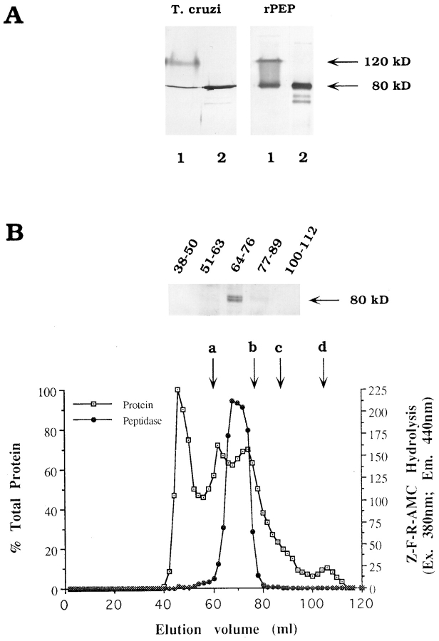
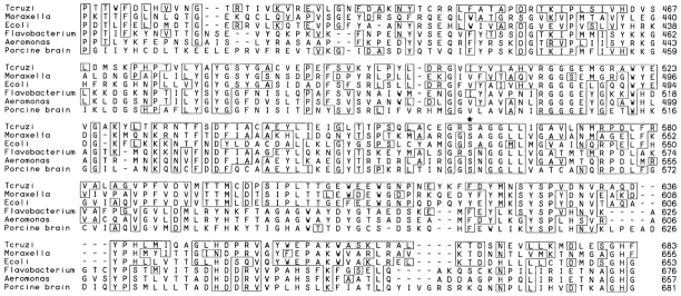
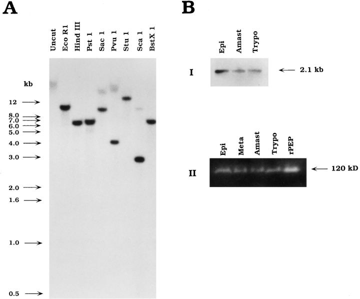
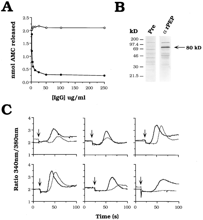
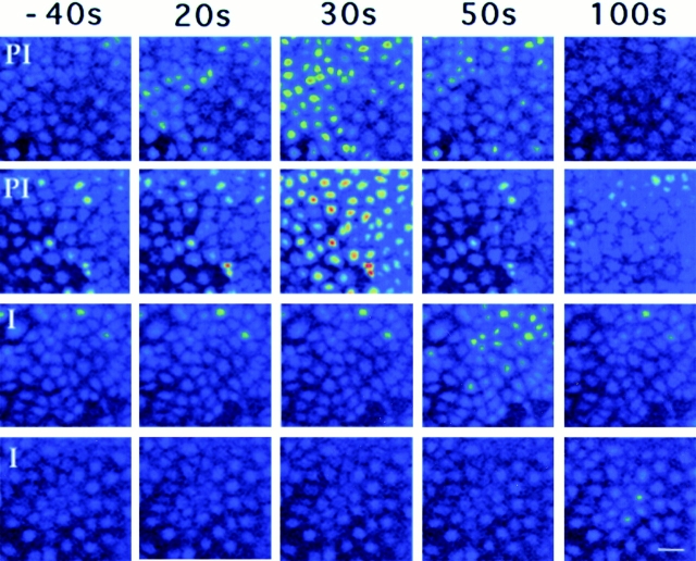
An early event in the Trypanosoma cruzi cell invasion process, the recruitment of host lysosomes, led us to investigate the involvement of signal transduction. Infective trypomastigotes were found to contain a soluble Ca2+-signaling activity for mammalian cells that is sensitive to protease inhibitors. Inhibitor and substrate utilization profiles were used to purify a candidate peptidase for involvement in this process, from which we isolated a full-length cDNA clone. The sequence revealed a novel enzyme, denominated T. cruzi oligopeptidase B, which is homologous to members of the prolyl oligopeptidase family of serine hydrolases, known to participate in the maturation of biologically active peptides. The T. cruzi oligopeptidase B was expressed as a fully active product in Escherichia coli, and antibodies to the recombinant enzyme inhibited both peptidase activity and Ca2+ signaling induced in normal rat kidney cells by trypomastigote extracts. Our data suggest that the T. cruzi oligopeptidase B participates in processing events in the cytoplasm of the parasites, generating a factor with Ca2+-signaling activity for mammalian cells.


Figures

Figure 1
Figure 1
Nucleotide sequence of cloned cDNA and deduced amino acid sequence of the T. cruzi oligopeptidase B. The deduced amino acid sequence is shown below the nucleotide sequence. Both are numbered on the right. Amino acid sequences obtained for tryptic peptides of the purified peptidase (underlined). The stop codons that limit the open reading frame (bold); polyadenylation signals are highlighted (double underline). These sequence data are available from GenBank under accession number U69897.
Figure 2
Figure 2
Molecular size determination of native and recombinant T. cruzi oligopeptidase B. (A) Immunoblots of oligopeptidase B partially purified from epimastigotes (T. cruzi, lanes 1 and 2) or 0.6 μg purified recombinant peptidase (rPEP, lanes 1 and 2) with guinea pig polyclonal anti–oligopeptidase B IgG (25 μg/ml) followed by acid phosphatase–conjugated anti–guinea pig IgG. Samples were reduced with 5% β-mercaptoethanol and kept at either 25°C (lanes 1) or heated for 5 min at 100°C (lanes 2) before SDS-PAGE. (B) FPLC fractionation of a trypomastigote soluble extract on Sephacryl-300. Proteins were eluted in PBS2+ while A280 nm was continuously monitored (open squares). The peak absorbance value (fraction 41) was taken as 100% and the baseline as 0% protein. Peptidase activity (black circles) was assayed in 20 μl aliquots of each fraction using ZFR-AMC as a substrate, as described in Materials and Methods. Fractions containing peptidase activity (64-76) were pooled, concentrated, reduced, boiled for 5 min, separated by SDS-PAGE, and immunoblotted with anti–oligopeptidase B guinea pig IgG as described above. The elution of molecular mass standards is indicated by letters (a) catalase = 232 kD; (b) aldolase = 158 kD; (c) albumin = 67 kD; (d) ribonuclease A = 13.7 kD.
Figure 3
Figure 3
The T. cruzi oligopeptidase B is homologous to members of the prolyl oligopeptidase family of serine hydrolases. Residues 412– 683 of the T. cruzi oligopeptidase B (top) are aligned with Moraxella lacunata (residues 385–655; Yoshimoto et al., 1995) and Escherichia coli protease II (residues 384– 653; Kanatani et al., 1991), and prolyl oligopeptidases from Flavobacterium meningosepticum (residues 409– 676; Yoshimoto et al., 1991), Aeromonas hydrophila (residues 390–657; Kanatani et al., 1993), and porcine brain (residues 403–681; Rennex et al., 1991). Identical residues are boxed, and the active site serine residue included in the consensus sequence GxSxGGzz is denoted by an asterisk (*).
Figure 4
Figure 4
The T. cruzi oligopeptidase B gene is present as a single copy in the genome and expressed in all life cycle stages of the parasite. (A) Southern blot of T. cruzi genomic DNA digests probed with the full-length oligopeptidase B cDNA labeled with [32P]dCTP. The blot was washed to a final stringency of 0.6× SSC at 60°C and exposed to film for 5 d. (B) Expression of the oligopeptidase B gene in life cycle stages of T. cruzi. (I) Amplification of homologous sequences from total parasite RNA by RT-PCR. PCR products were separated on agarose, blotted to nylon, and probed with the fulllength peptidase cDNA, revealing a single band of 2.1 kb in three life cycle stages of T. cruzi. (II) Detection of peptidase activity in parasite soluble extracts separated by SDS-PAGE. Total soluble extract was prepared from each parasite life cycle stage, and 3 × 10⁷ parasite equivalents were loaded in each lane. Oligopeptidase B activity was detected directly in a gel overlay assay as described in the Materials and Methods. Active purified recombinant peptidase (0.6 μg; rPEP) migrated with an apparent molecular mass of 120 kD, similar to oligopeptidase B activity expressed in each developmental stage of T. cruzi: epimastigotes (Epi), metacyclics (Meta), amastigotes (Amast), and trypomastigotes (Trypo).
Figure 5
Figure 5
Antibodies to the recombinant peptidase inhibit peptidase activity and Ca2+ signaling in mammalian cells by trypomastigote extracts. (A) Oligopeptidase B activity in TSE pretreated with increasing concentrations of preimmune rabbit IgG (open circles) or immune rabbit IgG against rPEP (black circles). Purified IgG ranging from 0.75–500 μg/ml was added to TSE followed by a 1-h incubation at 4°C. Hydrolysis of the Z-Phe-Arg-AMC oligopeptidase B substrate was measured at 37°C for 10 min, as described in Materials and Methods. The values for nmol AMC released are plotted as a function of IgG concentration ([IgG] μg/ml). (B) Total trypomastigote soluble extract (6 × 10⁶ parasite equivalents) was heated to 100°C in Laemmli sample buffer in the presence of 5% β2-mercaptoethanol before separation on a 10% SDS-PAGE gel and immunoblotting with 5 μg/ml αrPEP (αrPEP) or 5 μg/ml IgG prepared from the preimmune rabbit serum (Pre) as described in the Materials and Methods. The arrow indicates the 80-kD oligopeptidase B. (C) Effect of anti-rPEP on the Ca2+-signaling activity of TSE on NRK fibroblasts. TSE was preincubated with 200 μg/ml preimmune IgG (black line) or anti-rPEP IgG (grey line) for 1 h at 4°C. The ability of TSE to induce [Ca2+]i transients in NRK cells loaded with the Ca2+-sensitive dye fura-2/AM was measured in a spectrofluorometric assay, as described in Materials and Methods. Results are plotted as the ratio of the fluorescence emission of fura-2/AM at 510 nm after alternating excitation at 340 nm/380 nm against time (s). TSE was added to the cells at the points indicated by the arrow. The results of six independent experiments are shown.
Figure 6
Figure 6
Antibodies to the recombinant T. cruzi oligopeptidase B delay the onset of [Ca2+]i transients in NRK cells. Time-lapse fluorescence microscopy images of NRK cells loaded with the Ca2+-sensitive dye fluo-3/AM. Images shown were acquired 40 s before, and 20, 30, 50, and 100 s after exposure to TSE. In the two experiments shown on the top of the figure, TSE was preincubated for 1 h at 4°C with 200 μg/ml preimmune rabbit IgG (PI); in the two experiments shown at the bottom, preincubation was carried out with 200 μg/ml anti-rPEP immune IgG (I). Fluorescence intensity is pseudocolored, with the highest [Ca2+]i shown in red and the lowest in blue. Bar, 10 μm.
Figure 7
Figure 7
The T. cruzi oligopeptidase B is localized in the cytoplasm of the parasites. Ultrathin cryosections of aldehyde-fixed T. cruzi were labeled with affinitypurified guinea pig anti–oligopeptidase B IgG followed by protein A–10 nm gold. The subcellular localization of oligopeptidase B in trypomastigotes (A) is similar to that of amastigotes (B). No labeling is detected in the nucleus (N), kinetoplast (K), mitochondrion (M), Golgi complex (G), on the parasite surface (S), or in the flagellar pocket (FP). Bar, 0.5 μm.

References

    1. Andrews NW, Hong KS, Robbins ES, Nussenzweig V. Stagespecific surface antigens expressed during the morphogenesis of vertebrate forms of Trypanosoma cruzi. . Exp Parasitol. 1987;64:474–484. - PubMed
    1. Ashall F. Characterization of an alkaline peptidase of Trypanosoma cruziand other trypanosomatids. Mol Biochem Parasitol. 1990;38:77–87. - PubMed
    1. Barr SC, Han WP, Andrews NW, Lopez JW, Ball BA, Pannabecker TL, Gilmour RF., Jr A factor from Trypanosoma cruzi induces repetitive cytosolic free Ca2+transients in isolated primary canine cardiac myocytes. Infect Immun. 1996;64:1770–1777. - PMC - PubMed
    1. Barrett AJ, Rawlings ND. Oligopeptidases, and the emergence of the prolyl oligopeptidase family. Biol Chem Hoppe-Seyler. 1992;373:353–360. - PubMed
    1. Bliska JB, Galan JE, Falkow S. Signal transduction in the mammalian cell during bacterial attachment and entry. Cell. 1993;73:903–920. - PubMed

Publication types

MeSH terms

Associated data