. 1997 Feb 10;136(3):621-31.
doi: 10.1083/jcb.136.3.621.

Small, membrane-bound, alternatively spliced forms of ankyrin 1 associated with the sarcoplasmic reticulum of mammalian skeletal muscle

Affiliations

Small, membrane-bound, alternatively spliced forms of ankyrin 1 associated with the sarcoplasmic reticulum of mammalian skeletal muscle

D Zhou et al. J Cell Biol. .

Abstract

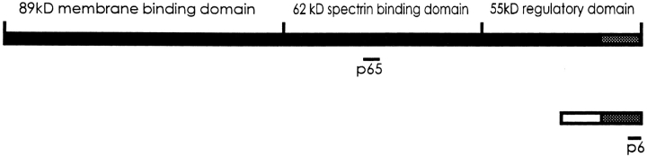
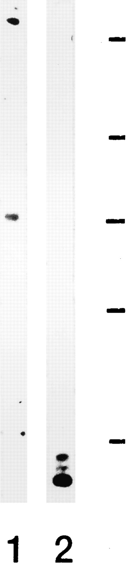
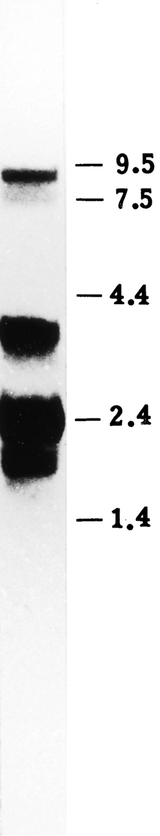
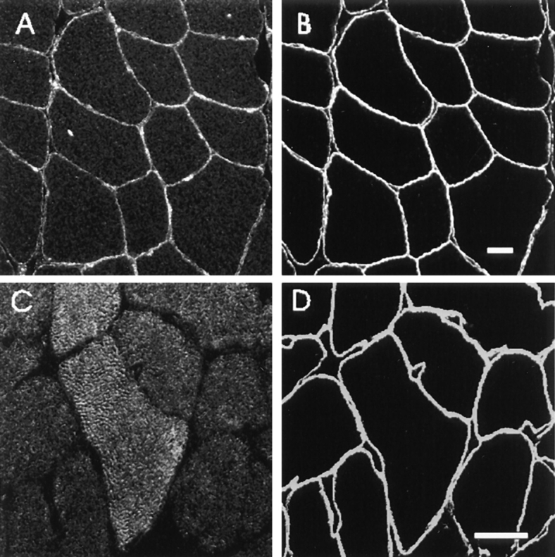
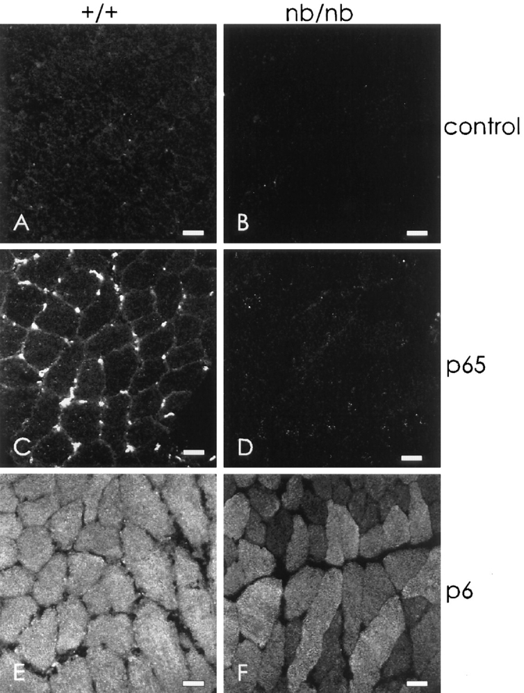
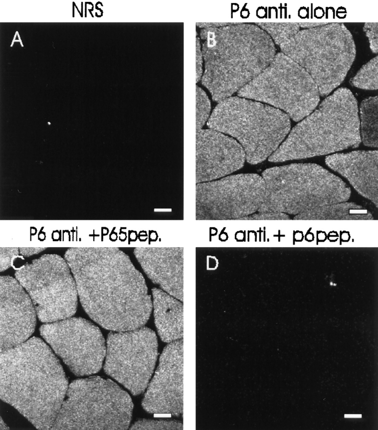
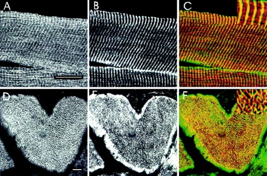
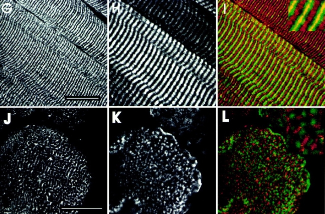
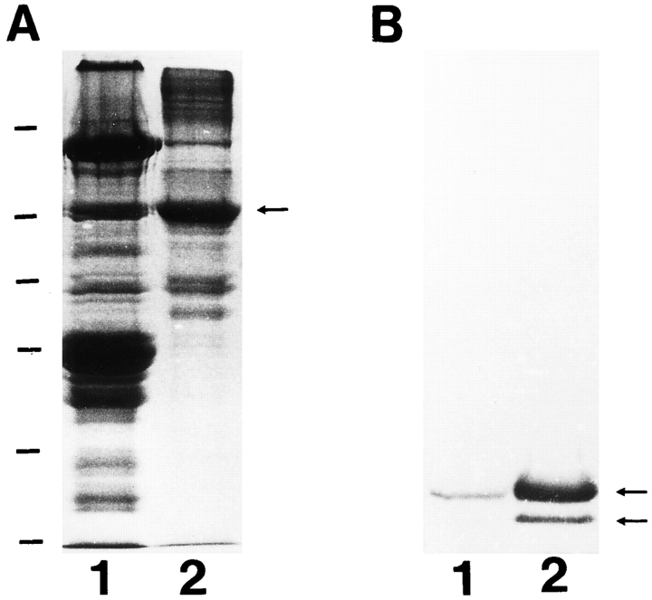
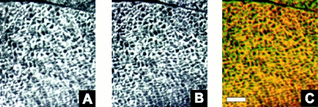
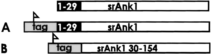
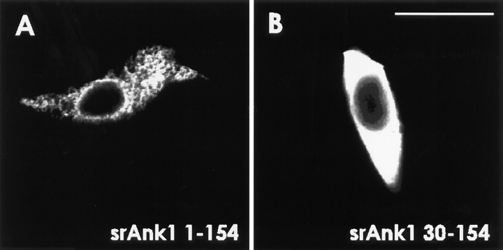
We have recently found that the erythroid ankyrin gene, Ank1, expresses isoforms in mouse skeletal muscle, several of which share COOH-terminal sequence with previously known Ank1 isoforms but have a novel, highly hydrophobic 72-amino acid segment at their NH2 termini. Here, through the use of domain-specific peptide antibodies, we report the presence of the small ankyrins in rat and rabbit skeletal muscle and demonstrate their selective association with the sarcoplasmic reticulum. In frozen sections of rat skeletal muscle, antibodies to the spectrin-binding domain (anti-p65) react only with a 210-kD Ank1 and label the sarcolemma and nuclei, while antibodies to the COOH terminus of the small ankyrin (anti-p6) react with peptides of 20 to 26 kD on immunoblots and decorate the myoplasm in a reticular pattern. Mice homozygous for the normoblastosis mutation (gene symbol nb) are deficient in the 210-kD ankyrin but contain normal levels of the small ankyrins in the myoplasm. In nb/nb skeletal muscle, anti-p65 label is absent from the sarcolemma, whereas anti-p6 label shows the same distribution as in control skeletal muscle. In normal skeletal muscle of the rat, anti-p6 decorates Z lines, as defined by antidesmin distribution, and is also present at M lines where it surrounds the thick myosin filaments. Immunoblots of the proteins isolated with rabbit sarcoplasmic reticulum indicate that the small ankyrins are highly enriched in this fraction. When expressed in transfected HEK 293 cells, the small ankyrins are distributed in a reticular pattern resembling the ER if the NH2-terminal hydrophobic domain is present, but they are uniformly distributed in the cytosol if this domain is absent. These results suggest that the small ankyrins are integral membrane proteins of the sarcoplasmic reticulum. We propose that, unlike the 210-kD form of Ank1, previously localized to the sarcolemma and believed to be a part of the supporting cytoskeleton, the small Ank1 isoforms may stabilize the sarcoplasmic reticulum by linking it to the contractile apparatus.


Figures

Figure 2
Figure 2
Diagrammatic representation of the small, alternatively spliced ankyrin of skeletal muscle relative to the large, erythroid ankyrin. The three major domains of a typical erythroid ankyrin are indicated. The region of the small ankyrin shared with the larger protein is shown by a shaded bar. The novel region containing a stretch of hydrophobic amino acids at the NH2 terminus is shown as an open bar. The locations of the epitopes of the two antibodies, anti-p65 and anti-p6, are underlined. The long isoform depicted in this figure is Cb14/11 from Birkenmeier et al., 1993.
Figure 3
Figure 3
Imunoblotting of Ank1 in skeletal muscle by peptide-specific antibodies. Proteins in a homogenate of rat skeletal muscle were separated by SDS-PAGE and transferred to a nitrocellulose membrane. Strips of the membrane from the same gel were incubated with antibodies to the spectrinbinding domain of Ank1 (anti-p65; lane 1) and to the COOH-terminal region of the small ankyrins (anti-p6; lane 2), followed by alkaline phosphatase-conjugated secondary antibodies. The molecular mass standards are shown to the right of lane 2 by dashes (from top to bottom, in kD): 200, 97, 68, 43, and 29.
Figure 1
Figure 1
Northern blot analysis of erythroid ankyrin in rat skeletal muscle. Aliquots containing 10 μg of skeletal muscle mRNA were fractionated on a 1% agarose gel and transferred to a nylon membrane. The membrane was hybridized with full-length cDNA probes for erythroid ankyrin. The standards (in kb) are shown to the right. The results show the presence of three predominant transcripts at 2.0, 2.4, and 3.5 kb, all of which are too small to encode the typical ankyrin containing membrane-binding, spectrin-binding, and regulatory domains. An additional transcript at 9.0 kb and a minor transcript at 7.5 kb, also detected in the blots, are long enough to encode such an ankyrin.
Figure 4
Figure 4
Distribution of Ank1 proteins in skeletal muscle. Cross sections of adult rat diaphragm were labeled with the polyclonal p65 antibody to the spectrin-binding domain of Ank1 (A) and the p6 antibody to the small ankyrins (C). The sections were doublelabeled with monoclonal antibodies to syntrophin to mark the sarcolemma (B and D). The bound antibodies were detected with rhodamine-conjugated goat anti–rabbit IgG and FITC-conjugated goat anti–mouse IgG secondary antibodies. The results show that the 210-kD (and perhaps the 70-kD) ankyrin of skeletal muscle is distributed at the sarcolemma and nuclei while the small ankyrins are inside the muscle fibers. Bars, 20 μm.
Figure 6
Figure 6
The small Ank1 isoforms in skeletal muscle are unaffected by the nb/nb mutation. Cross sections of diaphragms from wild-type (A, C, and E) and nb/nb (B, D, and F) mice were labeled with nonimmune rabbit serum (A and B), p65 antibodies to the spectrin-binding domain of ankyrin 1 (C and D), and p6 antibodies to the small ankyrins (E and F), followed by rhodamineconjugated secondary antibodies. Samples were taken from unperfused animals, so erythrocytes remaining in the capillaries could react with the antibodies if they contained the appropriate antigens. The results show that skeletal muscle fibers in the nb/nb mouse selectively lack the 210-kD Ank1 at the sarcolemma but are not deficient in the small myoplasmic forms. Bars, 20 μm.
Figure 5
Figure 5
Specificity of antibodies to the small ankyrins assessed by immunofluorescence. Cross sections of rat diaphragm were labeled with nonimmune rabbit serum (A), p6 antibody to the small ankyrins (B), p6 antibodies preincubated with a 100-fold molar excess of irrelevant peptide (C), and the antibodies preincubated with a 100-fold molar excess of the antigenic peptide (D), followed by rhodamine-conjugated secondary antibodies. All the settings used to obtain these images on the laser scanning confocal microscope were identical. C and D show adjacent serial sections. Bars, 20 μm.
Figure 7
Figure 7
The small skeletal muscle ankyrins surround contractile structures at the Z and M lines. Cross sections (D–F and J–L) and longitudinal sections (A–C and G–I) of rat diaphragm were double labeled with polyclonal p6 antibodies to the small ankyrins (A, D, G, and J) and monoclonal antibodies to desmin (B and E) or myosin (H and K), followed by rhodamine-conjugated goat anti–rabbit IgG and FITC-conjugated goat anti–mouse IgG. To compare the paired antibodies, confocal microscopic images from rhodamine and fluorescein channels were overlain (C, F, I, and L). In the overlays, desmin and myosin are shown in green and the small ankyrins in red; regions containing both the small ankyrins and myosin or desmin are shown in yellow. Higher magnification views of selected regions are shown as inserts. The small ankyrins were found not only at Z lines (depicted in yellow in C and F) but also at the M lines (depicted by yellow in I), where it surrounds myosin in the thick filaments (L). Bars, 20 μm.
Figure 7
Figure 7
The small skeletal muscle ankyrins surround contractile structures at the Z and M lines. Cross sections (D–F and J–L) and longitudinal sections (A–C and G–I) of rat diaphragm were double labeled with polyclonal p6 antibodies to the small ankyrins (A, D, G, and J) and monoclonal antibodies to desmin (B and E) or myosin (H and K), followed by rhodamine-conjugated goat anti–rabbit IgG and FITC-conjugated goat anti–mouse IgG. To compare the paired antibodies, confocal microscopic images from rhodamine and fluorescein channels were overlain (C, F, I, and L). In the overlays, desmin and myosin are shown in green and the small ankyrins in red; regions containing both the small ankyrins and myosin or desmin are shown in yellow. Higher magnification views of selected regions are shown as inserts. The small ankyrins were found not only at Z lines (depicted in yellow in C and F) but also at the M lines (depicted by yellow in I), where it surrounds myosin in the thick filaments (L). Bars, 20 μm.
Figure 8
Figure 8
The small ankyrins of skeletal muscle are highly enriched in purified sarcoplasmic reticulum. Protein (50 μg) from total homogenate of rabbit skeletal muscle (lanes 1) and from purified sarcoplasmic reticulum (lanes 2) were separated by SDSPAGE (17% acrylamide gel) and either stained with Coomassie blue (A) or transferred to nitrocellulose filters and blotted with p6 antibodies to the small ankyrins (B). (A) Coomassie blue staining of the gel. The arrow indicates the position of the Ca2+-ATPase. (B) Immunoblotting. Arrows indicate the small ankyrins. Molecular mass standards, shown to the left of A, are, from top to bottom, (in kD): 200, 97, 68, 43, 29, and 18.
Figure 9
Figure 9
Comparison of the distribution of the small ankyrins and the SERCA1 ATPase in rat skeletal muscle. Frozen cross sections (5 μm) of the extensor digitorum longus muscle of the rat were prepared and double-labeled by immunofluorescence with anti-p6 antibodies to the small ankyrins and monoclonal antibodies to the SERCA1 ATPase of fast twitch muscle fibers, as described in Materials and Methods. Images were obtained by confocal microscopy. (A) SERCA ATPase, visualized with fluoresceinated anti-mouse IgG. (B) Small ankyrins, visualized with tetramethylrhodamine-conjugated anti-rabbit IgG. (C) Computer overlay of the images in A and B, in which structures labeled by both antibodies appear yellow. There is extensive coincidence of the two labels. Bar, 6 μm.
Figure 10
Figure 10
Small ankyrins expressed in transfected HEK 293 cells with or without the NH2-terminal hydrophobic domain. cDNA sequences encoding amino acids 1–154 or 30– 154 of the small ankyrins were obtained by RT-PCR and cloned into pcDNA3.1 HisA, which introduces an NH2-terminal FLAG tag. Plasmid DNA was introduced as a calcium phosphate precipitate into HEK 293 cells. 1 d later, cells were fixed, permeabilized, and labeled with anti-p6. Cells expressing the fulllength, tagged protein (srAnk1 1–154) were labeled in a reticular pattern in the cytoplasm (A), whereas cells expressing the truncated version of the protein that lacked the hydrophobic NH2-terminal sequence (srAnk1 30–154) were labeled uniformly in the cytoplasm (B). Identical results were obtained when transfected cells were labeled with anti-FLAG (data not shown). Bar, 20 μm.
Figure 10
Figure 10
Small ankyrins expressed in transfected HEK 293 cells with or without the NH2-terminal hydrophobic domain. cDNA sequences encoding amino acids 1–154 or 30– 154 of the small ankyrins were obtained by RT-PCR and cloned into pcDNA3.1 HisA, which introduces an NH2-terminal FLAG tag. Plasmid DNA was introduced as a calcium phosphate precipitate into HEK 293 cells. 1 d later, cells were fixed, permeabilized, and labeled with anti-p6. Cells expressing the fulllength, tagged protein (srAnk1 1–154) were labeled in a reticular pattern in the cytoplasm (A), whereas cells expressing the truncated version of the protein that lacked the hydrophobic NH2-terminal sequence (srAnk1 30–154) were labeled uniformly in the cytoplasm (B). Identical results were obtained when transfected cells were labeled with anti-FLAG (data not shown). Bar, 20 μm.

References

    1. Bennett V. Purification of an active proteolytic fragment of the membrane attachment site for human erythrocyte spectrin. J Biol Chem. 1978;253:2292–2299. - PubMed
    1. Bennett V, Davis J. Erythrocyte ankyrin: immunoreactive analogues are associated with mitotic structures in cultured cells and with the microtubules in brain. Proc Natl Acad Sci USA. 1981;78:7550–7554. - PMC - PubMed
    1. Bennett V, Gilligan DM. The spectrin-based membrane skeleton and micron-scale organization of the plasma membrane. Annu Rev Cell Biol. 1993;9:27–66. - PubMed
    1. Birkenmeier CS, White RA, Peters LL, Hall EJ, Lux SE, Barker JE. Complex patterns of sequence variation and multiple 5′ and 3′ ends are found among transcripts of the erythroid ankyrin gene. J Biol Chem. 1993;268:9533–9540. - PubMed
    1. Bodine DM, Birkenmeier CS, Barker JE. Spectrin deficient inherited hemolytic anemias in the mouse: characterization by spectrin synthesis and mRNA activity in reticulocytes. Cell. 1984;37:721–729. - PubMed

Publication types