. 1997 Feb 10;136(3):717-27.
doi: 10.1083/jcb.136.3.717.

Threshold levels of fluid shear promote leukocyte adhesion through selectins (CD62L,P,E)

Affiliations

Threshold levels of fluid shear promote leukocyte adhesion through selectins (CD62L,P,E)

M B Lawrence et al. J Cell Biol. .

Erratum in

  • J Cell Biol 1997 Apr 7;137(1):261

Abstract

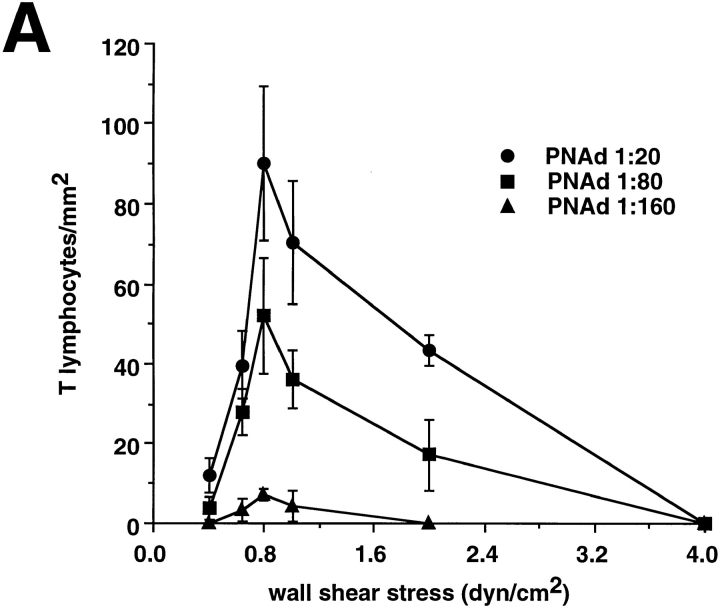
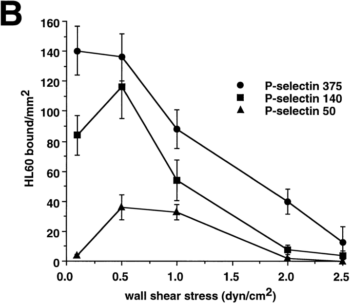
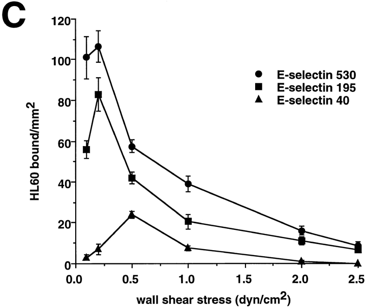
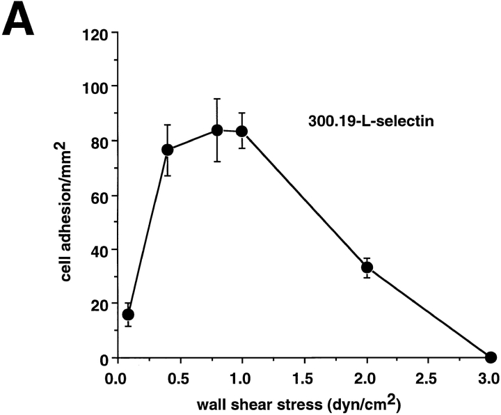
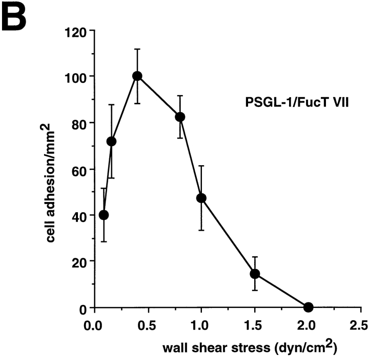
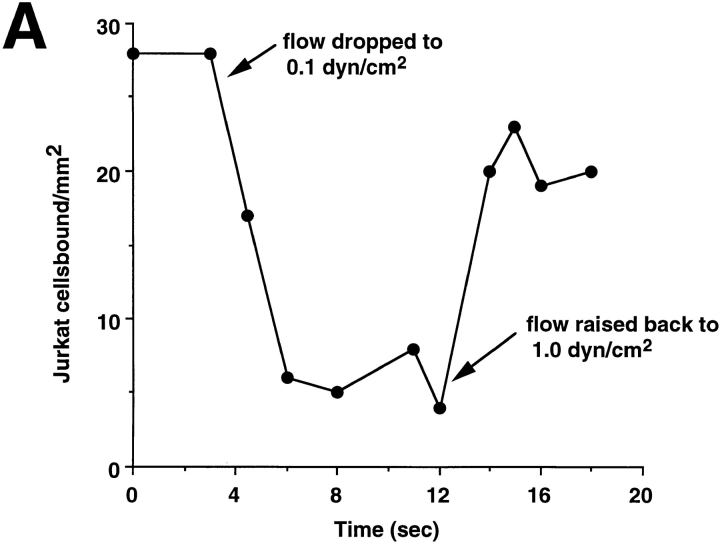
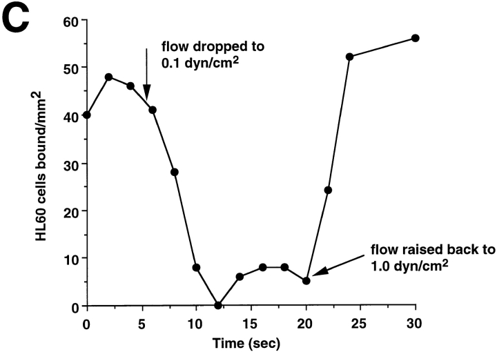
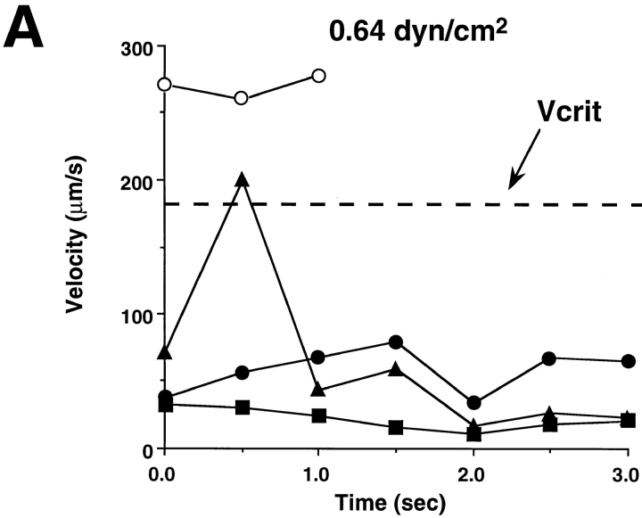
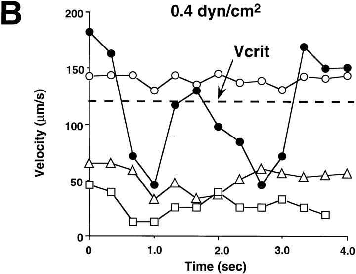
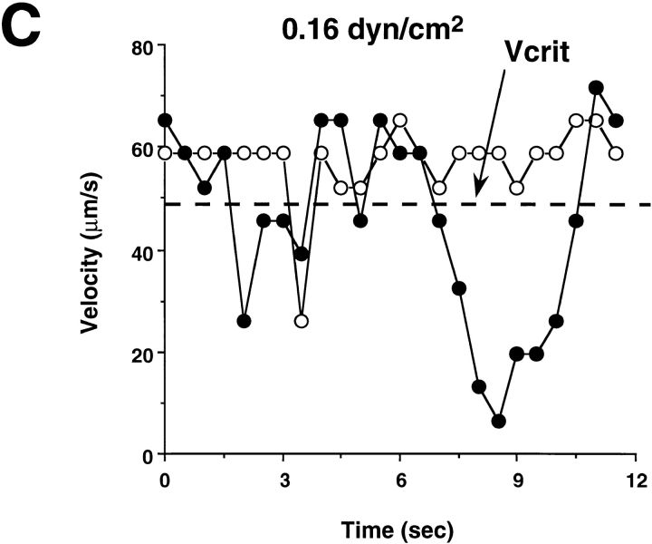
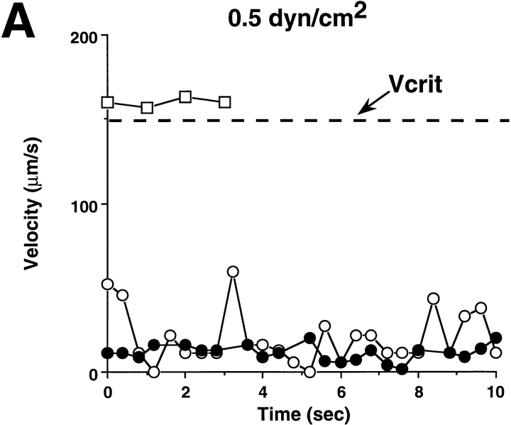
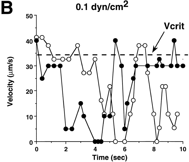
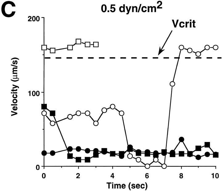
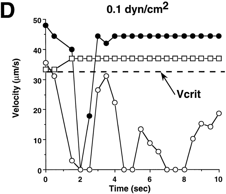
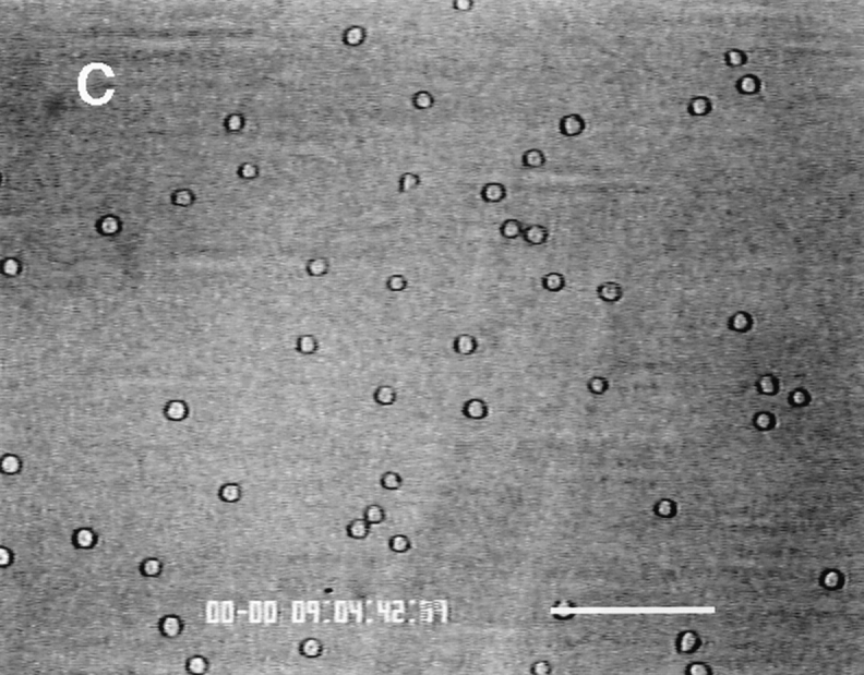
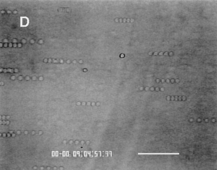
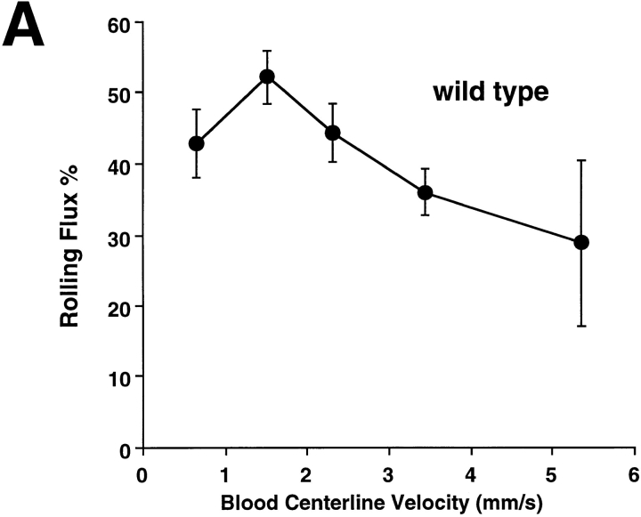
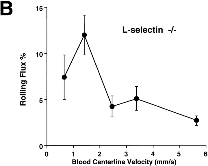
Leukocyte adhesion through L-selectin to peripheral node addressin (PNAd, also known as MECA-79 antigen), an L-selectin ligand expressed on high endothelial venules, has been shown to require a minimum level of fluid shear stress to sustain rolling interactions (Finger, E.B., K.D. Puri, R. Alon, M.B. Lawrence, V.H. von Andrian, and T.A. Springer. 1996. Nature (Lond.). 379:266-269). Here, we show that fluid shear above a threshold of 0.5 dyn/cm2 wall shear stress significantly enhances HL-60 myelocyte rolling on P- and E-selectin at site densities of 200/microm2 and below. In addition, gravitational force is sufficient to detach HL-60 cells from P- and E-selectin substrates in the absence, but not in the presence, of flow. It appears that fluid shear-induced torque is critical for the maintenance of leukocyte rolling. K562 cells transfected with P-selectin glycoprotein ligand-1, a ligand for P-selectin, showed a similar reduction in rolling on P-selectin as the wall shear stress was lowered below 0.5 dyn/cm2. Similarly, 300.19 cells transfected with L-selectin failed to roll on PNAd below this level of wall shear stress, indicating that the requirement for minimum levels of shear force is not cell type specific. Rolling of leukocytes mediated by the selectins could be reinitiated within seconds by increasing the level of wall shear stress, suggesting that fluid shear did not modulate receptor avidity. Intravital microscopy of cremaster muscle venules indicated that the leukocyte rolling flux fraction was reduced at blood centerline velocities less than 1 mm/s in a model in which rolling is mediated by L- and P-selectin. Similar observations were made in L-selectin-deficient mice in which leukocyte rolling is entirely P-selectin dependent. Leukocyte adhesion through all three selectins appears to be significantly enhanced by a threshold level of fluid shear stress.


Figures

Figure 1
Figure 1
Biphasic pattern of leukocyte adhesion through L-, P-, and E-selectin under flow conditions. (A) T lymphocytes were perfused over surface immobilized PNAd at wall shear stresses ranging from 0.4 to 4.0 dyn/cm2. Time of flow was adjusted to control for cell flux though the flow chamber (see Materials and Methods). PNAd was adsorbed at the indicated dilutions (see Materials and Methods). Adhesions were defined as cells remaining attached to the substrate for a minimum of 0.4 s (12 frames of videotape). Data points represent the means (±SEM) of three independent determinations. (B) HL-60 cells were perfused over immobilized purified P-selectin at the indicated wall shear stresses and P-selectin site densities (sites/μm2). (C) HL-60 cells were perfused over immobilized purified E-selectin at the indicated wall shear stresses and E-selectin site densities (sites/μm2). Data points represent means (±SEM) of rolling HL-60 cells averaged over 12–20 fields of view.
Figure 1
Figure 1
Biphasic pattern of leukocyte adhesion through L-, P-, and E-selectin under flow conditions. (A) T lymphocytes were perfused over surface immobilized PNAd at wall shear stresses ranging from 0.4 to 4.0 dyn/cm2. Time of flow was adjusted to control for cell flux though the flow chamber (see Materials and Methods). PNAd was adsorbed at the indicated dilutions (see Materials and Methods). Adhesions were defined as cells remaining attached to the substrate for a minimum of 0.4 s (12 frames of videotape). Data points represent the means (±SEM) of three independent determinations. (B) HL-60 cells were perfused over immobilized purified P-selectin at the indicated wall shear stresses and P-selectin site densities (sites/μm2). (C) HL-60 cells were perfused over immobilized purified E-selectin at the indicated wall shear stresses and E-selectin site densities (sites/μm2). Data points represent means (±SEM) of rolling HL-60 cells averaged over 12–20 fields of view.
Figure 2
Figure 2
Attachment in flow of L-selectin transfectants to PNAd (A) and PSGL-1/FucT VII transfectants to P-selectin (B). 300.19 cells transfected with L-selectin were perfused over isolated PNAd (1:40) immobilized on plastic at wall shear stresses ranging from 0.1 to 4.0 dyn/cm2. K562 cells transfected with PSGL-1 and FucT VII were perfused over isolated P-selectin (140 sites/μm2) at wall shear stresses ranging from 0.1 to 4.0 dyn/cm2. Transfectants forming rolling adhesions for 0.4 s or longer were counted as adherent. Binding of both transfected cell lines to their respective substrates was 100% EDTA dependent. Figure represents the mean of two independent experiments for each cell type. Each point represents the average number (±SEM) of cells bound in a minimum of 15 fields of view.
Figure 2
Figure 2
Attachment in flow of L-selectin transfectants to PNAd (A) and PSGL-1/FucT VII transfectants to P-selectin (B). 300.19 cells transfected with L-selectin were perfused over isolated PNAd (1:40) immobilized on plastic at wall shear stresses ranging from 0.1 to 4.0 dyn/cm2. K562 cells transfected with PSGL-1 and FucT VII were perfused over isolated P-selectin (140 sites/μm2) at wall shear stresses ranging from 0.1 to 4.0 dyn/cm2. Transfectants forming rolling adhesions for 0.4 s or longer were counted as adherent. Binding of both transfected cell lines to their respective substrates was 100% EDTA dependent. Figure represents the mean of two independent experiments for each cell type. Each point represents the average number (±SEM) of cells bound in a minimum of 15 fields of view.
Figure 3
Figure 3
Changes in shear dynamically regulate selectin mediated rolling. (A) Jurkat T lymphoma cells (which express L-selectin [Lawrence et al., 1995]) were perfused over immobilized PNAd (1:40 dilution) at 1.0 dyn/cm2 wall shear stress before the flow was reduced to 0.1 dyn/cm2 using an electronically controlled syringe pump. Individual Jurkat T lymphocytes were tracked frame by frame on the video monitor. The criteria for adhesion was a rolling velocity of less than the Vcrit (see Materials and Methods) of a noninteracting lymphocyte flowing at 0.1 dyn/cm2. Flow was raised back to 1.0 dyn/cm2 and the numbers of interacting Jurkat cells counted as a function of time. Data shown is representative of four experiments. For B and C, the assay procedure was identical, except that HL-60 cells were used in place of Jurkat cells to assess the dynamic adhesion of leukocytes with (B) P-selectin (140 sites/μm2), and (C) E-selectin (195 sites/μm2).
Figure 3
Figure 3
Changes in shear dynamically regulate selectin mediated rolling. (A) Jurkat T lymphoma cells (which express L-selectin [Lawrence et al., 1995]) were perfused over immobilized PNAd (1:40 dilution) at 1.0 dyn/cm2 wall shear stress before the flow was reduced to 0.1 dyn/cm2 using an electronically controlled syringe pump. Individual Jurkat T lymphocytes were tracked frame by frame on the video monitor. The criteria for adhesion was a rolling velocity of less than the Vcrit (see Materials and Methods) of a noninteracting lymphocyte flowing at 0.1 dyn/cm2. Flow was raised back to 1.0 dyn/cm2 and the numbers of interacting Jurkat cells counted as a function of time. Data shown is representative of four experiments. For B and C, the assay procedure was identical, except that HL-60 cells were used in place of Jurkat cells to assess the dynamic adhesion of leukocytes with (B) P-selectin (140 sites/μm2), and (C) E-selectin (195 sites/μm2).
Figure 4
Figure 4
Effect of wall shear stress on duration of L-selectin– mediated rolling interactions of leukocytes. Using videomicroscopy, the instantaneous velocity of flowing cells was determined by cell tracking. A single field of view was chosen, and the position of individual cells was tracked on a video monitor. All arrests or tethering events that lasted at least 1/15 s were counted as adhesive interactions. In A, the velocities of four Jurkat T lymphocyte cells were determined at 0.64 dyn/cm2 wall shear stress on PNAd at 1:60 dilution (see Materials and Methods). In B, wall shear stress was 0.4 dyn/cm2, and in C, wall shear stress was 0.16 dyn/cm2.
Figure 4
Figure 4
Effect of wall shear stress on duration of L-selectin– mediated rolling interactions of leukocytes. Using videomicroscopy, the instantaneous velocity of flowing cells was determined by cell tracking. A single field of view was chosen, and the position of individual cells was tracked on a video monitor. All arrests or tethering events that lasted at least 1/15 s were counted as adhesive interactions. In A, the velocities of four Jurkat T lymphocyte cells were determined at 0.64 dyn/cm2 wall shear stress on PNAd at 1:60 dilution (see Materials and Methods). In B, wall shear stress was 0.4 dyn/cm2, and in C, wall shear stress was 0.16 dyn/cm2.
Figure 5
Figure 5
Effect of wall shear stress on duration of P- and E-selectin–mediated rolling interactions of leukocytes. HL-60 cells were perfused over P- or E-selectin–coated substrates at the indicated wall shear stresses. For P-selectin substrates (140 sites/μm2, A and B), wall shear stress was 0.5 and 0.1 dyn/cm2, respectively. For E-selectin substrates (195 sites/μm2, C and D), wall shear stress was 0.5 and 0.1 dyn/ cm2, respectively.
Figure 5
Figure 5
Effect of wall shear stress on duration of P- and E-selectin–mediated rolling interactions of leukocytes. HL-60 cells were perfused over P- or E-selectin–coated substrates at the indicated wall shear stresses. For P-selectin substrates (140 sites/μm2, A and B), wall shear stress was 0.5 and 0.1 dyn/cm2, respectively. For E-selectin substrates (195 sites/μm2, C and D), wall shear stress was 0.5 and 0.1 dyn/ cm2, respectively.
Figure 6
Figure 6
Sequence of photomicrographs of HL-60 cells rolling on either P- or E-selectin before and after flowrate was lowered. A suspension of HL-60 cells was perfused over the selectin-coated substrate at 1.0 dyn/cm2 wall shear stress with the flow chamber in the normal orientation before being inverted along the axis of flow (see Materials and Methods). Images in each panel consist of five superimposed video frames (0.1 s apart) to show the multiple images of fast-moving nonadherent HL-60 cells compared to slow-rolling, adherent HL-60 cells. (A) HL-60 cells are rolling on P-selectin (140 sites/μm2) at 1.0 dyn/cm2 wall shear stress 30 s after the chamber was inverted. (B) 15 s after wall shear stress was lowered to 0.1 dyn/cm2, showing that all but one HL-60 cell have released from the surface. (C) HL-60 cells are rolling on E-selectin (195 sites/μm2) at 1.0 dyn/cm2 wall shear stress 40 s after inversion of flow chamber. (D) 14 s after wall shear stress was lowered to 0.1 dyn/cm2, showing that all but one HL-60 cell have released from the surface. In B and D, HL-60 cells released or detached from the substrate after the drop in wall shear stress are free to fall away from the wall of the flow chamber under the influence of gravity sedimentation and therefore are prevented from reattaching. Bar, 100 μm.
Figure 6
Figure 6
Sequence of photomicrographs of HL-60 cells rolling on either P- or E-selectin before and after flowrate was lowered. A suspension of HL-60 cells was perfused over the selectin-coated substrate at 1.0 dyn/cm2 wall shear stress with the flow chamber in the normal orientation before being inverted along the axis of flow (see Materials and Methods). Images in each panel consist of five superimposed video frames (0.1 s apart) to show the multiple images of fast-moving nonadherent HL-60 cells compared to slow-rolling, adherent HL-60 cells. (A) HL-60 cells are rolling on P-selectin (140 sites/μm2) at 1.0 dyn/cm2 wall shear stress 30 s after the chamber was inverted. (B) 15 s after wall shear stress was lowered to 0.1 dyn/cm2, showing that all but one HL-60 cell have released from the surface. (C) HL-60 cells are rolling on E-selectin (195 sites/μm2) at 1.0 dyn/cm2 wall shear stress 40 s after inversion of flow chamber. (D) 14 s after wall shear stress was lowered to 0.1 dyn/cm2, showing that all but one HL-60 cell have released from the surface. In B and D, HL-60 cells released or detached from the substrate after the drop in wall shear stress are free to fall away from the wall of the flow chamber under the influence of gravity sedimentation and therefore are prevented from reattaching. Bar, 100 μm.
Figure 7
Figure 7
Dependence of in vivo leukocyte rolling flux fraction on vessel centerline blood velocity. (A) Leukocyte rolling in 151 venules of the cremaster muscle of 12 wild-type mice after surgical exteriorization at t = 0 min. Mean ± SEM of leukocyte rolling flux fraction grouped in velocity classes spanning 1 mm/s (5–16 venules per class). (B) Leukocyte rolling flux in 54 venules of five L-selectin–deficient mice after surgical exteriorization. Rolling under these conditions is entirely P-selectin dependent as shown by antibody–blocking studies (Ley et al., 1995). Mean ± SEM, class width 1 mm/s, highest class all venules with velocities >4 mm/s.
Figure 7
Figure 7
Dependence of in vivo leukocyte rolling flux fraction on vessel centerline blood velocity. (A) Leukocyte rolling in 151 venules of the cremaster muscle of 12 wild-type mice after surgical exteriorization at t = 0 min. Mean ± SEM of leukocyte rolling flux fraction grouped in velocity classes spanning 1 mm/s (5–16 venules per class). (B) Leukocyte rolling flux in 54 venules of five L-selectin–deficient mice after surgical exteriorization. Rolling under these conditions is entirely P-selectin dependent as shown by antibody–blocking studies (Ley et al., 1995). Mean ± SEM, class width 1 mm/s, highest class all venules with velocities >4 mm/s.

References

    1. Alon R, Hammer DA, Springer TA. Lifetime of the P-selectincarbohydrate bond and its response to tensile force in hydrodynamic flow. Nature (Lond) 1995a;374:539–542. - PubMed
    1. Alon R, Kassner PD, Carr MW, Finger EB, Hemler ME, Springer TA. The integrin VLA-4 supports tethering and rolling in flow on VCAM-1. J Cell Biol. 1995b;128:1243–1253. - PMC - PubMed
    1. Arbones ML, Ord DC, Ley K, Ratech H, Curry-Maynard C, Otten G, Capon DJ, Tedder TF. Lymphocyte homing and leukocyte rolling and migration are impaired in L-selectin-deficient mice. Immunity. 1994;1:247–260. - PubMed
    1. Asako H, Kurose I, Wolf R, Defrees S, Zheng ZL, Phillips ML, Paulson JC, Granger DN. Role of H1-receptors and P-selectin in histamine-induced leukocyte rolling and adhesion in postcapillary venules. J Clin Invest. 1994;93:1508–1515. - PMC - PubMed
    1. Bargatze RF, Kurk S, Butcher EC, Jutila MA. Neutrophils roll on adherent neutrophils bound to cytokine-induced endothelial cells via L-selectin on the rolling cells. J Exp Med. 1994;180:1785–1792. - PMC - PubMed

Publication types

MeSH terms