. 1997 Feb 10;136(3):729-43.
doi: 10.1083/jcb.136.3.729.

Targeted disruption of decorin leads to abnormal collagen fibril morphology and skin fragility

Affiliations

Targeted disruption of decorin leads to abnormal collagen fibril morphology and skin fragility

K G Danielson et al. J Cell Biol. .

Abstract

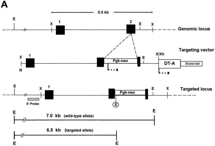
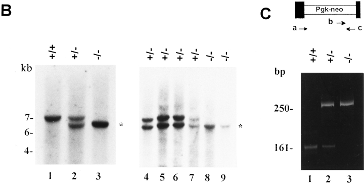
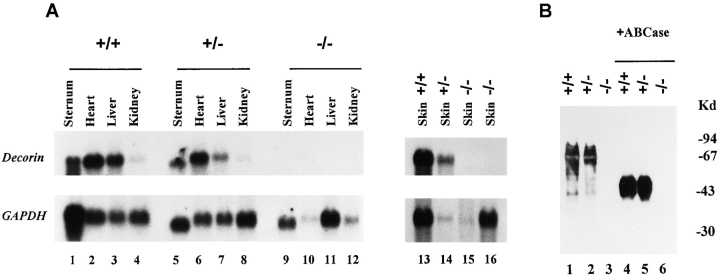
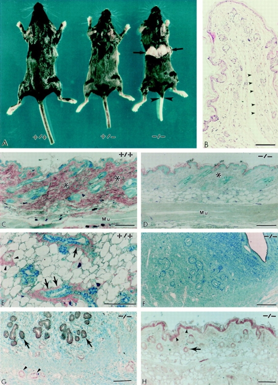
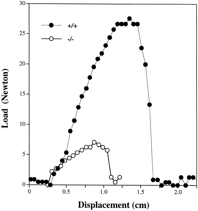
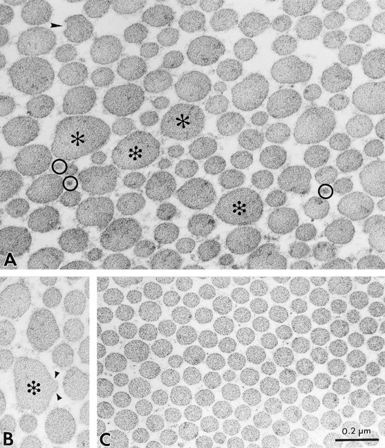
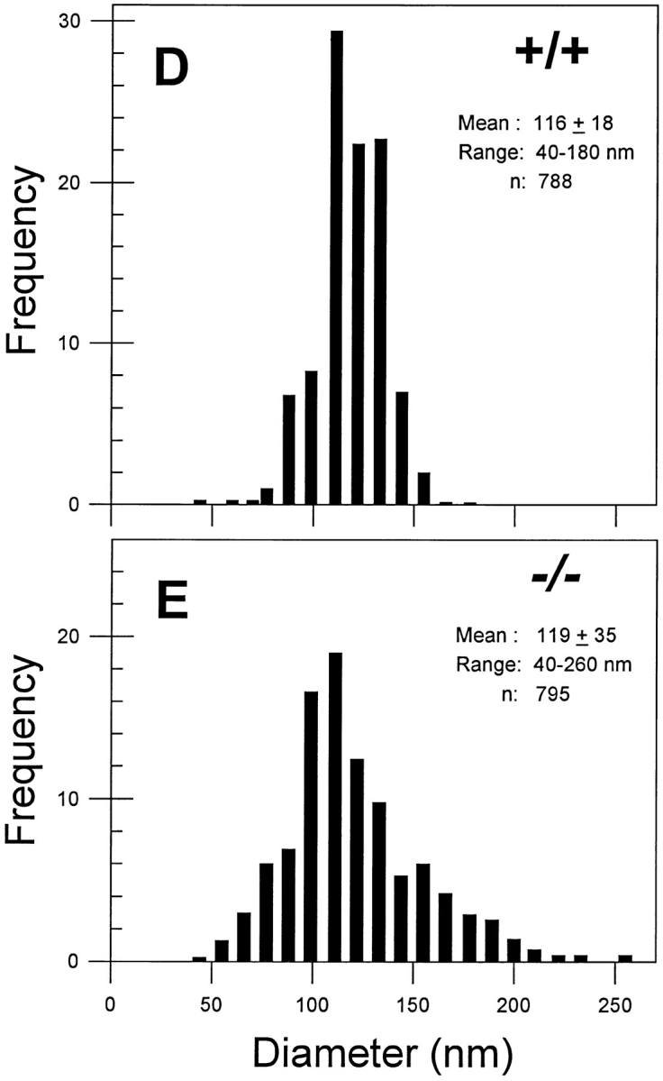
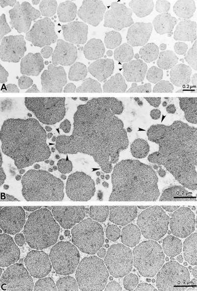
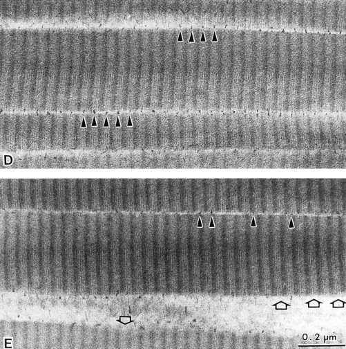
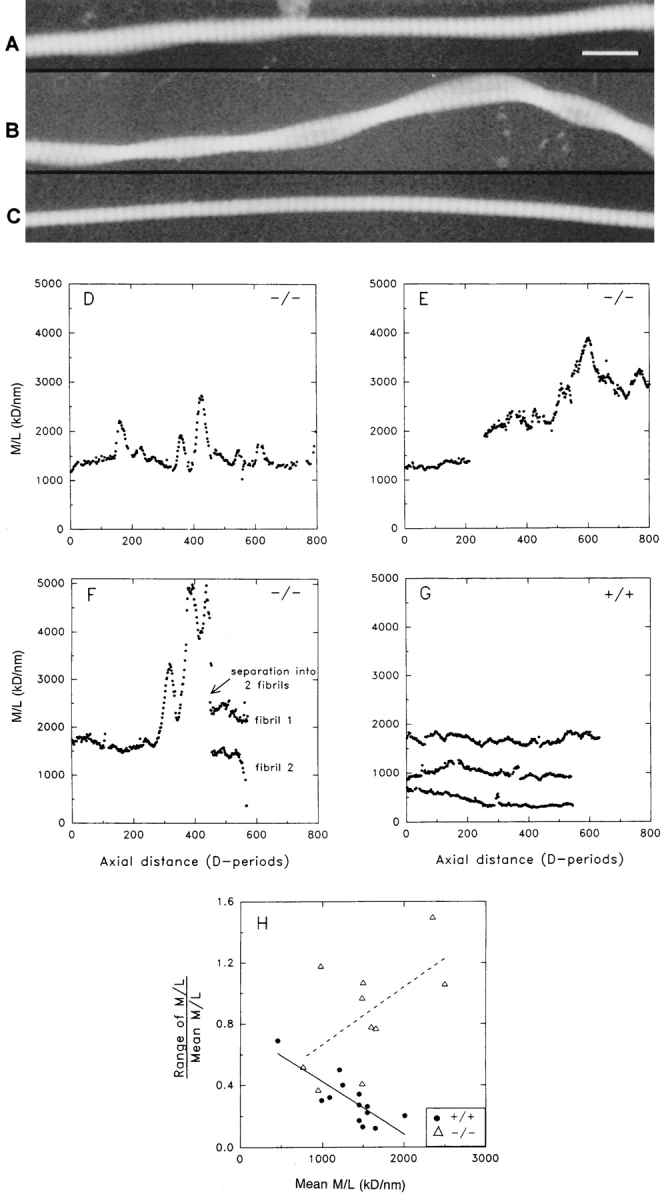
Decorin is a member of the expanding group of widely distributed small leucine-rich proteoglycans that are expected to play important functions in tissue assembly. We report that mice harboring a targeted disruption of the decorin gene are viable but have fragile skin with markedly reduced tensile strength. Ultrastructural analysis revealed abnormal collagen morphology in skin and tendon, with coarser and irregular fiber outlines. Quantitative scanning transmission EM of individual collagen fibrils showed abrupt increases and decreases in mass along their axes. thereby accounting for the irregular outlines and size variability observed in cross-sections. The data indicate uncontrolled lateral fusion of collagen fibrils in the decorindeficient mice and provide an explanation for the reduced tensile strength of the skin. These findings demonstrate a fundamental role for decorin in regulating collagen fiber formation in vivo.


Figures

Figure 1
Figure 1
Disruption of the decorin gene locus in mouse ES cells and generation of decorin-deficient mice. (A) Targeting strategy. A 5.5-kb XbaI genomic fragment encoding exons 1 and 2 (filled boxes, not in scale) was used to construct the targeting vector. The predicted structure of the disrupted allele (bottom panel) shows the 5′ probe used to detect the diagnostic allele of 6.5 kb in contrast with the wild-type allele of 7.0 kb. This was due to the presence of a new EcoRI site in the Pgk-neo cassette. Abbreviations for restriction endonucleases: E, EcoRI; X, XbaI; N, NotI; Xh, XhoI. (B) Southern blot analysis of tail DNA isolated from two separate litters of mice including wild-type (+/ +), heterozygous (+/−), and homozygous (−/−) animals. Lanes 1–3 are from 3-mo-old animals derived from the breeding of two heterozygous mice, while lanes 4–9 are from newborn animals derived from the breeding of a heterozygous male and a homozygous female. The targeted allele of 6.5 kb is labeled by an asterisk. The DNA was separated on a 0.75% agarose gel, transferred to a nitrocellulose filter, and hybridized under high stringency to a PCRgenerated probe 5′ to the targeting vector. The size of molecular weight markers is indicated in the left margin in kb. (C) PCR detection of the targeted allele using primers specific for exon 2 (a and c, top scheme) or Pgk-neo (primer b). By using primer a and c, a fragment of 161 bp was identified in the wild-type (lane 1) and heterozygous animal (lane 2). However, the combination of primer b and c gave rise to a larger fragment of 250 bp, encompassing the 3′ end of the neo cassette and the 3′ end of exon 2, which was detected only in the heterozygous (lane 2) and homozygous (lane 3) animals. The products were separated on a 6% nonreducing polyacrylamide gel and stained with ethidium bromide. The size in bp is indicated in the left margin.
Figure 1
Figure 1
Disruption of the decorin gene locus in mouse ES cells and generation of decorin-deficient mice. (A) Targeting strategy. A 5.5-kb XbaI genomic fragment encoding exons 1 and 2 (filled boxes, not in scale) was used to construct the targeting vector. The predicted structure of the disrupted allele (bottom panel) shows the 5′ probe used to detect the diagnostic allele of 6.5 kb in contrast with the wild-type allele of 7.0 kb. This was due to the presence of a new EcoRI site in the Pgk-neo cassette. Abbreviations for restriction endonucleases: E, EcoRI; X, XbaI; N, NotI; Xh, XhoI. (B) Southern blot analysis of tail DNA isolated from two separate litters of mice including wild-type (+/ +), heterozygous (+/−), and homozygous (−/−) animals. Lanes 1–3 are from 3-mo-old animals derived from the breeding of two heterozygous mice, while lanes 4–9 are from newborn animals derived from the breeding of a heterozygous male and a homozygous female. The targeted allele of 6.5 kb is labeled by an asterisk. The DNA was separated on a 0.75% agarose gel, transferred to a nitrocellulose filter, and hybridized under high stringency to a PCRgenerated probe 5′ to the targeting vector. The size of molecular weight markers is indicated in the left margin in kb. (C) PCR detection of the targeted allele using primers specific for exon 2 (a and c, top scheme) or Pgk-neo (primer b). By using primer a and c, a fragment of 161 bp was identified in the wild-type (lane 1) and heterozygous animal (lane 2). However, the combination of primer b and c gave rise to a larger fragment of 250 bp, encompassing the 3′ end of the neo cassette and the 3′ end of exon 2, which was detected only in the heterozygous (lane 2) and homozygous (lane 3) animals. The products were separated on a 6% nonreducing polyacrylamide gel and stained with ethidium bromide. The size in bp is indicated in the left margin.
Figure 2
Figure 2
Absence of decorin mRNA and protein in animals harboring a homozygous disruption of the decorin gene. (A) Northern blot analysis using total RNA prepared from selected tissues as indicated of wild-type (+/+), heterozygous (+/−), and homozygous (−/−) adult animals. High stringency hybridization was performed using cDNAs encoding the full-length decorin or the housekeeping gene GAPDH as 32P-labeled probes (49). Notice the lack of decorin transcripts in the Dcn−/− animals (lanes 9–12, 15, and 16). (B) Immunoblot analysis of guanidine HCl extracts of tail tissues before or after chondroitinase ABC (+ABCase) digestion using an anti-decorin antibody. Notice the presence of a proteoglycan centering around 94 kD and the presence of a 45-kD unprocessed protein core in the wild-type (lane 1) and heterozygous animal (lane 2); no immunoreactive material was detected in the homozygous animal (lane 3). After chondroitinase ABC digestion, the high molecular mass proteoglycan was converted into two broad bands of 45–50 kD (lanes 4 and 5). Decorin-specific epitopes were detected with a polyclonal antibody (LF-113) directed against a synthetic decorin peptide from the amino-terminal region spanning residues 36–49 of the mouse protein (16).
Figure 3
Figure 3
The phenotype of decorin-deficient mice reveals thinning and fragility of the skin. (A) Gross photograph of wild-type (+/+), heterozygous (+/−), and homozygous (−/−) littermates. Notice the sharp rupture of the back skin in the −/− animal (arrows) and the detachment of the tail skin (arrowheads) that occurred during cervical dislocation. The skin fragility was never observed in either +/+ or +/− animals (n > 300). B is a cross-section of the detached tail skin. Notice the sharp and bloodless line of rupture (arrowheads) along the deeper dermis. Immunohistochemical analysis of skin and skeletal muscle (C and D), mammary gland (E), and uterus (F) from wild-type (+/+) and decorin-deficient (−/−) animals using the LF-113 anti-decorin antibody. Notice the intense immunoreactivity in the dermis of a wild-type animal (C, asterisks) and the fine immunoreactivity on either side of the skeletal muscle (Mu). In contrast, the −/− dermis (D, asterisk), the subcutaneous and perimysial connective tissues, are totally unreactive as are the uterine wall and mucosa (F). As expected, the anti-decorin antibody labeled specifically the adventitia of small blood vessels (E, arrowheads) and the myoepithelial cells and fine connective tissue surrounding mammary ducts (E, arrows). Immunodetection of biglycan using an anti-peptide (LF-106) antibody in uterus (G) and skin (H) from Dcn−/− mice. Notice the “normal” expression of biglycan in the endometrium (G, arrows) and in the intramural small blood vessels (G, arrowheads). As expected, in skin, both the epidermis (H, arrowheads) and the follicular epithelium (H, arrow) were labeled by the anti-biglycan antibodies. Sections were reacted with LF-113 or LF-106 antisera at 1: 1,000 dilution, and then visualized with peroxidase-conjugated IgG (1:200) followed by counterstaining with 0.2% methylene blue. Bar, 100 μm.
Figure 4
Figure 4
The skin of decorin-deficient mice has reduced tensile strength. Freshly excised, dumbbell-shaped samples (4 × 2 cm) of dorsal skin oriented parallel to spine from +/+ (•) or −/− (○) animals were generated on a plastic template, thereby producing skin specimens with a narrow central region (1 cm2) where failure would occur by avoiding stress concentration at the jaws of the grip. The samples were gripped into a tensile testing machine (model 4Z387A; Comten Industries) and stretched to tensile failure at a constant strain rate of 1 mm/s, equivalent to 10%/s. A data collecting system linked to a Macintosh Quadra computer (Apple Computer Inc., Cupertino, CA) was used to generate load/displacement curves. Peak of curve represents point of failure. Tensile strength was computed from maximal load at failure divided by the initial cross-sectional area of the specimen in the reduced test area. Six age-matched animals were analyzed with two specimens per animal. Data points represent a typical experiment from one animal each.
Figure 5
Figure 5
Ultrastructural analysis of skin from the decorin-deficient mice reveals abnormal collagen fibrillogenesis. (A–C) Transmission electron micrographs of dermal collagen from Dcn−/− (A and B) and Dcn+/+ (C) animals. Notice the presence of larger (>200 nm) and irregular fibrils (A and B, asterisks) and the coexistence of smaller (30–40 nm) fibrils (A, circles) in the Dcn−/−. Note also the presence of coarser fibrils exhibiting lateral fusion to an adjacent tapered segment (B, arrowheads). In contrast, collagen from the wild-type mouse (C) showed a more compact and uniform pattern of fibril diameter and distribution. The Dcn+/− collagen pattern was identical to the Dcn+/+ animals (not shown). (D and E) Distribution of collagen fibril diameter in dermal collagen from Dcn+/+ (D) and Dcn−/− (E) animals. Notice that, although the mean diameter is not significantly different between the groups, the range and distribution is quite different in the Dcn−/− animals. Heterozygous animals had a distribution and an average collagen diameter (118 ± 14; n = 1,220) nearly identical to the wild type (not shown). Bar, 0.2 μm.
Figure 5
Figure 5
Ultrastructural analysis of skin from the decorin-deficient mice reveals abnormal collagen fibrillogenesis. (A–C) Transmission electron micrographs of dermal collagen from Dcn−/− (A and B) and Dcn+/+ (C) animals. Notice the presence of larger (>200 nm) and irregular fibrils (A and B, asterisks) and the coexistence of smaller (30–40 nm) fibrils (A, circles) in the Dcn−/−. Note also the presence of coarser fibrils exhibiting lateral fusion to an adjacent tapered segment (B, arrowheads). In contrast, collagen from the wild-type mouse (C) showed a more compact and uniform pattern of fibril diameter and distribution. The Dcn+/− collagen pattern was identical to the Dcn+/+ animals (not shown). (D and E) Distribution of collagen fibril diameter in dermal collagen from Dcn+/+ (D) and Dcn−/− (E) animals. Notice that, although the mean diameter is not significantly different between the groups, the range and distribution is quite different in the Dcn−/− animals. Heterozygous animals had a distribution and an average collagen diameter (118 ± 14; n = 1,220) nearly identical to the wild type (not shown). Bar, 0.2 μm.
Figure 6
Figure 6
Decorin-deficient mice reveal abnormal collagen in tail tendon and a decrease in collagen-associated proteoglycans. (A) Lower power view of cross-sectional area of adult tail tendon collagen from Dcn−/− reveals abnormal shape and multiple lateral fusions. Higher power view of Dcn−/− tendons shows bizarre, gigantic fibrils (>660 nm in largest diameter) with multiple lateral fusions (B, arrowheads), in contrast with the more uniform collagen fibrils of the wild-type animals (C) with an average diameter of ∼200 nm. Transmission electron micrographs of longitudinally sectioned collagen fibrils from Dcn+/+ (D) and Dcn−/− (E) tendon, after fixation in the presence of 0.05% cuprolinic blue and 0.3 M MgCl2, and staining with uranyl acetate to visualize the collagen binding pattern. Notice that the typical, 67-nm periodicity of the type I collagen is maintained in the Dcn−/− mice (E). However, numerous d bands are not occupied by proteoglycan granules (E, spaces between arrowheads and unfilled arrowheads), in contrast with the wild type where nearly every d band is occupied (D). Bar 0.2 μm.
Figure 6
Figure 6
Decorin-deficient mice reveal abnormal collagen in tail tendon and a decrease in collagen-associated proteoglycans. (A) Lower power view of cross-sectional area of adult tail tendon collagen from Dcn−/− reveals abnormal shape and multiple lateral fusions. Higher power view of Dcn−/− tendons shows bizarre, gigantic fibrils (>660 nm in largest diameter) with multiple lateral fusions (B, arrowheads), in contrast with the more uniform collagen fibrils of the wild-type animals (C) with an average diameter of ∼200 nm. Transmission electron micrographs of longitudinally sectioned collagen fibrils from Dcn+/+ (D) and Dcn−/− (E) tendon, after fixation in the presence of 0.05% cuprolinic blue and 0.3 M MgCl2, and staining with uranyl acetate to visualize the collagen binding pattern. Notice that the typical, 67-nm periodicity of the type I collagen is maintained in the Dcn−/− mice (E). However, numerous d bands are not occupied by proteoglycan granules (E, spaces between arrowheads and unfilled arrowheads), in contrast with the wild type where nearly every d band is occupied (D). Bar 0.2 μm.
Figure 7
Figure 7
Mass mapping of collagen fibrils isolated from the skin of decorin-deficient animals reveals pronounced nonuniformity in the axial mass distributions with repeated bulges. (A–C) Typical dark-field STEM images of isolate unstained fibrils from mouse skin. A and B show typical images for fibrils from a decorin-deficient mouse. A shows a single fibril with distinct bulging at two locations over 60 D-periods (1 D = 67 nm) length. B is consistent with two fibrils twisted together in close contact but not fused. C shows a typical fibril from control skin with uniform diameter along its length. (D–G) Axial mass distributions of single fibrils obtained by measurements from STEM images. A measurement of mass per unit length was made every two D-periods along the entire available length of each fibril. Missing points occur where parts of a fibril were clearly contaminated. (D–F) Typical axial mass distribution profiles from the decorin-deficient sample or (G) from wild-type animals. The decorin-deficient sample shows pronounced mass peaks in the fibrial axial mass distribution, which are not found in the control. (H) Scatter plot of the relative variation in M/L along single fibrils against the corresponding mean M/L. First order regression lines are shown superimposed on each set of data. The data from the decorin-deficient samples show an overall increase in the relative intrafibrillar variation in M/L with increasing lateral size (Mean M/L); this contrasts with the control sample that shows the opposite trend. Bar, 200 μm.

References

    1. Andrikopoulos K, Liu X, Keene DR, Jaenisch R, Ramirez F. Targeted mutation in the col5a2gene reveals a regulatory role for type V collagen during matrix assembly. Nat Genet. 1995;9:31–36. - PubMed
    1. Baribault H, Oshima RG. Polarized and functional epithelia can form after the targeted inactivation of both mouse keratin 8 alleles. J Cell Biol. 1991;115:1675–1684. - PMC - PubMed
    1. Baribault H, Price J, Miyai K, Oshima RG. Mid-gestational lethality in mice lacking keratin 8. Genes & Dev. 1993;7:1191–1202. - PubMed
    1. Baribault H, Penner J, Iozzo RV, Wilson-Heiner M. Colorectal hyperplasia and inflammation in keratin 8-deficient FVB/N mice. Genes & Dev. 1994;8:2964–2973. - PubMed
    1. Bavinton JH, Peters DE, Ramshaw AM. A morphologic study of a mild form of ovine dermatosparaxis. J Invest Dermatol. 1985;84:391–395. - PubMed

Publication types