

Synthesis and luminescence spectral characterization of long-lifetime lipid metal-ligand probes
- PMID: 9025912
- PMCID: PMC6906605
- DOI: 10.1006/abio.1996.9869
Synthesis and luminescence spectral characterization of long-lifetime lipid metal-ligand probes
Erratum in
- Anal Biochem 1997 May 1;247(2):465
Abstract
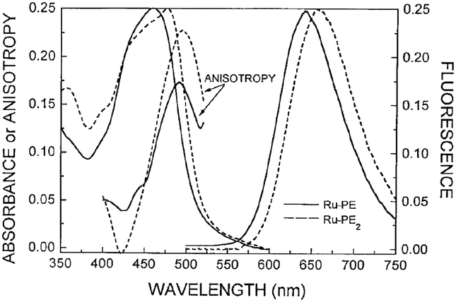
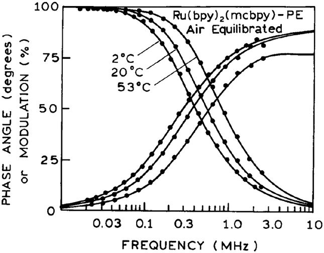
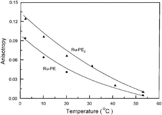
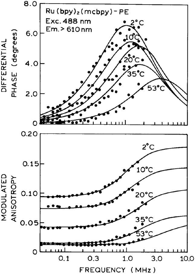
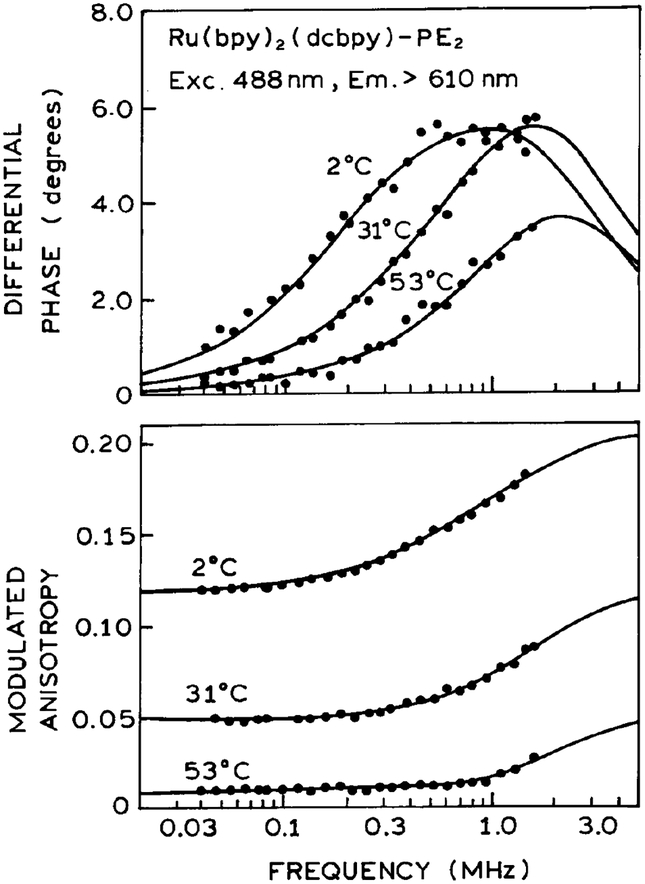
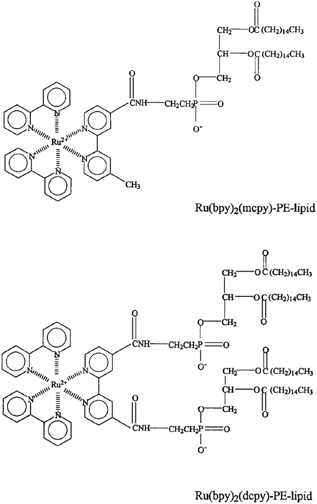
We synthesized phospholipid analogues of phosphatidyl ethanolamine which contains a ruthenium metal-ligand complex (MLC) covalently bound to the amino group. Two analogues were synthesized, containing either one (Ru-PE) or two (Ru-PE2) lipid molecules covalently linked to the MLC by the amino group of the lipid. These MLC-lipid probes display intensity decay times from 682 to 357 ns, depending on temperature. Importantly, the luminescence MLC groups display polarized emission, enabling their use for studies of membrane dynamics. The long intensity decay times allowed measurement of the overall rotation correlation time of lipid vesicles to several microseconds. The spectral properties of the model membranes containing Ru-PE or Ru-PE2 were independent of the probe-to-lipid molar ratio from 1:20 to 1:100, suggesting minimal tendency for probe-probe interactions. These MLC-lipid probes can be expected to have numerous applications in studies of membrane dynamics on the microsecond timescale.
Figures
References
-
- Stubbs CD, and Williams BW (1992) in Topics in Fluores cence Spectroscopy, Vol. 3, Biochemical Applications (Lakowicz JR, Ed.), pp. 231–271, Plenum, New York.
-
- Dewey TG (Ed.) (1991) in Biophysical and Biochemical Aspects of Fluorescence Spectroscopy, p. 294, Plenum, New York.
-
- Davenport L, Knutson JR, and Brand L (1989) in Subcellu lar Biochemistry, (Harris JR, and Etemadi AH, Eds.), pp. 145–188, Plenum, New York.
-
- Shinitzky M, Dianoux AC, Gitler C, and Weber G (1971) Biochemistry 10, 2106–2113. - PubMed
-
- Cogen U, Shinitzky M, Weber G, and Nishida T (1973) Bio chemistry 12, 521–528. - PubMed
Publication types
MeSH terms
Substances
Grants and funding
LinkOut - more resources
Full Text Sources
Other Literature Sources
Miscellaneous
