. 1997 Mar 1;17(5):1633-41.
doi: 10.1523/JNEUROSCI.17-05-01633.1997.

Ectopic alpha2-adrenoceptors couple to N-type Ca2+ channels in axotomized rat sensory neurons

Affiliations

Ectopic alpha2-adrenoceptors couple to N-type Ca2+ channels in axotomized rat sensory neurons

F A Abdulla et al. J Neurosci. .

Abstract

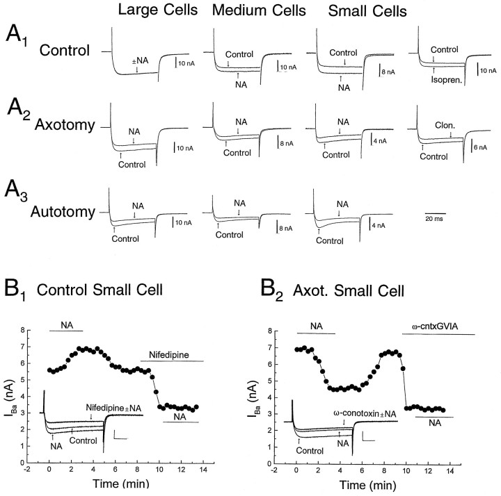
Dorsal root ganglion (DRG) neurons from control rats or from rats in which the sciatic nerve had been sectioned were studied by whole-cell recording techniques. Noradrenaline (10-100 micro;M) activated beta-adrenoceptors and increased L-type Ca2+ channel current in control DRG cells, but this had little effect on excitability (the number of action potentials generated by a pulse of current at rheobasic strength). By contrast, in cells from nerve-damaged animals, noradrenaline activated alpha2-adrenoceptors, suppressed N-type Ca2+ channel current, and increased excitability. In axotomized cells, it also reduced total outward current recorded at +70 mV. Because noradrenaline did not affect total outward current recorded in the presence of the Ca2+ channel blocker Cd2+ (0. 5-1 mM), its effects on excitability may result from reduction of Ca2+-sensitive K+-conductance(s) following suppression of N-type Ca2+ channel current. The strongest effects of noradrenaline were seen in small cells and in cells from animals that exhibited autotomy, a self-mutilatory behavior that can accompany peripheral nerve damage. Because many of these small DRG cells may be involved in the transmission of nociceptive information, changes in coupling between Ca2+ channels and adrenoceptors may contribute to the generation of the ectopic sensory nerve activity that has been implicated in the etiology of neuropathic pain.


Figures

Fig. 1.
Fig. 1.
Effect of noradrenaline on the excitability of rat DRG neurons. Each cell was depolarized with a 200 msec pulse of current at rheobasic strength (current trace not shown) in the absence and presence of 10 μm noradrenaline (NA). Shown are typical recordings from large, medium, and small cells:A1, control animals;A2, axotomized animals;A3, animals exhibiting autotomy.B, Graphic summary of data collected from all cells (i.e., those responding and those insensitive to NA). The excitatory effects of NA (number of spikes discharged in response to a 200 msec pulse of rheobasic current) are seen most clearly in small cells and in cells from animals that exhibit autotomy. Error bars represent SEM. *p < 0.025, **p < 0.002, ***p < 0.001 (paired t test for number of spikes before and after NA application to each cell).
Fig. 2.
Fig. 2.
Effects of NA and Cd2+ on spike width of DRG cells from (A) control animals, (B) axotomized animals, and (C) axotomized animals that exhibited autotomy. The three columns of typical data records illustrate effects of 10 μmNA on spike width in large, medium, and small cells.D, Lack of effect of 1 mm Cd2+on spike width of a control large cell and reduction of spike width observed in a medium and in a small cell.
Fig. 3.
Fig. 3.
Effects of adrenergic drugs, Cd2+, and dihydropyridines on spike width of DRG cells from control animals, axotomized animals, and axotomized animals that exhibited autotomy.A, Increase in AP duration of small control cells induced by 2 μm BAY K 8644 or 10 μmisoprenaline (Isopren.). B, Decrease in spike width of small axotomized cells induced by 1 μmnifedipine (Nifedipine) or 10 μm clonidine (Clon.). C, Occlusion by 1 mmCd2+ of the increase of AP duration invoked in a medium control cell by 10 μm NA. Left trace shows initial NA-induced increase in spike width; right tracewas obtained after washout of NA; Cd2+ decreased AP duration, and subsequent application of NA was unable to restore or increase AP duration. D, Occlusion by 1 mmCd2+ of the decrease of AP duration invoked in a small axotomized cell by 10 μm NA. Left traceshows initial NA-induced decrease in spike width; right trace was obtained after washout of NA; Cd2+decreased AP duration, but subsequent application of NA was unable to invoke a further decrease.
Fig. 4.
Fig. 4.
Increases in excitability invoked by blocking Ca2+ channels with Cd2+ or by activation of α2-adrenoceptors. A, Increase in excitability of a large cell from an axotomized animal induced by 1 mm Cd2+. B, Antagonism by 1 μm yohimbine (Yoh.) of NA-induced enhancement of excitability in a large cell from an animal that exhibited autotomy. Both cells were stimulated with a depolarizing current pulse at rheobasic strength.
Fig. 5.
Fig. 5.
Effects of adrenergic drugs and Ca2+channel blockers on Ba2+ currents of DRG cells from (A1) control animals, (A2) axotomized animals, and (A3) axotomized animals that exhibited autotomy. Shown are effects of 10 μm NA on IBa evoked from a holding potential of −90 mV and recorded at −10 mV for each of the three experimental groups. Note NA-induced potentiation ofIBa in control cells and attenuation of the current in cells from the axotomized and axotomized-autotomy groups. NA exerts the most profound potentiation or suppression ofIBa in small cells, and suppression ofIBa is most pronounced in cells from animals that exhibit autotomy. Potentiation of IBain a small control cell by isoprenaline (Isopren., 10 μm) and suppression in a small cell from an axotomized animal by clonidine (Clon., 10 μm) also are illustrated in A1 andA2, respectively. Calibration (20 msec) refers to all records inA1,A2, andA3.B1, Time course of potentiation of IBa by 10 μm NA in a small control cell. Subsequent superfusion of 2 μm nifedipine suppresses IBa,L and prevents NA-induced potentiation. Data points indicate peak IBaafter a step to −10 mV from a holding potential of −60 mV. Sample data records are shown in the inset; calibration is 3 nA/10 msec. B2, Time course of suppression of IBa in a small cell from an axotomized animal by 10 μm NA and further suppression ofIBa,N by 1 μm ω-cntxGVIA. NA fails to affect the current recorded in the presence of the toxin. Voltage commands are as in B1. Sample data records are shown in inset; calibration is 4 nA/10 msec.
Fig. 6.
Fig. 6.
Lack of effects of NA on outward currents in DRG cells from control animals and suppression of Ca2+-sensitive portion of current in cells from axotomized animals. A1,A2,A3, Lack of effect of 10 μm NA on total outward current recorded in a small cell at +70 mV from a holding potential of −90 mV in the absence and in the presence of 1 mm Cd2+. A1, Graphic representation of the time course of the experiment; each point represents the amplitude of the outward current response to a test pulse to +70 mV evoked once every 20 sec. A2, Superimposed current responses recorded in the presence and absence of NA.A3, Superimposed responses recorded in Cd2+ in the presence and absence of NA.B1,B2, B3, Reduction of Ca2+-sensitive component of outward current in an axotomized small cell by 10 μm NA (voltage commands as inA). B1, Graphic representation of the time course of the experiment; note that NA only suppresses the outward current before the addition of 1 mmCd2+. B2, B3, Superimposed data records to illustrate action of NA and its occlusion by Cd2+.C1,C2,C3, Pharmacology of reduction of outward current in an axotomized small cell by 10 μm NA (voltage commands as in A and B).C1, Graphic representation of the time course of the experiment; note that NA does not suppress the outward current in the presence of yohimbine (1 μm).C2,C3, Superimposed data records to illustrate antagonism of the action of NA by yohimbine. Calibration (20 nA/20 msec) in B refers to both B andC. All voltage traces were omitted for clarity.

References

    1. Akins PT, McCleskey EW. Characterization of potassium currents in adult rat sensory neurons and modulation by opioids and cyclic AMP. Neuroscience. 1993;56:759–769. - PubMed
    1. Bessou P, Perl ER. Response of cutaneous sensory units with unmyelinated fibres to noxious stimuli. J Neurophysiol. 1969;32:1025–1043. - PubMed
    1. Bonica JJ. Causalgia and other reflex sympathetic dystrophies. In: Bonica JJ, editor. The management of pain. Lea and Febiger; Philadelphia: 1990. pp. 220–243.
    1. Coderre TJ, Grimes RW, Melzak R. Deafferentation and chronic pain in animals: an evaluation of evidence suggesting autotomy is related to pain. Pain. 1986;26:61–84. - PubMed
    1. Cox DH, Dunlap K. Pharmacological discrimination of N-type from L-type calcium current and its selective modulation by neurotransmitters. J Neurosci. 1992;12:906–914. - PMC - PubMed

Publication types

LinkOut - more resources