Comparative Study
. 1997 Mar 1;17(5):1734-47.
doi: 10.1523/JNEUROSCI.17-05-01734.1997.

Tension distribution of single motor units in multitendoned muscles: comparison of a homologous digit muscle in cats and monkeys

Affiliations
Comparative Study

Tension distribution of single motor units in multitendoned muscles: comparison of a homologous digit muscle in cats and monkeys

M H Schieber et al. J Neurosci. .

Abstract

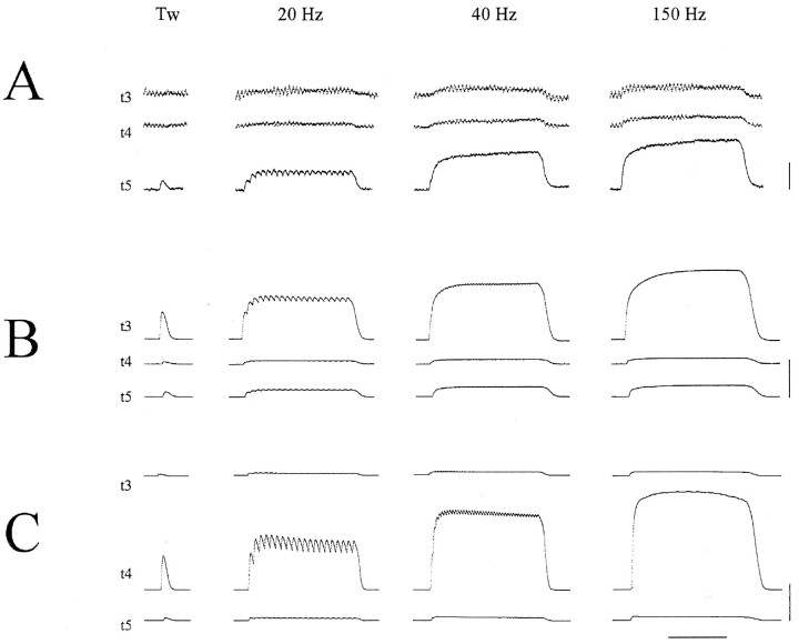
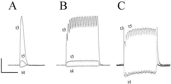
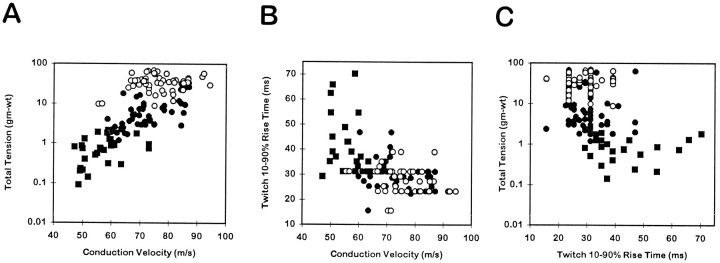
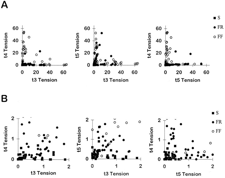
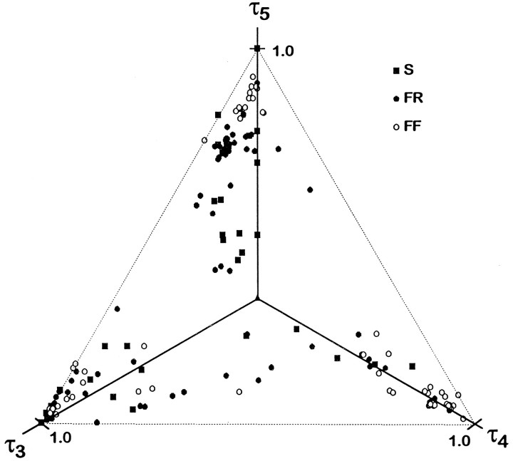
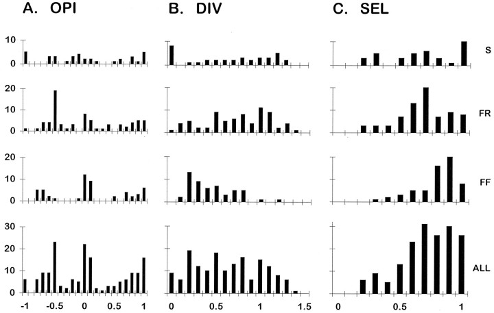
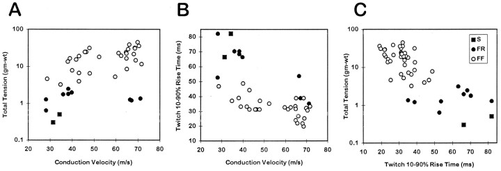
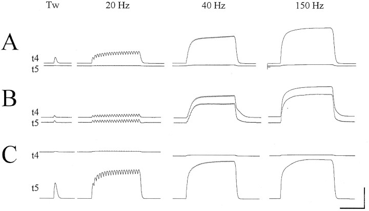
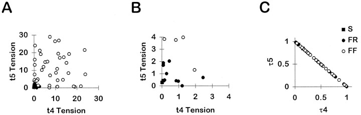
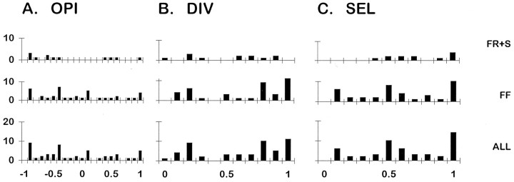
To determine whether single motor units (MUs) in multitendoned muscles distribute tension to multiple tendons or instead focus tension selectively on a single tendon, we examined the distribution of tension generated by single MUs in the cat extensor digitorum lateralis (EDLat), and in its macaque homolog, the extensor digiti quarti et quinti (ED45). General properties of MUs (maximal tetanic tension, axonal conduction velocity, and twitch rise time) were similar in these muscles to those reported for other limb muscles in cats and monkeys. Most cat EDLat MUs were found to exert tension rather selectively on one of the three tendons of the muscle. Fast fatigable MUs were slightly but significantly more selective than fast fatigue-resistant and slow MUs. In contrast, and contrary to expectation, the macaque ED45 contained a lower proportion of MUs that exerted tension selectively on one of the two tendons of the muscle, and a higher proportion of relatively nonselective MUs. These findings suggest that the cat EDLat may consist of three functional subdivisions, each acting preferentially on a different tendon, whereas the macaque ED45 is more likely to function as a single multitendoned muscle.


Figures

Fig. 1.
Fig. 1.
Three cat EDLat MUs. For each MU, simultaneous records of the tension on the tendon to digit 3 (t3), on the tendon to digit 4 (t4), and on the tendon to digit 5 (t5) are shown during single twitches and during tetanic stimulation at 20, 40, and 150 Hz. A, The MU shown was classified as type S, and exerted most of its tension on t5.B, The MU shown was classified as type FR, and exerted most of its tension on t3. C, The MU shown was classified as type FF, and exerted most of its tension on t4. Note the sag of the t4 tension of this MU after the first few stimuli of the 20 Hz tetanus. Such sag during 20 Hz tetani is characteristic of type FF MUs, and is not seen in type S or type FR MUs (A orB). All three MUs were from the same preparation. Horizontal calibration bar at bottom represents 0.5 sec; vertical calibration bars at right represent 0.2 gm-wt in A, 0.5 gm-wt in B, and 20 gm-wt inC.
Fig. 2.
Fig. 2.
Unloading of an EDLat tendon by a MU. Simultaneous tension records of EDLat’s three tendons are shown during a single twitch (A) and during tetanic stimulation at 20 Hz (B, C) of a single MU that unloaded the t4 tendon while exerting tension on the t3 and t5 tendons. In the single twitch (A) and in the high gain record (C), tension in the t4 tendon can be seen to decrease with each increase in tension in the other two tendons. Horizontal calibration bar = 0.5 sec; vertical calibration bar = 1.51 gm-wt in A, 2.36 gm-wt in B, 0.30 gm-wt in C.
Fig. 3.
Fig. 3.
Contractile properties of EDLat MUs. Scatter plots are shown of total tetanic tension during a 150 Hz tetanus (logarithmic scale) versus conduction velocity (A), twitch 10–90% rise time versus conduction velocity (B), and total tension versus twitch rise time (C). Significant correlations were found for all three relationships. Different symbols represent MUs of different physiological types: slow (▪), fast fatigue-resistant (•), or fast fatigable (○). Although the ranges of different MU types overlap considerably, significant differences related to MU type were found for all three variables.
Fig. 4.
Fig. 4.
EDLat MU tension on two of three tendons.A, Scatter plots of the tension exerted by each MU on the tendon to digit 4 (t4) versus the tendon to digit 3 (t3; left), on the tendon to digit 5 (t5) versus t3 (middle), and on t4 versus t5 (right). Most points in each plot lie close to one axis, indicating that the MU exerted tension relatively selectively on that tendon. B, Scatter plots of the same data at an expanded scale to show the data from small MUs. Symbols as in Figure 3.
Fig. 5.
Fig. 5.
Fractional tension of EDLat MUs on all three tendons. Plotted in three dimensions, the fractional tensions of EDLat MUs all lie in the same plane (τ3 + τ4 + τ5 = 1). The axes, therefore, have been rotated such that all points lie in the plane of the page. This plot reveals that rather than being scattered randomly over the possible area of an equilateral triangle (dashed lines), points representing most MUs lie close to one corner, indicating that they exerted tension preferentially on one tendon. Symbols as in Figure 3.
Fig. 6.
Fig. 6.
Histograms of OPI, DIV, and SEL indices for EDLat MUs. For each index, separate histograms are shown for EDLat MUs of each physiological type [slow (S), fast fatigue resistant (FR), and fast fatigable (FF)], as well as for all types together (ALL). Horizontal axis labels at bottomindicate the upper limits of histogram bins; for example, the rightmost bin of SEL histograms, labeled 1.0, includes values from 0.901 to 1.000.
Fig. 7.
Fig. 7.
Contractile properties of macaque ED45 MUs. Scatter plots are shown of total tension during a 150 Hz tetanus (logarithmic scale) versus conduction velocity (A), twitch 10–90% rise time versus conduction velocity (B), and total tension versus twitch rise time (C). Significant correlations were found for all three relationships. Symbols as in Figure 3.
Fig. 8.
Fig. 8.
Three monkey ED45 MUs. For each MU, simultaneous records of the tension on the tendon to digit 4 (t4) and on the tendon to digit 5 (t5) are shown during a single twitch and during tetanic stimulation at 20, 40, and 150 Hz. A, The MU shown exerted tension selectively on t4 (OPI = 0.923; DIV = 0.149; SEL = 0.923); B, the MU exerted equivalent tension on both t4 and t5 (OPI = 0.031; DIV = 0.999; SEL = 0.031); C, the MU exerted tension selectively on t5 (OPI = −0.960; DIV = 0.078; SEL = 0.960). Horizontal calibration bar = 0.5 sec; vertical calibration bar = 12.5 gm-wt in A and C, 5 gm-wt inB.
Fig. 9.
Fig. 9.
Scatter plots of ED45 MU tension. A, Scatter plots of the tension exerted by each MU on the tendon to digit 4 (t4) versus the tendon to digit 5 (t5). Although many points in each plot lie close to one axis, indicating that those MUs exerted tension relatively selectively on that tendon, many other points lie well between the axes, indicating that those MUs exerted comparable tension on both t4 and t5.B, Scatter plots of the same data at an expanded scale to show the data from small MUs. C, Plotted in two dimensions, the fractional tensions of ED45 MUs all lie on the same line (τ4 + τ5 = 1). Some points lie near one of the axes, and therefore represent MUs that exerted tension rather selectively on that tendon. But many points are spread along the line, indicating that many MUs exerted comparable fractions of their tension on both t4 and t5. Symbols as in Figure 3.
Fig. 10.
Fig. 10.
Histograms of OPI, DIV, and SEL indices for ED45 MUs. For each index, separate histograms are shown for slow plus fast fatigue-resistant MUs (S+FR), for fast fatigable MUs (FF), and for all types together (ALL). Labels on the horizontal axes at bottom indicate the upper limit of each histogram bin.

References

    1. Balice-Gordon RJ, Thompson WJ. The organization and development of compartmentalized innervation in rat extensor digitorum longus muscle. J Physiol. 1988;398:211–231. - PMC - PubMed
    1. Botterman BR, Iwamoto GA, Gonyea WJ. Classification of motor units in flexor carpi radialis muscle of the cat. J Neurophysiol. 1985;54:676–690. - PubMed
    1. Burke RE, Levine DN, Tsairis P, Zajac FE., III Physiological types and histochemical profiles in motor units of the cat gastrocnemius. J Physiol (Lond) 1973;234:723–748. - PMC - PubMed
    1. Burke RE. Selective recruitment of motor units. In: Humphrey DR, Freund H-J, editors. Motor control: concepts and issues. Wiley; New York: 1995. pp. 5–21.
    1. Burke RE, Tsairis P. Anatomy and innervation ratios in motor units of cat gastrocnemius. J Physiol (Lond) 1973;234:749–765. - PMC - PubMed

Publication types

LinkOut - more resources