. 1997 Mar 1;17(5):1880-90.
doi: 10.1523/JNEUROSCI.17-05-01880.1997.

Selective roles for hippocampal, prefrontal cortical, and ventral striatal circuits in radial-arm maze tasks with or without a delay

Affiliations

Selective roles for hippocampal, prefrontal cortical, and ventral striatal circuits in radial-arm maze tasks with or without a delay

S B Floresco et al. J Neurosci. .

Abstract

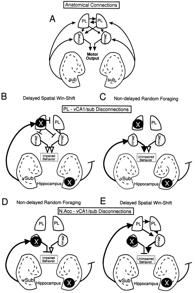
The hippocampus, the prefrontal cortex, and the ventral striatum form interconnected neural circuits that may underlie aspects of spatial cognition and memory. In the present series of experiments, we investigated functional interactions between these areas in rats during the performance of delayed and nondelayed spatially cued radial-arm maze tasks. The two-phase delayed task consisted of a training phase that provided rats with information about where food would be located on the maze 30 min later during a test phase. The single-phase nondelayed task was identical to the test phase of the delayed task, but in the absence of a training phase rats lacked previous knowledge of the location of food on the maze. Transient inactivation of the ventral CA1/subiculum (vSub) by a bilateral injection of lidocaine disrupted performance on both tasks. Lidocaine injections into the vSub on one side of the brain and the prefrontal cortex on the other transiently disconnected these two brain regions and significantly impaired foraging during the delayed task but not the nondelayed task. Transient disconnections between the vSub and the nucleus accumbens produced the opposite effect, disrupting foraging during the nondelayed task but not during the delayed task. These data suggest that serial transmission of information between the vSub and the prefrontal cortex is required when trial-unique, short-term memory is used to guide prospective search behavior. In contrast, exploratory goal-directed locomotion in a novel situation not requiring previously acquired information about the location of food is dependent on serial transmission between the hippocampus and the nucleus accumbens. These results indicate that different aspects of spatially mediated behavior are subserved by separate, distributed limbic-cortical-striatal networks.


Figures

Fig. 1.
Fig. 1.
Diagrams of the delayed spatial win-shift (SWSh) and the random foraging (RF) eight-arm radial-maze tasks.A, The delayed SWSh task consists of a training and a test phase. During the training phase, 4 of 8 arms on a radial maze are randomly blocked, and the 4 remaining open arms are baited. Once the animal has retrieved the 4 pieces of food from the open arms, it is removed from the maze for a delay (ranging from 5 to 30 min). After the delay, the animal is placed back onto the maze for the test phase. The arms that were blocked previously are now open and baited. The rat must remember which arms were previously blocked and enter them to receive the food reward. B, The nondelayed RF task consists of one phase. Four arms are randomly baited each day. The optimal foraging strategy entails entering the arms in a nonrepetitive manner. Unlike the test phase of the delayed SWSh task, the animal has no previous knowledge of the location of food at the beginning of a nondelayed RF trial.
Fig. 2.
Fig. 2.
Histology from the bilateral vSub inactivation experiments. Location of cannulae tips (black circles) for all rats used for data analysis in Experiment 1. Plates are computer-generated adaptations from Swanson (1992) that were modified to resemble those from Paxinos and Watson (1986).Numbers beside each slide correspond to millimeters from bregma.
Fig. 3.
Fig. 3.
The effects of bilateral inactivation of the vSub on performance of delayed and nondelayed radial-arm maze tasks.A, Number of errors (mean ± SEM) made by rats on the day before the first injection (open bar) and after infusions of saline (hatched bar) and lidocaine (black bar) into the vSub before the nondelayed random foraging task. **p < 0.001 relative to saline and day previous. Inset shows number of revisits to baited arms and nonbaited arms on saline (hatched bar) and lidocaine (black bar) injection days. B, Number of errors (mean ± SEM) made by rats during the test phase on the day before the first injection (open bar) and after infusions of saline (hatched bar) and lidocaine (black bar) into the vSub before the training phase of the delayed spatial win-shift task. Inset shows number of errors made during the training phase on the day before the first injection (open bar) and on saline (hatched bar) and lidocaine (black bar) injection days.C, Number of errors (mean ± SEM) made by rats during the test phase on the day before the first injection (open bar) and after infusions of saline (hatched bar) and lidocaine (black bar) into the vSub before the test phase of the delayed spatial win-shift task. **p < 0.001 relative to saline and day previous.Inset shows number of across-phase errors (cross-hatched bar) and within-phase errors (stripped bar) made by rats on lidocaine injection days. **p < 0.01.
Fig. 4.
Fig. 4.
The effects of PL–vSub disconnections on performance of a radial-arm maze test battery. A, Nondelayed random foraging. Number of errors (mean ± SEM) made by rats on the day before the first injection (open bar), after unilateral infusions of saline into both the PL and the vSub (hatched bar), unilateral infusions of lidocaine (Lido) into the PL and contralateral saline in the vSub (gray bar), unilateral infusions of Lido into the vSub and contralateral saline into the PL (stripped bar), and unilateral Lido into the vSub and contralateral Lido into the PL (disconnection; black bar) before the nondelayed RF task. B, Delayed spatial win-shift. Number of errors (mean ± SEM) made by rats on the day before the first injection (open bar), after unilateral infusions of saline into both the PL and the vSub (hatched bar), unilateral infusions of lidocaine (Lido) into the PL and contralateral saline in the vSub (gray bar), unilateral infusions of Lido into the vSub and contralateral saline into the PL (stripped bar), and unilateral Lido into the vSub and contralateral Lido into the PL (disconnection; black bar) before the test phase of the delayed SWSh task. **p < 0.001 versus all other treatment conditions.Inset shows number of across-phase (cross-hatched bar) versus within-phase (horizontal-stripped bar) errors made by rats during Lido/Lido (disconnection) injection days. C, Location of cannulae tips (black circles) for all rats used for data analysis receiving PL–vSub disconnections before either the nondelayed RF task or the delayed SWSh task. Plates are computer-generated adaptations from Swanson (1992) that were modified to resemble those from Paxinos and Watson (1986).Numbers beside each plate correspond to millimeters from bregma. For clarity, C represents the location of cannulae tips on sides that received infusions on disconnection injection days. All animals received infusions of either lidocaine or saline in each hemisphere.
Fig. 5.
Fig. 5.
The effects of N.Acc.–vSub disconnections on performance of a radial-arm maze test battery. A, Nondelayed random foraging. Number of errors (mean ± SEM) made by rats on the day before the first injection (open bar), after unilateral infusions of saline into both the N.Acc. and the vSub (hatched bar), unilateral infusions of lidocaine (Lido) into the N.Acc. and contralateral saline in the vSub (gray bar), unilateral infusions of Lido into the vSub and contralateral saline into the N.Acc. (stripped bar), and unilateral Lido into the vSub and contralateral Lido into the N.Acc. (disconnection; black bar) before the nondelayed RF task. **p < 0.001 versus all other treatment conditions. Inset shows number of reentries to baited arms and nonbaited arms on saline/saline (hatched bar) and Lido/Lido disconnection (black bar) injection days. B, Delayed spatial win-shift. Number of errors (mean ± SEM) made by rats on the day before the first injection (open bar), after unilateral infusions of saline into both the N.Acc. and the vSub (hatched bar), unilateral infusions of lidocaine (Lido) into the N.Acc. and contralateral saline in the vSub (gray bar), unilateral infusions of Lido into the vSub and contralateral saline into the N.Acc. (stripped bar), and unilateral Lido into the vSub and contralateral Lido into the N.Acc. (disconnection;black bar) before the delayed SWSh task.C, Location of cannulae tips (black circles) for all rats used for data analysis receiving N.Acc.–vSub disconnections before either the nondelayed RF task or the delayed SWSh task. Plates are computer-generated adaptations fromSwanson (1992) that were modified to resemble those from Paxinos and Watson (1986). Numbers beside each plate correspond to millimeters from bregma. For clarity, C represents the location of cannulae tips on sides that received infusions on disconnection injection days. All animals received infusions of either lidocaine or saline in each hemisphere.
Fig. 6.
Fig. 6.
Diagram of the anatomical connections investigated in the present study between the vSub, the PL, and the N.Acc. × represents the location of the unilateral inactivations to the vSub and PL or N.Acc. for each task. Solid arrows represent intact pathways. Open arrows represent pathways that are not blocked but do not carry the relevant spatial information because of a concomitant lidocaine-induced lesion upstream of this pathway.T-symbols represent blocked, nonfunctional pathways. A, An overview of the ipsi- and contralateral connections between the three brain regions. Note the unilateral projections from vSub to the forebrain and the contralateral projections between the PL and its connections to the N.Acc.B, Proposed route of information transfer between the vSub and PL during the delayed SWSh task. By disconnecting the PL–vSub pathway, information cannot be processed by the PL to generate appropriate responses after a delay, thereby disrupting appropriate output (impairment). C, Proposed route of information transfer between the vSub and the PL during the nondelayed RF task. By disconnecting the PL–vSub pathway, information is still able to access the N.Acc., thereby allowing for appropriate output (no impairment).D, Proposed route of information transfer between the vSub and N.Acc. during the nondelayed RF task. Disconnection of the N.Acc.–vSub pathway prevents the flow of information from the vSub through the N.Acc. to motor output centers (impairment).E, Proposed route of information transfer between the vSub and N.Acc. during the delayed SWSh task. Information from the vSub may be routed primarily through the PL and subsequently to the N.Acc. Thus, even though the pathway from the vSub to the N.Acc. is disconnected, spatial information may still be transferred from the unanesthetized vSub to the ipsilateral PL and subsequently routed to the contralateral N.Acc., allowing for appropriate output (no impairment).

References

    1. Albert DJ, Madryga FJ. An examination of the functional spread of 4 μl of slowly infused lidocaine. Behav Neural Biol. 1980;29:378–384. - PubMed
    1. Amaral DG, Witter MP. Hippocampal formation. In: Paxinos G, editor. The rat nervous system, 2nd Ed. Academic; San Diego: 1994. pp. 443–494.
    1. Barnes CA, McNaughton BL, Mizumori SJ, Leonard BW, Lin LH. Comparison of spatial and temporal characteristics of neuronal activity in sequential stages of hippocampal processing. Prog Brain Res. 1990;83:287–300. - PubMed
    1. Brog JS, Salyapongse A, Deutch A, Zahm DS. The pattern of afferent innervation of the core and shell in the “accumbens” part of the ventral striatum: immunohistochemical detection of retrogradely transported fluoro-gold. J Comp Neurol. 1993;338:255–278. - PubMed
    1. Conde F, Maire-Lepoivre E, Audinat E, Crepel F. Afferent connections of the medial frontal cortex of the rat. II. Cortical and subcortical afferents. J Comp Neurol. 1995;325:567–593. - PubMed

Publication types

MeSH terms

LinkOut - more resources