. 1997 Feb 17;185(4):663-72.
doi: 10.1084/jem.185.4.663.

IgE enhances mouse mast cell Fc(epsilon)RI expression in vitro and in vivo: evidence for a novel amplification mechanism in IgE-dependent reactions

Affiliations

IgE enhances mouse mast cell Fc(epsilon)RI expression in vitro and in vivo: evidence for a novel amplification mechanism in IgE-dependent reactions

M Yamaguchi et al. J Exp Med. .

Abstract

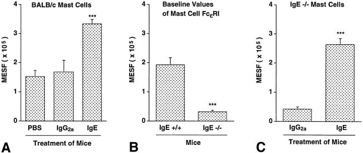
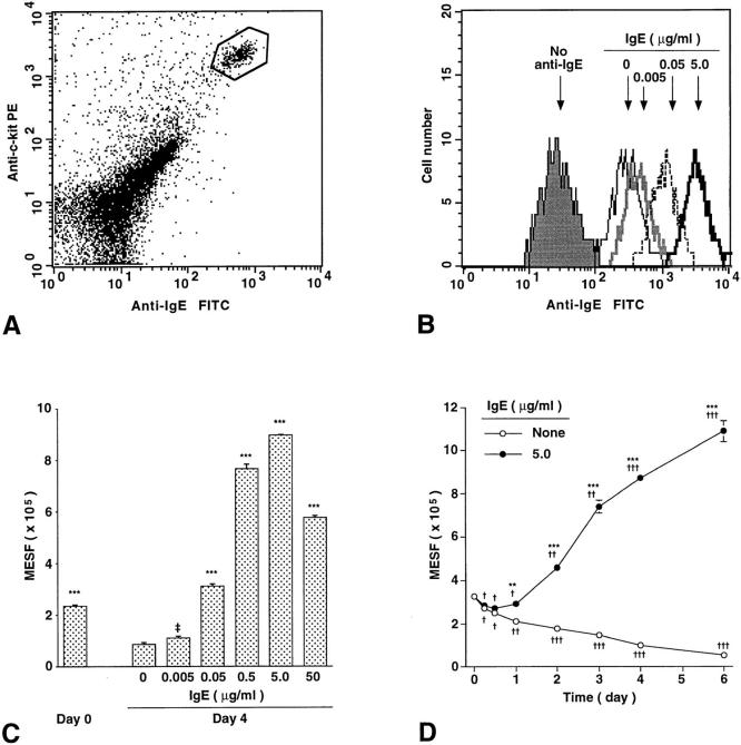
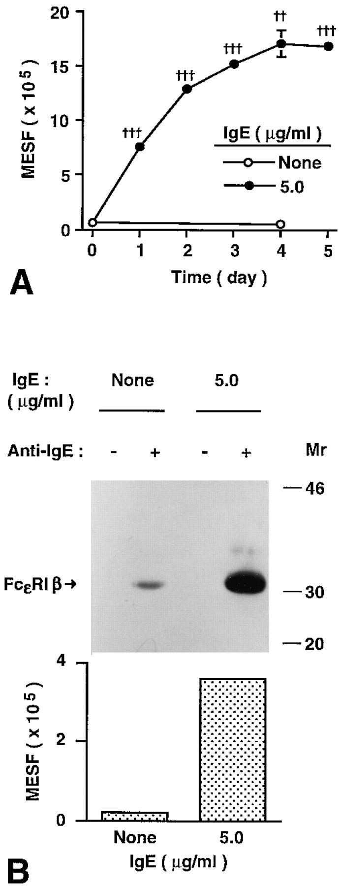
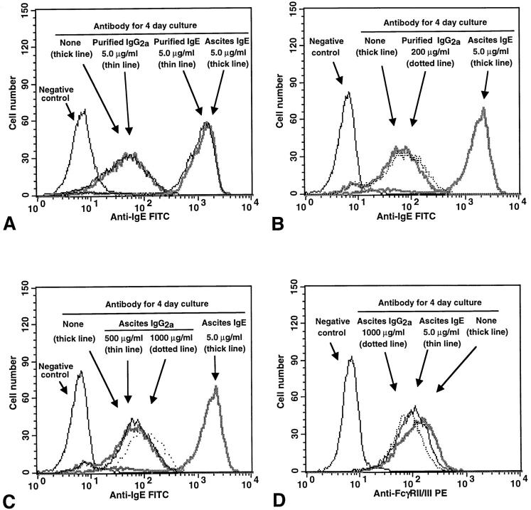
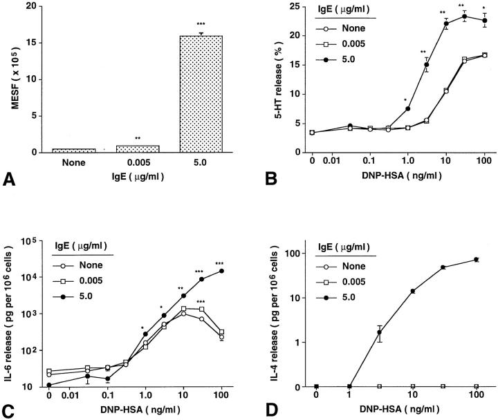
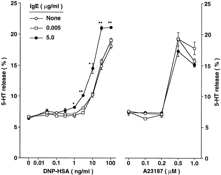
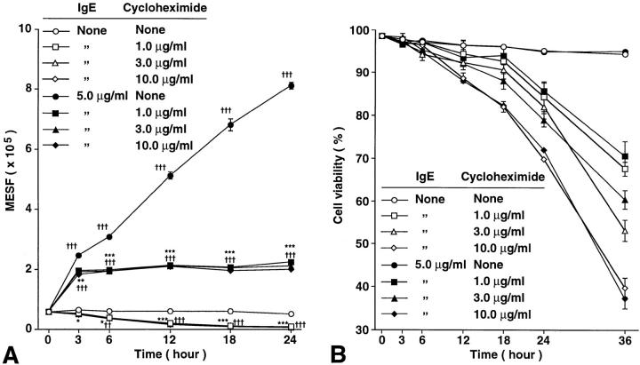
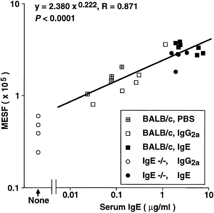
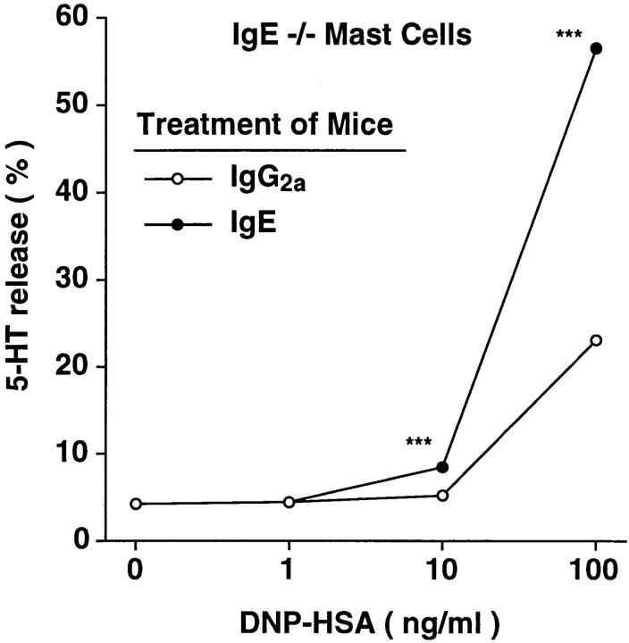
The binding of immunoglobulin E (IgE) to high affinity IgE receptors (Fc(epsilon)RI) expressed on the surface of mast cells primes these cells to secrete, upon subsequent exposure to specific antigen, a panel of proinflammatory mediators, which includes cytokines that can also have immunoregulatory activities. This IgE- and antigen-specific mast cell activation and mediator production is thought to be critical to the pathogenesis of allergic disorders, such as anaphylaxis and asthma, and also contributes to host defense against parasites. We now report that exposure to IgE results in a striking (up to 32-fold) upregulation of surface expression of Fc(epsilon)RI on mouse mast cells in vitro or in vivo. Moreover, baseline levels of Fc(epsilon)RI expression on peritoneal mast cells from genetically IgE-deficient (IgE -/-) mice are dramatically reduced (by approximately 83%) compared with those on cells from the corresponding normal mice. In vitro studies indicate that the IgE-dependent upregulation of mouse mast cell Fc(epsilon)RI expression has two components: an early cycloheximide-insensitive phase, followed by a later and more sustained component that is highly sensitive to inhibition by cycloheximide. In turn, IgE-dependent upregulation of Fc(epsilon)RI expression significantly enhances the ability of mouse mast cells to release serotonin, interleukin-6 (IL-6), and IL-4 in response to challenge with IgE and specific antigen. The demonstration that IgE-dependent enhancement of mast cell Fc(epsilon)RI expression permits mast cells to respond to antigen challenge with increased production of proinflammatory and immunoregulatory mediators provides new insights into both the pathogenesis of allergic diseases and the regulation of protective host responses to parasites.


Figures

Figure 7
Figure 7
IgE can regulate levels of peritoneal mast cell FcεRI expression in vivo. (A) FcεRI expression in peritoneal mast cells from BALB/c mice that had been treated for 4 d with a daily i.v. injection of 100 μg of affinity-purified IgE (n = 7) or IgG2a (n = 6) mAb, or vehicle (PBS) alone (n = 4). ***P <0.0001 or <0.005 versus values for PBS- or IgG2atreated mice, respectively. (B) Baseline levels of FcεRI expression on peritoneal mast cells from IgE +/+ (n = 8) and IgE −/− (n = 7) mice. ***P <0.0001 versus values for IgE +/+ cells. (C) FcεRI expression in peritoneal mast cells from IgE −/− mice treated with purified IgE mAb (n = 5) or purified IgG2a mAb (n = 4) as in (A). ***P <0.0001 versus values for cells from IgG2a-treated mice. All values in (A–C) are mean ± SEM.
Figure 1
Figure 1
IgE enhances mouse peritoneal mast cell FcεRI expression in vitro in a time- and dose-dependent manner. (A) Identification of peritoneal mast cells (gated, in box) by flow cytometry. Freshly isolated peritoneal cells of retired breeder BALB/c mice were stained for the expression of surface IgE receptor and c-kit. The gated cells were sorted and confirmed microscopically as 100% mast cells. (B–D) Regulation of surface FcεRI expression on peritoneal mast cells by ascites IgE ex vivo. (B) Peritoneal cells isolated from BALB/c mice were cultured for 4 d without IgE or with IgE at 0.005, 0.05, or 5.0 μg/ml, incubated with excess IgE at 4°C for 50 min to saturate mast cell FcεRI, and then mast cell surface FcεRI expression was analyzed by flow cytometry. The background fluorescence intensity of mast cells stained only for c-kit is also shown (no anti-IgE). (C) Dose-dependent effect of IgE on surface FcεRI expression on peritoneal mast cells. Peritoneal cells from 5 BALB/c mice were combined, cultured with or without IgE for 4 d, and analyzed by flow cytometry for surface FcεRI. MESF, molecules of equivalent soluble fluorochrome units (see Materials and Methods). All data are mean ± SEM (n = 3); ‡P = 0.076, ***P <0.0001 versus values for cells cultured without IgE for 4 d. (D) Time course of changes in mast cell surface FcεRI expression in peritoneal cells (combined from 8 BALB/c mice) cultured with or without IgE (5.0 μg/ml). All data are mean ± SEM (n = 3); **P <0.001, ***P <0.0001 versus values at the same time point for cells cultured without IgE; P <0.05, †† P <0.001, ††† P <0.0001 versus baseline (day 0) value.
Figure 2
Figure 2
IgE enhances FcεRI expression in mouse bone marrow–derived cultured mast cells (BMCMCs) in vitro. (A) Kinetics of IgE-induced changes in surface FcεRI expression in BMCMCs. BMCMCs (always >95% purity at 4–5 wk of culture) from BALB/c (IgE +/+) mice were cultured with or without ascites IgE (at 5.0 μg/ml) for the indicated times. BMCMCs were stained only for FcεRI (with FITC–IgE) and were analyzed by flow cytometry using a different instrument setting from that in Fig. 1. All data are mean ± SEM (n = 3); ††P <0.001, ††† P <0.0001 versus value at day 0. (B) Upregulation of surface FcεRI protein by IgE. BMCMCs from IgE −/− mice were cultured with or without ascites IgE at 5 μg/ml for 21 h. After sensitization with IgE at 10 μg/ ml for 30 min at 4°C to saturate surface FcεRI, cells (n = 2) were analyzed by flow cytometry for FcεRI expression (see Materials and Methods) and by immunoprecipitation with anti-IgE and Western blot (2.5 × 10⁷ BMCMCs per lane) to assess levels of surface-expressed FcεRI β chain. The difference of detected FcεRI expression between cells incubated with or without IgE was 14fold (by flow cytometry) and 17-fold (by Western blot), respectively.
Figure 3
Figure 3
Effects of various mouse IgE or IgG2a mAb preparations on surface expression of (A–C) FcεRI or (D) FcγRII/III in BMCMCs. BALB/c BMCMCs were cultured with or without various concentrations of mouse IgE or IgG2a mAb, either as diluted ascites preparations (Ascites IgE or IgG2a) or as affinity-purified mAb preparations (Purified IgE or IgG2a), for 4 d. Negative control in (A–C), cells cultured for 4 d without IgE or IgG2a and then incubated with FITC–anti-IgE without prior sensitization with IgE. (A) Incubation with either purified or ascites IgE mAb (at 5.0 μg IgE/ml) resulted in an equivalent increase in the FcεRI expression of the cells, whereas cells incubated with either purified IgG2a (at 5.0 μg/ml) or no antibody (none) exhibited similar low levels of staining. (B and C) Incubation with high concentrations of purified (B) or ascites (C) IgG2a resulted in little or no increase in BMCMC FcεRI expression, in comparison with positive control BMCMCs that had been incubated with IgE at 5.0 μg/ml. (D) Incubation for 4 d with IgE or IgG2a had little or no effect on the expression of FcγRII/III by BMCMCs. Negative control in (D), cells cultured for 4 d without IgE or IgG2a and then incubated with a rat IgG2b mAb of irrelevant specificity, instead of the rat 2.4G2 anti–mouse FcγRII/III mAb, before staining with the PE– anti-rat IgG antibody. The same results were obtained with ascites IgE (shown) or purified IgE (not shown).
Figure 4
Figure 4
IgE-induced enhancement of mouse BMCMC FcεRI expression increases the ability of these cells to release mediators in response to IgE and specific antigen. Effects of IgE-induced changes in BMCMC FcεRI expression (A) on IgE- and antigen-dependent release of (B) serotonin (5-HT), (C) IL-6, and (D) IL-4. BALB/c BMCMCs were cultured without IgE or with ascites IgE at 0.005 or 5.0 μg/ml for 4 d. All values are mean ± SEM (n = 3). (A–D) *P <0.05, **P <0.005, ***P <0.0001 versus corresponding values for cells cultured without IgE. Compared with values at 0 DNP–HSA for cells that had been incubated with the same concentration of IgE for 4 d, significant (P <0.01) release of 5-HT first occurred at 1.0 versus 3 ng/ml DNP–HSA for cells incubated with IgE at 5.0 versus 0.005 or 0 ng/ml, respectively. For IL-6 release, the corresponding values were 0.3 ng/ml DNP–HSA for cells incubated with IgE at 5.0 or 0.005 μg/ml versus 1 ng/ml DNP–HSA for cells incubated without IgE. Detectable release of IL–4 occurred at 3.0 ng/ml DNP– HSA for cells incubated with IgE at 5.0 μg/ml, but not at all in cells incubated with IgE at 0.005 or 0 ng/ml. Similar results were observed in another experiment using IgE −/− BMCMCs.
Figure 5
Figure 5
Serotonin (5-HT) release from BMCMCs exhibiting different levels of surface expression of FcεRI, after challenge with various concentrations of either specific antigen (DNP–HSA) or the calcium ionophore, A23187. WBB6F1 +/+ BMCMCs were cultured without IgE or with ascites IgE at 0.005 or 5.0 μg/ml for 4 d before passive sensitization and antigen challenge or challenge with A23187. Zero in the A23187 experiment refers to cells incubated without A23187 but in the highest concentration of vehicle used for these studies (DMSO at 0.1%). *P <0.05, **P <0.001, versus corresponding values for cells cultured without IgE. The data shown (mean ± SEM; n = 3 per point) are representative of the results obtained in two separate experiments using WBB6F1 +/+ BMCMCs.
Figure 6
Figure 6
Time course of changes in (A) the baseline or IgE-induced levels of FcεRI expression, and (B) cell viability, as assessed by Trypan blue staining, of WBB6F1+/+ BMCMCs cultured with or without mouse IgE mAb (at 5.0 μg/ml) and with or without various concentrations of cycloheximide (see Materials and Methods). All data are mean ± SEM (n = 3). (A) *P <0.05, **P <0.001, ***P <0.0001 versus values at the same timepoint for cells cultured with the same concentration of IgE, but without cycloheximide; P <0.05, †† P <0.001, ††† P <0.0001 versus initial baseline (zero h) value. (B) Compared with values at the same timepoint for cells cultured with the same concentration of IgE, but without cycloheximide, a significant (P <0.05) reduction in cell viability first occurred at 18, 12, or 6 h for cells cultured with cycloheximide at 1, 3, or 10 μg/ml, respectively. Similar results were observed in two experiments using BALB/c BMCMCs or Cl.MC/C57.1 cloned mast cells (18).
Figure 8
Figure 8
Correlation of serum IgE concentration at sacrifice and surface FcεRI expression of peritoneal mast cells in the mice from the experiments depicted in Fig. 7 (A and C). The genotypes of the mice, and the treatment which they received (PBS, IgE, or IgG2a, i.v. daily for 4 d) are depicted in the symbol key in the figure. Note that the correlation coefficient was calculated based on all data except those from IgG2a-treated IgE −/− mice, which were devoid of serum IgE; as a result, n = 22.
Figure 9
Figure 9
Upregulation of secretory function of IgE −/− peritoneal mast cells after administration of IgE in vivo. Peritoneal cells were combined from two of the IgE −/− mice that had been treated with affinity-purified IgG2a mAb and four of the IgE −/− mice that had been treated with purified IgE mAb from Fig. 7 C. IgE- and antigen-induced 5-HT release was determined for each group of cells (n = 3 per point) as in Fig. 4 B. ***P <0.0001 versus corresponding values for cells from IgG2atreated mice.

Similar articles

Cited by

References

    1. Ishizaka T, Ishizaka K. Activation of mast cells for mediator release through IgE receptors. Prog Allergy. 1984;34:188–235. - PubMed
    1. Jarrett, E.E.E., and H.R.P. Miller. 1982. Production and activities of IgE in helminth infection. In Progress in Allergy. Vol. 31: Immunity and Concomitant Immunity in Infectious Diseases. P. Kallós, volume editor. S. Karger, Basel. 178–233. - PubMed
    1. Matsuda H, Watanabe N, Kiso Y, Hirota S, Ushio H, Kannan Y, Azuma M, Koyama H, Kitamura Y. Necessity of IgE antibodies and mast cells for manifestation of resistance against larval Haemaphysalis longicornisticks in mice. J Immunol. 1990;144:259–262. - PubMed
    1. Bochner BS, Lichtenstein LM. Anaphylaxis. N Engl J Med. 1991;324:1785–1790. - PubMed
    1. Ravetch JV, Kinet J-P. Fc receptors. Annu Rev Immunol. 1991;9:457–492. - PubMed

Publication types