. 1997 Feb 17;185(4):795-800.
doi: 10.1084/jem.185.4.795.

NKG2A complexed with CD94 defines a novel inhibitory natural killer cell receptor

Affiliations

NKG2A complexed with CD94 defines a novel inhibitory natural killer cell receptor

A G Brooks et al. J Exp Med. .

Abstract

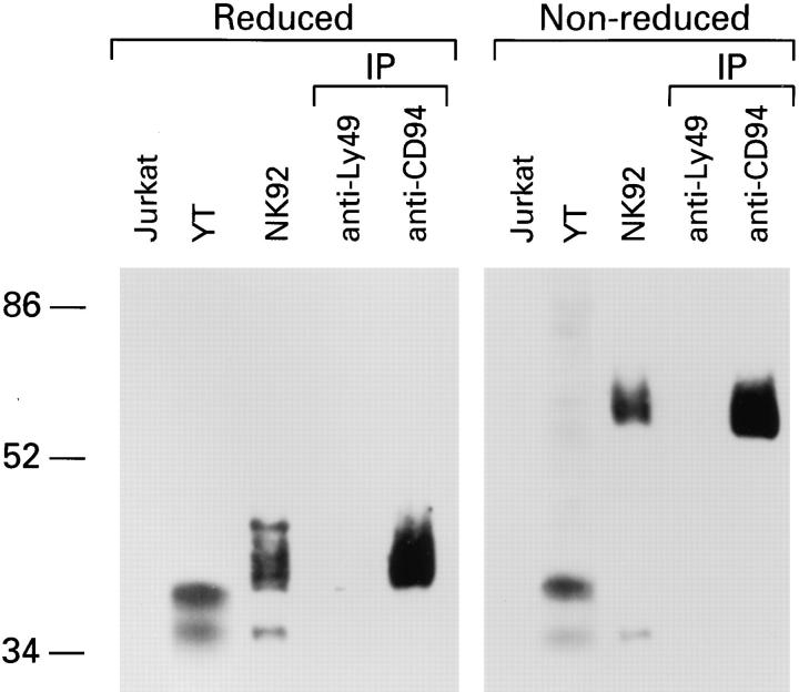
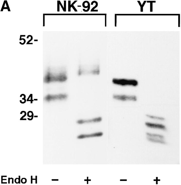
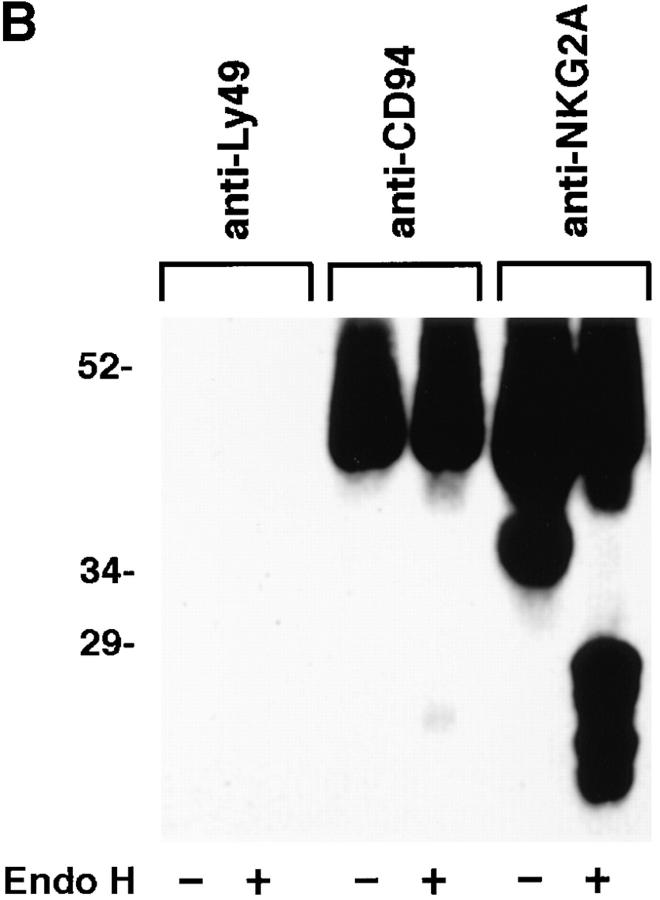
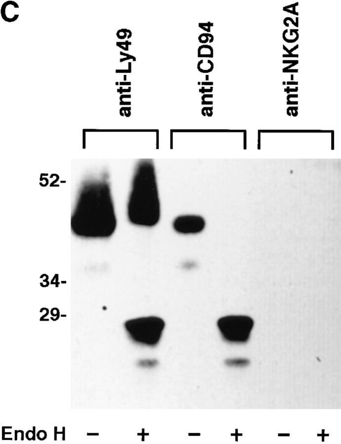
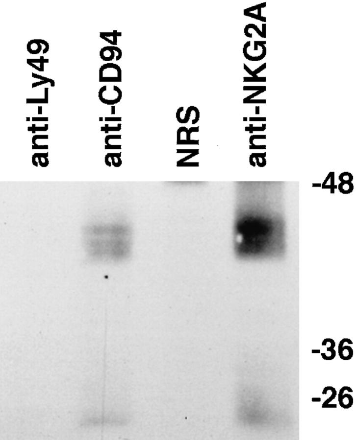
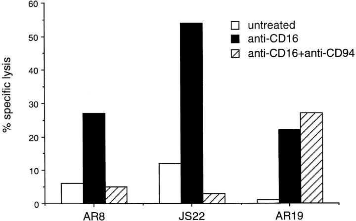
CD94 is a C-type lectin expressed by natural killer (NK) cells and a subset of T cells. Blocking studies using anti-CD94 mAbs have suggested that it is a receptor for human leukocyte antigen class I molecules. CD94 has recently been shown to be a 26-kD protein covalently associated with an unidentified 43-kD protein(s). This report shows that NKG2A, a 43-kD protein, is covalently associated with CD94 on the surface of NK cells. Cell surface expression of NKG2A is dependent on the association with CD94 as glycosylation patterns characteristic of mature proteins are found only in NKG2A that is associated with CD94. Analysis of NK cell clones showed that NKG2A was expressed in all NK cell clones whose CD16-dependent killing was inhibited by cross-linking CD94. The induction of an inhibitory signal is consistent with the presence of two immunoreceptor tyrosine-based inhibitory motifs (V/LXYXXL) on the cytoplasmic domain of NKG2A. Similar motifs are found on Ly49 and killer cell inhibitory receptors, which also transmit negative signals to NK cells.


Figures

Figure 1
Figure 1
NKG2A is covalently associated with CD94. Lysates from either Jurkat, YT or NK-92 cells, together with anti-CD94 or control (antiLy49) immunoprecipitates (IP) from NK-92 were separated by SDS-PAGE under reducing or non-reducing conditions and analyzed by Western blot with anti-NKG2A peptide-specific Ab.
Figure 3
Figure 3
NKG2A fails to mature in the absence of CD94. (A) Whole cell lysates of NK-92 or YT were either digested with Endoglycosidase Hf (+) or mock digested (−), separated by SDS-PAGE under reducing conditions and analyzed by Western blot with anti-NKG2A peptide-specific Ab. (B) Anti-CD94, anti-NKG2A or control (anti-Ly49), immunoprecipitates from NK-92 cells were either digested with Endoglycosidase Hf, (+) or mock digested (−), separated by SDS-PAGE under reducing conditions and analyzed by Western blot with anti-NKG2A peptide-specific Ab. (C) NK-92 cell lysates were exhaustively precleared with control (anti-Ly49), anti-CD94 or anti-NKG2A Ab. The remaining lysates were divided in two and either digested with Endoglycosidase Hf, (+) or mock digested (−), separated by SDS-PAGE under reducing conditions and analyzed by Western blot with antiNKG2A peptide Ab. The immunoblots in B and C were overexposed to detect trace amounts of immature CD94-associated NKG2A in B and to emphasize that no mature NKG2A could be detected in NK-92 lysates after removal of CD94 associated NKG2A in C.
Figure 3
Figure 3
NKG2A fails to mature in the absence of CD94. (A) Whole cell lysates of NK-92 or YT were either digested with Endoglycosidase Hf (+) or mock digested (−), separated by SDS-PAGE under reducing conditions and analyzed by Western blot with anti-NKG2A peptide-specific Ab. (B) Anti-CD94, anti-NKG2A or control (anti-Ly49), immunoprecipitates from NK-92 cells were either digested with Endoglycosidase Hf, (+) or mock digested (−), separated by SDS-PAGE under reducing conditions and analyzed by Western blot with anti-NKG2A peptide-specific Ab. (C) NK-92 cell lysates were exhaustively precleared with control (anti-Ly49), anti-CD94 or anti-NKG2A Ab. The remaining lysates were divided in two and either digested with Endoglycosidase Hf, (+) or mock digested (−), separated by SDS-PAGE under reducing conditions and analyzed by Western blot with antiNKG2A peptide Ab. The immunoblots in B and C were overexposed to detect trace amounts of immature CD94-associated NKG2A in B and to emphasize that no mature NKG2A could be detected in NK-92 lysates after removal of CD94 associated NKG2A in C.
Figure 2
Figure 2
NKG2A/CD94 heterodimers are expressed on the cell surface of polyclonal NK cells. Cell surface proteins of polyclonal NK cells were labeled with biotin and immunoprecipitated with anti-CD94, anti-NKG2A peptide-specific Ab, anti-Ly49 mAb or, normal rabbit serum (NRS). Immunoprecipitated proteins were reduced and separated on SDS-PAGE, transferred to PVDF, detected with streptavidin conjugated-HRP and visualized with enhanced chemiluminescence.
Figure 4
Figure 4
CD94 cross-linking inhibits CD16-mediated lysis of NKG2A positive NK cell clones. 51Cr-labeled P815 target cells were either preincubated with anti-CD16 (anti-CD16), or anti-CD16 and anti-CD94 (anti-CD16 + anti-CD94) or complete medium (untreated) prior to incubation with the NKG2A positive NK cell clones AR8 and JS22 or the NKG2A negative NK cell clone, AR19 for 4 h. 51Cr released was measured in a γ counter and the percentage of specific lysis calculated.

References

    1. Trinchieri G. Biology of natural killer cells. Adv Immunol. 1989;47:187–376. - PMC - PubMed
    1. Shimizu Y, DeMars R. Demonstration by class I gene transfer that reduced susceptibility of human cells to natural killer cell-mediated lysis is inversely correlated with HLA class I antigen expression. Eur J Immunol. 1989;19:447–451. - PubMed
    1. Storkus WJ, Alexander J, Payne JA, Dawson JR, Cresswell P. Reversal of natural killing susceptibility in target cells expressing transfected class I HLA genes. Proc Natl Acad Sci USA. 1989;86:2361–2364. - PMC - PubMed
    1. Liao NS, Bix M, Zijlstra M, Jaenisch R, Raulet D. MHC class I deficiency: susceptibility to natural killer (NK) cells and impaired NK activity. Science (Wash DC) 1991;253:199–202. - PubMed
    1. Smith HR, Karlhofer FM, Yokoyama WM. Ly-49 multigene family expressed by IL-2-activated NK cells. J Immunol. 1994;153:1068–1079. - PubMed