. 1997 Feb 18;94(4):1080-5.
doi: 10.1073/pnas.94.4.1080.

Native-like structure of a protein-folding intermediate bound to the chaperonin GroEL

Affiliations

Native-like structure of a protein-folding intermediate bound to the chaperonin GroEL

M S Goldberg et al. Proc Natl Acad Sci U S A. .

Abstract

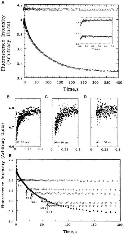
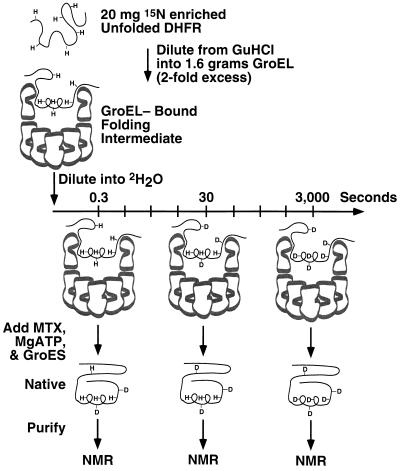
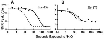
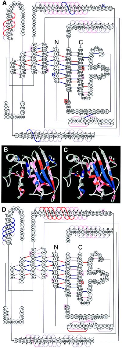
The chaperonin GroEL binds nonnative proteins in its central channel through hydrophobic interactions and initiates productive folding in this space underneath bound co-chaperone, GroES, in the presence of ATP. The questions of where along the folding pathway a protein is recognized by GroEL, and how much structure is present in a bound substrate have remained subjects of discussion, with some experiments suggesting that bound forms are fully unfolded and others suggesting that bound species are partially structured. Here we have studied a substrate protein, human dihydrofolate reductase (DHFR), observing in stopped-flow fluorescence experiments that it can rapidly bind to GroEL at various stages of folding. We have also analyzed the structure of the GroEL-bound protein using hydrogen-deuterium exchange and NMR spectroscopy. The pattern and magnitude of amide proton protection indicate that the central parallel beta-sheet found in native DHFR is present in a moderately stable state in GroEL-bound DHFR. Considering that the strands are derived from distant parts of the primary structure, this suggests that a native-like global topology is also present. We conclude that significant native-like structure is present in protein-folding intermediates bound to GroEL.


Figures

Figure 1
Figure 1
(A) Tryptophan fluorescence intensity changes of DHFR during refolding from 6 M urea in the absence (▵) or presence (○) of GroEL; the Inset shows the behavior for the first 0.5 sec. The data were fit to a sum of three exponentials using a nonlinear least-squares fitting program available from the SAS institute (Cary, NC). The lines reflecting these fits are shown. (BE) Tryptophan fluorescence of refolding DHFR following addition of GroEL at various times after the initiation of folding. Injections after 30, 50, and 100 msec are shown independently in BD, respectively. Injections after 5 (▵), 20 (+), 30 (○), 50 (□), and 60 (▿) sec are shown in E, as is a trace in the absence of GroEL (▴). The small differences between the intensity of the DHFR–GroEL complexes and the intensity of DHFR at the same folding times reflects the apparent cancellation of the decrease expected from the 2-fold dilution in the second mixing step by an increase due to addition of chaperone (50-fold excess by weight).
Figure 2
Figure 2
Illustration of the procedure used to prepare samples for NMR analysis of amide exchange rates of GroEL-bound DHFR. The top ring of GroEL is shown as a cross-section. Amide protons protected from exchange are labeled as “H,” while those which exchange with solvent deuterons are labeled as “D.” After each of 10 time points, DHFR is refolded to the native state from which further exchange is negligible, separated from GroEL, and placed in an NMR spectrometer to measure the proton occupancy of each amide.
Figure 3
Figure 3
Representative amide exchange data for residues Leu-159 (A) and Ile-175 (B). NMR peak volumes are plotted as a function of time of exposure of the GroEL-bound folding intermediate to 2H2O. The time scale is logarithmic. The solid lines are the result of nonlinear least-squares fitting of the data to a single exponential plus a constant. The dashed lines indicate the exchange rates calculated for the same amide in a random coil environment. Protection factors, defined by the ratio of the two rate constants, are 24 for Leu-159 and 2 for Ile-175.
Figure 4
Figure 4
Location of protected amides in GroEL-bound and native DHFR. (A) Diagram of hydrogen bonds in DHFR, colored according to the degree of protection in the GroEL-bound state. Blue represents protection factors >10, red between 5 and 10, pink <5 or unmeasurable, and gray hydrogen bonds that are inferred from the crystal structures but not observed by NMR. Probes that do not form hydrogen bonds directly with backbone or side chain acceptors are shown in boxes whose color reflects the degree of protection. Hydrogen bonds were identified from the x-ray structures of DHFR (27, 28) using the program hbplus (29). (B) Ribbon diagram of native DHFR (28), displaying the protection factors of the GroEL-bound state, colored as in A. (C and D) Ribbon (C) and hydrogen bond diagrams (D) displaying the protection factors measured for native DHFR. Blue represents protection factors >9 × 10⁶, red between 2.5 × 10⁶ and 9 × 10⁶, and pink <2.5 × 10⁶.

References

    1. Weissman J S, Hohl C M, Kovalenko O, Kashi Y, Chen S, Braig K, Saibil H R, Fenton W A, Horwich A L. Cell. 1995;83:577–587. - PubMed
    1. Mayhew M, da Silva A C, Martin J, Erdjument-Bromage H, Tempst P, Hartl F U. Nature (London) 1996;379:420–426. - PubMed
    1. Weissman J S, Rye H S, Fenton W A, Beechem J M, Horwich A L. Cell. 1996;84:481–490. - PubMed
    1. Braig K, Otwinowski Z, Hegde R, Boisvert D C, Joachimiak A, Horwich A L, Sigler P B. Nature (London) 1994;371:578–586. - PubMed
    1. Landry S J, Gierasch L M. Annu Rev Biophys Biomol Struct. 1994;23:645–669. - PubMed

Publication types