. 1997 Feb 18;94(4):1361-5.
doi: 10.1073/pnas.94.4.1361.

Highly purified CD25- resting T cells cannot be infected de novo with HIV-1

Affiliations

Highly purified CD25- resting T cells cannot be infected de novo with HIV-1

C S Chou et al. Proc Natl Acad Sci U S A. .

Abstract

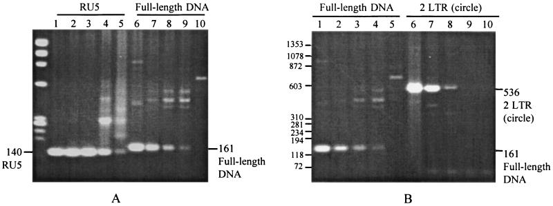
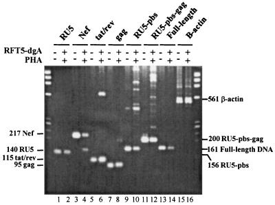
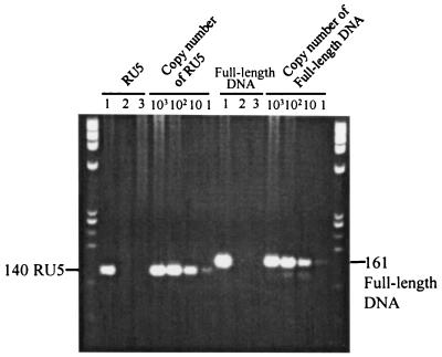
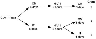
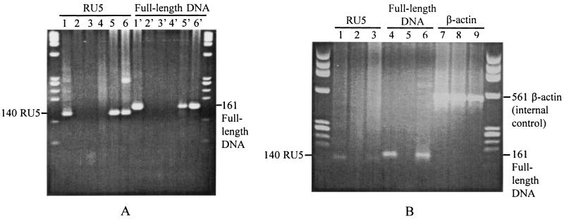
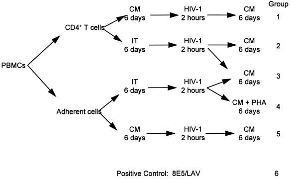
Previous studies have demonstrated that the expression of CD25 can distinguish CD25- latently infected cells from CD25+ cells actively producing virus. Our studies were designed to characterize the nature and stability of the viral genome in CD25- quiescent HIV-1-infected cells and to determine whether these cells could be infected de novo with HIV-1. Our results show that: (i) When unfractionated peripheral blood mononuclear cells are first infected with HIV-1 and the CD25- cells then isolated, the latter contain only incomplete DNA transcripts and no full-length DNA or 2-LTR circles. Phytohemagglutinin activation of these CD25- cells results in the generation of full-length viral DNA and p24 production. (ii) When CD25- CD4+ cells are first purified from peripheral blood mononuclear cells and then incubated with HIV-1, viral DNA cannot be detected, suggesting that these purified cells cannot be infected. Furthermore, CD25-adherent cells do not facilitate the infection of CD4+ CD25- T cells when they were present at the time of incubation with HIV-1. Taken together, these studies suggest either that (i) the CD25- cells containing incomplete DNA transcripts are derived from infected-activated CD25+ cells, which subsequently become CD25- or (ii) the presence of CD25+ cells is required for the infection of CD25- cells in vitro.


Figures

Figure 1
Figure 1
Analysis of HIV-1-infected unfractionated and CD25 resting PBMCs. (A) The proviral genome. PBMCs were infected on day 1 and then incubated with either CM or CM plus RFT5-dgA for 6 days. Following treatment, 106 viable cells were collected from unfractionated (A: lanes 1 and 6) or CD25 (B: lanes 5 and 10) PBMCs. From 102 to 104 unfractionated PBMCs (A) were added to 106 CD25 PBMCs (B) as follows: B + 0.01% A in lanes 4 and 9; B + 0.1% A in lanes 3 and 8; B + 1% A in lanes 2 and 7. The proviral genome in cells from each group was studied by intact-cell PCR as described. Lanes 1–5 depict HIV-1 early DNA transcripts (RU5) and lanes 6–10 show HIV-1 full-length DNA. (B) HIV-1 full-length DNA and 2-LTR circles. PBMCs were prepared as described in A. Lanes 1–5 show full-length DNA and lanes 6–10 show 2-LTR circles. The cell-free culture supernatant was collected from each group of cells and assayed for p24 antigen. p24 concentrations were as follows: lane 1, 36,680 pg/ml; lane 2, 153 pg/ml; lane 3, 22 pg/ml; lane 4, <5 pg/ml; lane 5, <5 pg/ml.
Figure 2
Figure 2
PHA activation of CD25 PBMCs. CD25 quiescent PBMCs were purified by treatment with RFT5-dgA for 6 days. Cells were then thoroughly washed to remove RFT5-dgA and cultured in CM containing 5 μg/ml PHA. Intact cells were harvested 6 days after PHA stimulation and subjected to intact-cell PCR as described. HIV-1-infected unfractionated PBMCs were used as controls and β-actin served as the internal control. PHA-stimulated HIV-1-infected CD25 quiescent cells are shown in lanes 2, 4, 6, 8, 10, 12, 14, and 16. Unfractionated HIV-1 infected control cells are shown in lanes 1, 3, 5, 7, 9, 11, 13, and 15.
Figure 3
Figure 3
Infection of CD4+ CD25 quiescent T lymphocytes with HIV-1. CD4+ CD25 quiescent T lymphocytes were isolated from fresh PBMCs by removing blood macrophages and natural killer cells, followed by positive selection of CD4+ cells, and elimination of CD25+ cells by treatment with RFT5-dgA as described. Purified quiescent CD4+ CD25 T cells were exposed to HIV-1 for 2 hr, washed thoroughly to remove excess virus, treated with trypsin to remove virions attached to the cell surface, and cultured in either CM (lane 2) or CM + RFT5-dgA (lane 3) and HIV-1-infected unfractionated CD4+ cells were included as controls. Six days later, cells were harvested for intact-cell PCR and cell-free culture supernatants were assayed for p24 production. (1: 17,740 ± 2, 470 pg/ml; both 2 and 3 were <5 pg/ml.) The protocol schematic was as follows:
Figure 3
Figure 3
Infection of CD4+ CD25 quiescent T lymphocytes with HIV-1. CD4+ CD25 quiescent T lymphocytes were isolated from fresh PBMCs by removing blood macrophages and natural killer cells, followed by positive selection of CD4+ cells, and elimination of CD25+ cells by treatment with RFT5-dgA as described. Purified quiescent CD4+ CD25 T cells were exposed to HIV-1 for 2 hr, washed thoroughly to remove excess virus, treated with trypsin to remove virions attached to the cell surface, and cultured in either CM (lane 2) or CM + RFT5-dgA (lane 3) and HIV-1-infected unfractionated CD4+ cells were included as controls. Six days later, cells were harvested for intact-cell PCR and cell-free culture supernatants were assayed for p24 production. (1: 17,740 ± 2, 470 pg/ml; both 2 and 3 were <5 pg/ml.) The protocol schematic was as follows:
Figure 4
Figure 4
(A) The role of adherent cells in acute HIV-1 infection. To determine whether the adherent cells present in PBMCs could facilitate the infection of CD4+ cells, different groups of cells were prepared and incubated with HIV-1 as described in the protocol schematic: (1) unfractionated CD4+ T cells (lanes 1 and 1′); (2) quiescent CD25 CD4+ T cells (lanes 2 and 2′); (3) CD25 adherent cells plus quiescent CD25 CD4+ T cells (lanes 3 and 3′); (4) CD25 adherent cells (lanes 4 and 4′); (5) unfractionated adherent cells (lanes 5 and 5′). Cells from each group were subjected to intact-cell PCR and lanes 1–6 show HIV-1 early transcripts (RU5) and lanes 1′–6′ show HIV-1 full-length DNA. The protocol schematic was as follows:
Figure
Figure
(B) The role of adherent cells in the infection of CD25 cells with HIV-1. Fresh PBMCs were prepared as described. PBMCs were cultured either in CM (lanes 1, 4, and 7) or in CM + RFT5-dgA (lanes 2, 5, and 8) for 6 days, and then exposed to HIV-1 for 2 hr and cultured in CM for 6 additional days, and then harvested and analyzed by intact-cell PCR. The 8E5/LAV cell line was used as positive control (lanes 3, 6, and 9). Lanes: 1–3, early transcripts (RU5); 4–6, full-length DNA; 7–9, β-actin as internal control.

References

    1. Borvak J, Chou C-S, Bell K, Van Dyke G, Zola H, Ramilo O, Vitetta E S. J Immunol. 1995;155:3196–3204. - PubMed
    1. Zack J A, Arrigo S J, Weitsman S R, Go A S, Haislip A, Chen I S Y. Cell. 1990;61:213–222. - PubMed
    1. Zack J A, Haislip A, Krogstad P, Chen I S Y. J Virol. 1992;66:1717–1725. - PMC - PubMed
    1. Stevenson M, Stanwick T L, Dempsey M P, Lamonica C A. EMBO J. 1990;9:1551–1560. - PMC - PubMed
    1. Spina C A, Guatelli J C, Richman D D. J Virol. 1995;69:2977–2988. - PMC - PubMed

Publication types