. 1997 Feb 18;94(4):1441-6.
doi: 10.1073/pnas.94.4.1441.

Initiation of liver growth by tumor necrosis factor: deficient liver regeneration in mice lacking type I tumor necrosis factor receptor

Affiliations

Initiation of liver growth by tumor necrosis factor: deficient liver regeneration in mice lacking type I tumor necrosis factor receptor

Y Yamada et al. Proc Natl Acad Sci U S A. .

Abstract

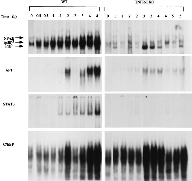
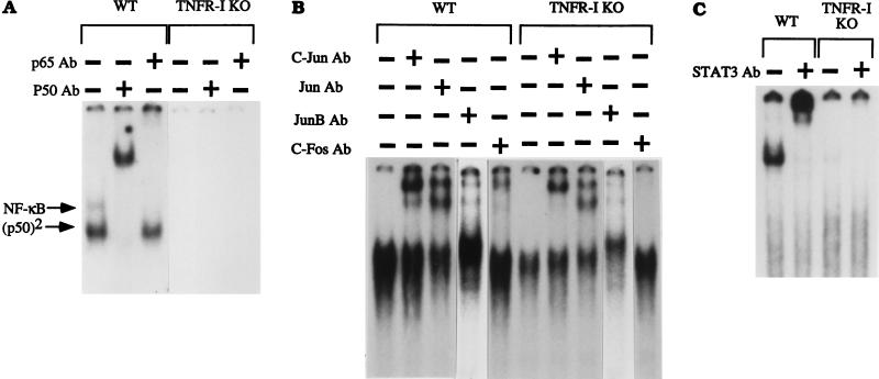
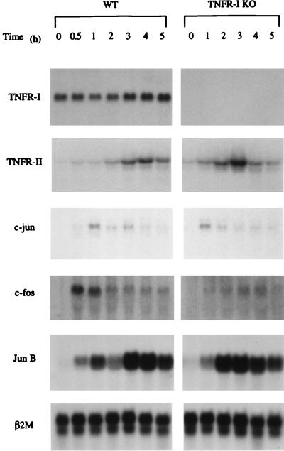
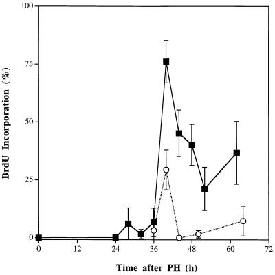
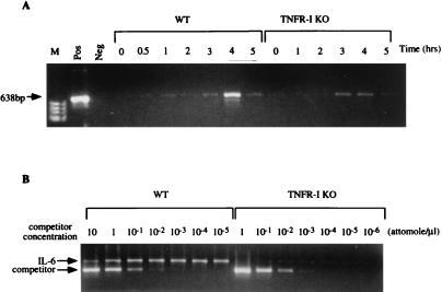
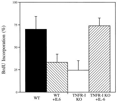
The mechanisms that initiate liver regeneration after resection of liver tissue are not known. To determine whether cytokines are involved in the initiation of liver growth, we studied the regeneration of the liver after partial hepatectomy (PH) in mice lacking type I tumor necrosis factor receptor (TNFR-I). DNA synthesis after PH was severely impaired in these animals, and the expected increases in the binding of the NF-kappaB and STAT3 transcription factors shortly after PH failed to occur. Binding of AP-1 after PH was decreased in TNFR-I knockout mice compared with animals with the intact receptor whereas C/EBP binding was not modified. Injection of interleukin 6 in TNFR-I-deficient animals 30 min before PH corrected the defect in DNA synthesis and restored STAT3 and AP-1 binding to normal levels but had no effect on NF-kappaB binding in the regenerating liver. The results indicate that TNF, signaling through the TNFR-I, can initiate liver regeneration and acts by activating an interleukin 6-dependent pathway that involves the STAT3 transcription factor.


Figures

Figure 1
Figure 1
Transcription factor binding after PH in wild-type (WT) and TNFR-I knockout mice (TNFR-I KO). Mice were partially hepatectomized and killed 0.5 and 5 h after the operation, as indicated at the top of the figure (two mice per time point). Nuclear extracts were prepared, and EMSAs were performed using 10 μg of nuclear protein in each lane. Reticulocyte lysate was used as a marker to determine the position of the p50/p65 NF-κB heterodimer (11). p50 homodimer position is indicated as (p50)2. There was variability in AP-1 binding in wild-type mice killed 2 h after PH. PHF, PH factor
Figure 2
Figure 2
Mobility-shift analysis of transcription factor binding using specific antibodies (Ab). Wild-type (WT) and TNFR-I knockout mice (TNFR-I KO) were partially hepatectomized and killed 4 h after the operation for NF-κB, STAT3, and AP-1 analysis. Nuclear extracts were incubated for 30 min with 32P-labeled probes followed by addition of 1 μl of the antibody (1 μg/μl) to 10 μg of extract. After incubation for 30 min, nuclear extracts (10 μg per lane) were analyzed by EMSA. The antibody used in each assay is indicated at the top of the figure. For each transcription factor, the first lane shows EMSA of the nuclear extract without antibody preincubation.
Figure 3
Figure 3
Expression of TNFR-I, TNFR-II, c-jun, c-fos, and Jun B mRNAs in wild-type (WT) and TNFR-I knockout mice (TNFR-I KO). Animals were partially hepatectomized and killed 0.5–5 h after operation. Samples (20 μg/lane) were separated by electrophoresis and hybridized with α32P-deoxycytidine 5′-triphosphate probes. β2-Microglobulin (β2M) mRNA (bottom row) was used as a loading control.
Figure 4
Figure 4
Hepatocyte DNA synthesis after PH in wild-type and TNFR-I knockout mice. Wild-type and TNFR-I knockout mice were partially hepatectomized and killed 24–68 h after the operation (three animals per time point) as indicated. All animals received an i.p. injection of 30 μg/g BrdUrd (BrdU) 2 h before killing. The ordinate shows the percentage of hepatocytes labeled by BrdUrd at the various time points. The bars indicate the SD of the means. ▪, Wild-type C57BL/6 mice; ○, TNFR-I knockout mice.
Figure 5
Figure 5
IL-6 mRNA expression after PH. (A) Wild-type (WT) and TNFR-I knockout (TNFR-I KO) mice were partially hepatectomized and killed 0.5–5 h after the operation, as indicated at the top of A. M, molecular weight marker lane; Pos, positive control for IL-6 mRNA (CLONTECH); Neg, mRNA not added for reverse transcription step. (B) Quantitation of IL-6 mRNA by competitive PCR in livers of wild-type and knockout mice 4 h after PH. Competitor concentrations are indicated at the top of B. The product sizes are 638 bp for IL-6 mRNA and 435 bp for the competitive fragment.
Figure 6
Figure 6
Effect of IL-6 on hepatocyte DNA synthesis in TNFR-I knockout mice (TNFR-I KO). Wild-type (WT) and TNFR-I mice were partially hepatectomized and killed 40 h after the operation. Three TNFR-I knockout animals and three wild-type mice received an s.c. injection of recombinant human IL-6 (1 mg/kg) 30 min before PH. All mice were injected with BrdUrd (BrdU) 2 h before killing (see Fig. 4). The ordinate shows the percentage of hepatocytes labeled by BrdUrd.
Figure 7
Figure 7
Effect of IL-6 on transcription factor binding in TNFR-I knockout mice. Wild-type and TNFR-I mice were partially hepatectomized and killed 3 h after PH. A group of knockout mice received an s.c. injection of IL-6 (see Fig. 5). Nuclear extracts (prepared from two animals in each group) were analyzed by EMSA as described in Fig. 1 (10 μg/lane). Lanes: 1, wild-type mice; 2, TNFR-I knockout mice; and 3, TNFR-I knockout mice injected with IL-6. The arrow on the right indicates the position of the STAT3 band.

References

    1. Fausto N, Webber E M. In: The Liver Biology and Pathobiology. Arias I, Boyer J, Fausto N, Jakoby W, Schachter D, Shafritz D, editors. New York: Raven; 1994. pp. 1059–1084.
    1. Bucher N L R. In: Liver Regeneration and Carcinogenesis: Molecular and Cellular Mechanisms. Jirtle R L, editor. San Diego: Academic; 1995. pp. 1–25.
    1. Fausto N, Laird A D, Webber E M. FASEB J. 1995;9:1527–1536. - PubMed
    1. Diehl A M, Rai R M. FASEB J. 1996;10:215–227. - PubMed
    1. Taub R. FASEB J. 1996;10:413–427. - PubMed

Publication types

MeSH terms

LinkOut - more resources