Comparative Study
. 1997 Feb 18;94(4):1453-8.
doi: 10.1073/pnas.94.4.1453.

A bacterial basic region leucine zipper histidine kinase regulating toluene degradation

Affiliations
Comparative Study

A bacterial basic region leucine zipper histidine kinase regulating toluene degradation

P C Lau et al. Proc Natl Acad Sci U S A. .

Abstract

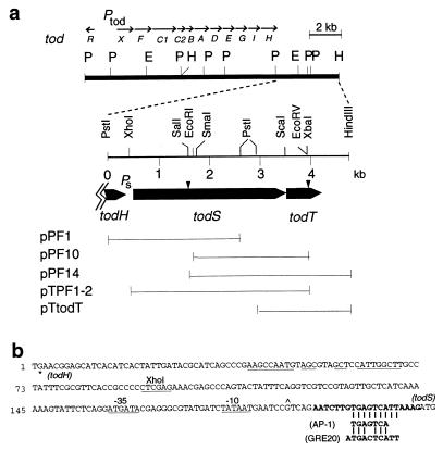
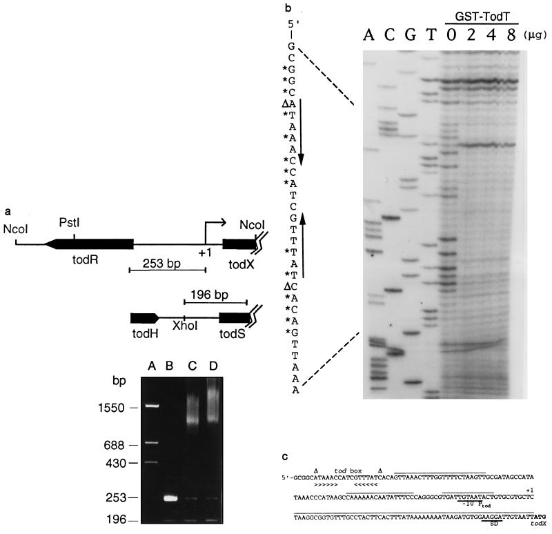
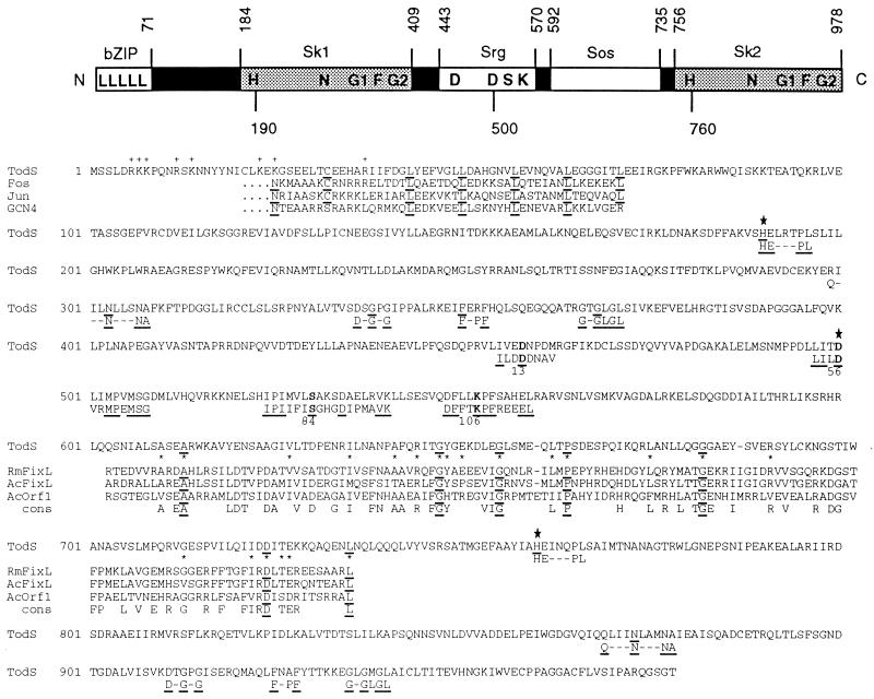
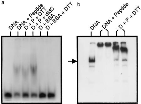
The two-component signal transduction pathways in bacteria use a histidine-aspartate phosphorelay circuit to mediate cellular changes in response to environmental stimuli. Here we describe a novel two-component todST system, which activates expression of the toluene degradation (tod) pathway in Pseudomonas putida F1. The todS gene is predicted to encode a sensory hybrid kinase with two unique properties--a basic region leucine zipper dimerization motif at the N terminus and a duplicated histidine kinase motif. Evidence from a synthetic peptide model suggests that TodS binds as a dimer to a pseudopalindromic sequence (5'-TGACTCA), which resembles the recognition sequence of the eukaryotic transcription factors Fos and Jun. These results provide additional evidence that bacteria and eukaryotes share common regulatory motifs. The todT gene product, a response regulator, was overproduced as a fusion protein in Escherichia coli, and the purified protein was found to bind specifically to a 6-bp palindromic DNA structure in the tod control region. The phosphorylated form of TodT appears to be the activator of tod structural genes. This is the first report of a two-component system that regulates aromatic metabolism in bacteria.


Figures

Figure 1
Figure 1
(a) Localization of todS and todT genes in PpF1 chromosome. The todXFC1C2BADEGIH operon encodes a membrane protein (TodX) and seven enzymes (TodABC1C2, TodD–TodI) for the total conversion of toluene to pyruvate and acetyl-CoA (8, 9). todR is a truncated LysR-type protein and has no apparent regulatory role in tod expression (9). E, EcoRI; P, PstI; H, HindIII. Ptod and Ps are promoter sites. Sites of kanamycin (Km) resistance gene disruptions are indicated (arrowheads). Plasmid derivatives in the todST region are described in the text. (b) Characteristics of the todHS noncoding region. ∗, stop codon of todH. An inverted repeat sequence is underlined. Promoter elements resembling the E. coli −10 (TATAAT) and −35 (TTGACA) sequences, and Pseudomonas rpoD-like consensus sequences are underlined. A transcriptional start site (∧) was determined in E. coli by primer extension analysis (data not shown). The 20-mer sequence (in boldface type) in its double-stranded form was used in the gel retardation assays as described in Fig. 5. The AP-1-binding sequence of Jun/Fos (10) and yeast GCN4 transcription factors (GRE20; ref. 11) are provided for comparison.
Figure 4
Figure 4
(a) Specific binding of TodT to todRX intergenic region. DNA fragments (253 bp and 196 bp) used in gel retardation assays (lanes B–D) are as marked. Lanes: A, DNA standard; B, no protein; C, 1 μg protein; D, 3 μg protein (see Materials and Methods). (b) DNase I footprint of TodT binding at the tod promoter. The complement of the sequence surrounding the protected region is written alongside; * and Δ indicate DNase I protected and hypersensitive sites, respectively; converging arrows indicate a 6-bp inverted repeat labeled as tod box in c, together with other sequence characteristics of the tod regulatory region. +1, transcription start site and −10 promoter element (Ptod) were as determined previously (4). SD, Shine–Dalgarno sequence; A+T rich sequence is overlined.
Figure 2
Figure 2
(Upper) A schematic domain structure of TodS and its amino acid sequence characteristics. bZIP is characterized by the repeating heptad leucines (L) and the preceding charged (+) residues (17). (Lower) A comparison with eukaryotic Fos, Jun, and GCN4 bZIP sequences is shown; in TodS an invariant asparagine (N) found among eukaryotic bZIP proteins is replaced by a lysine (K), but a conserved cysteine (C) at position −11 from the first leucine is present. Sk1 and Sk2 are two histidine kinase domains characterized by the conserved amino acid blocks known as H (HE—PL), N (Q—N—NA), G1 (D-G-G), F (F-PF), and G2 (G-GLGL) as compiled by Parkinson and Kofoid (4). These conserved residues in TodS are underlined. Srg contains the conserved DDSK residues (in boldface type) characteristic of bacterial response regulators; the conserved Ser (S) is often replaced by a Thr (T) (18). Shown below the boldface residues, for comparison, are the corresponding regions from the TodT sequence. The large asterisks indicate potential phosphorylation sites. The Sos domain is defined by amino acid sequence similarity with the oxygen-sensing domain of Rhizobium meliloti FixL (19) and related proteins from Azorhizobium caulinodans (AcORF1) (20). Invariant residues are underlined; cons, consensus residues. The top three similarity scores as defined by the blast program (ref. ; National Center for Biotechnology Information, Bethesda) for the Sk1 domain are: ORFX18 and PhoR of Bacillus subtilis (GenBank accession nos. L09228L09228 and M23549M23549, respectively); CpxA of E. coli [Protein Identification Resource (PIR) no. A29549A29549]; for Sk2: NodV and NswA of Bradyrhizobium japonicum (GenBank accession nos. M31765M31765 and Z22637Z22637, respectively); C4-dicarboxylate sensor kinase of Rhodobacter capsulatus (PIR no. S30288S30288); for Srg: PhoB of R. meliloti (GenBank accession no. M96261M96261), phosphate acceptor regulatory protein (CheY) of P. aeruginosa (GenBank accession no. X61231X61231), and PhoB of Klebsiella pneumoneae (GenBank accession no. M31794M31794).
Figure 3
Figure 3
Positive transactivation action of TodT by indole assay and essentiality of Asp-56. The plasmids pGSTtodT (a), pGSTtodT(D56N) (b), and their parental vector pGEX-4T-3 (c) were transformed individually into E. coli HB101(pDTG301). A transformant from each was streaked on a Luria–Bertani plate containing 1 mM indole and the appropriate antibiotics. The plate was incubated overnight at 37°C, followed by an overnight incubation at room temperature. The strain containing wild-type TodT but not the mutant TodT in pGSTtodT(D56N) produced indigo colonies.
Figure 5
Figure 5
Gel retardation assays showing DNA binding to a dimerized synthetic peptide. (a) The DNA substrate was a 32P-labeled double-stranded 20-mer, 5′-AATCTTGTGAGTCATTAAAG; the underlined portion is the eukaryotic AP-1 recognition sequence (see Fig. 1). P, peptide TodSbs; D, DNA samples; DTT, 10 mM dithiothreitol; BSA, bovine serum albumin; dIdC, poly(dI-dC) (Pharmacia). (b) The DNA substrate was a 32P-labeled 196-bp restriction fragment prepared by PCR amplification of the DNA region from an XhoI site in the todHS intergenic region to the nucleotides corresponding to the first leucine of the bZIP sequence. The arrow indicates the position of the 196-bp amplified DNA fragment. The minor faster migrating bands are unpurified PCR products. DNA binding was performed as described (6): 320 nM peptide/3 fmol of 20-mer/≈25 ng of PCR DNA (0.3 fmol). Binding was performed on ice for 30 min in buffer containing 20 mM Tris (pH 7.4), 2 mM MgCl2, 2 mM EDTA, 5 mM KCl, 0.1% Nonidet P-40. Mixtures were separated in 8% polyacrylamide gels in 0.5× Tris-borate EDTA buffer at 12°C.

References

    1. Zylstra G J, Gibson D T. In: Genetic Engineering: Principles and Methods. Setlow J K, editor. Vol. 13. New York: Plenum; 1991. pp. 183–203. - PubMed
    1. van der Meer J R, De Vos W M, Harayama S, Zehnder A J B. Microbiol Rev. 1992;56:677–694. - PMC - PubMed
    1. Finette B A, Gibson D T. Biocatalysis. 1988;2:29–37.
    1. Parkinson J S, Kofoid E C. Annu Rev Genet. 1992;26:71–112. - PubMed
    1. Hoch J A, Silhavy T J. Two-Component Signal Transduction. Washington, DC: Am. Soc. Microbiol.; 1995.

Publication types

MeSH terms

LinkOut - more resources