. 1997 Feb 18;94(4):1550-5.
doi: 10.1073/pnas.94.4.1550.

Amyloid precursor protein processing and A beta42 deposition in a transgenic mouse model of Alzheimer disease

Affiliations

Amyloid precursor protein processing and A beta42 deposition in a transgenic mouse model of Alzheimer disease

K Johnson-Wood et al. Proc Natl Acad Sci U S A. .

Abstract

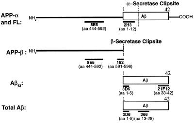
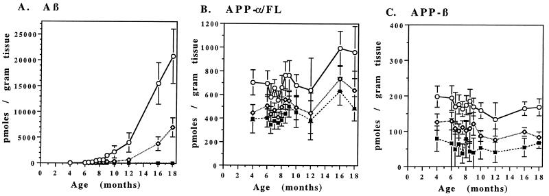
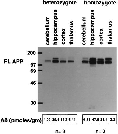
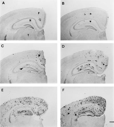
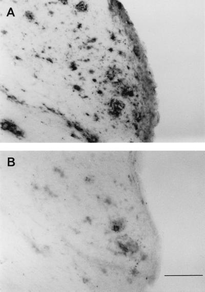
The PDAPP transgenic mouse, which overexpresses human amyloid precursor protein (APP717V-->F), has been shown to develop much of the pathology associated with Alzheimer disease. In this report, levels of APP and its amyloidogenic metabolites were measured in brain regions of transgenic mice between 4 and 18 months of age. While absolute levels of APP expression likely contribute to the rate of amyloid beta-peptide (Abeta) deposition, regionally specific factors also seem important, as homozygotic mice express APP levels in pathologically unaffected regions in excess of that measured in certain amyloid plaque-prone regions of heterozygotic mice. Regional levels of APP and APP-beta were nearly constant at all ages, while A beta levels dramatically and predictably increased in brain regions undergoing histochemically confirmed amyloidosis, most notably in the cortex and hippocampus. In hippocampus, A beta concentrations increase 17-fold between the ages of 4 and 8 months, and by 18 months of age are over 500-fold that at 4 months, reaching an average level in excess of 20 nmol of A beta per g of tissue. A beta1-42 constitutes the vast majority of the depositing A beta species. The similarities observed between the PDAPP mouse and human Alzheimer disease with regard to A beta42 deposition occurring in a temporally and regionally specific fashion further validate the use of the model in understanding processes related to the disease.


Figures

Figure 1
Figure 1
Immunoassays to forms of Aβ and APP described in the text. Immunoassays were prepared to measure APP-α/FL, APP-β (secreted APP ending at the methionine preceding the start of the Aβ region), and total Aβ. Antibodies are mouse mAbs except 192, which is an affinity-purified rabbit polyclonal antibody. Antibodies 192 and 21F12 are specific to fragments of APP and Aβ, respectively, with carboxyl termini as indicated. Antibody 3D6 is specific for Aβ1–5 and does not cross react with APP-α/FL. Methodologies used for the immunoassays are described in text.
Figure 2
Figure 2
Age-dependent changes in brain Aβ and APP levels in the PDAPP transgenic mice. PDAPP mice were sacrificed at the ages indicated and levels of Aβ (A), APP-β (B), and APP-α/FL (C) were determined in the cortex (⋄), hippocampus (○), and cerebellum (▪) by ELISA. Values represent the means ± SD of 9–14 animals.
Figure 3
Figure 3
Levels of APP and Aβ in brain regions of heterozygous and homozygous PDAPP transgenic mice. Two-month-old PDAPP mice were sacrificed, and the amount of APP-FL was measured by Western blot analysis using an APP carboxyl-terminal antibody (anti-6; ref. 6) in hippocampus, cortex, cerebellum, and thalamus of heterozygous and homozygous mice. Essentially similar results were obtained from Western blot analyses using other APP antibodies (human-specific 8E5 and 2H3; data not shown). The amount of Aβ in these same regions from eight heterozygous and three homozygous 2-month-old animals was determined by ELISA, and the average levels are listed.
Figure 4
Figure 4
Age-dependent increases in Aβ plaque burden in the PDAPP mouse. Aβ deposits in the opposite hemisphere of brains used for Aβ and APP ELISAs at 4 (A, arrow indicates deposit), 8 (B), 10 (C), 12 (D), 16 (E), and 18 (F) months of age. Brains are shown from mice with Aβ ELISA values that correspond to the mean of their age group. Deposition typically occurs in an age- and region-dependent manner, with early and heavy involvement of the frontal cortex (F) and hippocampus (H), while the underlying thalamus (T) is devoid of plaques. Arrows in D outline the outer molecular layer of the dentate gyrus, which contains terminals from the perforant pathway. [Bar (in F) = 500 μm.]
Figure 5
Figure 5
42 and Aβ40 immunohistochemical analysis in 18-month-old PDAPP mice. Aβ deposits in adjacent sections from an 18-month-old mouse visualized with antibodies specific for Aβ42 (A) and Aβ40 (B). [Bar (in B) = 100 μm.]

References

    1. Selkoe D. Annu Rev Neurosci. 1994;17:489–517. - PubMed
    1. Goate A, Chartier-Harlin M-C, Mullan M, Brown J, Crawford F, et al. Nature (London) 1991;349:704–706. - PubMed
    1. Chartier-Harlin M-C, Crawford F, Houlden H, Warren A, Hughes D, Fidani L, Goate A, Rossor M, Roques P, Hardy J, Mullan M. Nature (London) 1991;353:844–846. - PubMed
    1. Murrell J, Farlow M, Ghetti B, Benson M. Science. 1991;254:97–99. - PubMed
    1. Mullan M, Crawford F, Axelman K, Houlden H, Lilies L, Winblad B, Lannfelt L. Nat Genet. 1992;1:345–347. - PubMed

MeSH terms

Substances