. 1997 Feb 18;94(4):1591-6.
doi: 10.1073/pnas.94.4.1591.

Why do mitochondria synthesize fatty acids? Evidence for involvement in lipoic acid production

Affiliations

Why do mitochondria synthesize fatty acids? Evidence for involvement in lipoic acid production

H Wada et al. Proc Natl Acad Sci U S A. .

Abstract

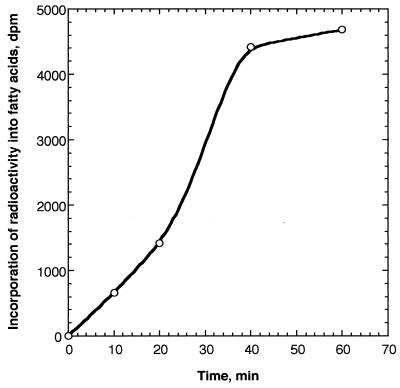
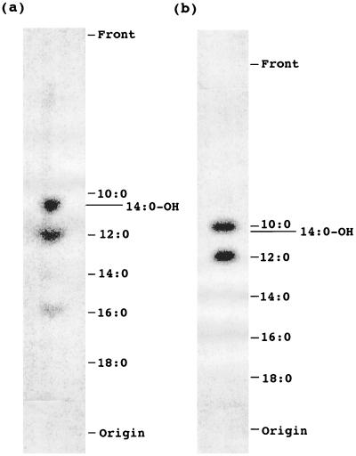
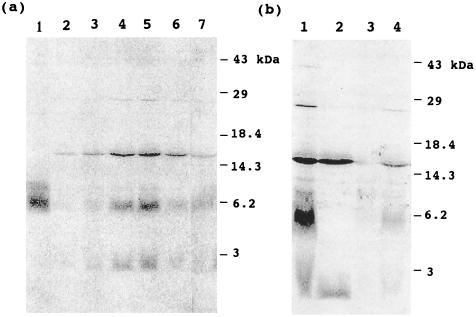
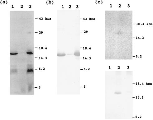
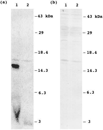
The function of acyl carrier protein (ACP) in mitochondria isolated from pea leaves has been investigated. When pea leaf mitochondria were labeled with [2-14C] malonic acid in vitro, radioactivity was incorporated into fatty acids, and, simultaneously, ACP was acylated. [1-14C]Acetate was much less effective as a precursor for fatty acid synthesis, suggesting that mitochondria do not possess acetyl-CoA carboxylase. The incorporation of radioactivity from [2-14C]malonate into fatty acids and the labeling of ACP were inhibited by cerulenin and required ATP and Mg2+. These findings indicate that plant mitochondria contain not only ACP, but all enzymes required for de novo fatty acid synthesis. Over 30% of the radioactive products from pea mitochondria labeled with [2-14C]malonate were recovered in H protein, which is a subunit of glycine decarboxylase and contains lipoic acid as an essential constituent. In similar experiments, the H protein of Neurospora mitochondria was also labeled by [2-14C]malonate. The labeling of pea H protein was inhibited by addition of cerulenin into the assay medium. Together, these findings indicate that ACP is involved in the de novo synthesis of fatty acids in plant mitochondria and that a major function of this pathway is production of lipoic acid precursors.


Figures

Figure 1
Figure 1
Time course of incorporation of [2-14C]malonate into fatty acids. Pea mitochondria (400 μg of protein) were labeled with [2-14C]malonic acid for the designated times, and radioactivity incorporated into fatty acids was determined by scintillation counting.
Figure 2
Figure 2
Analysis of saponifiable fatty acids synthesized de novo in pea mitochondria with [2-14C]malonic acid. Fatty acids synthesized in pea mitochondria in vitro were separated on reversed-phase TLC plates and analyzed by PhosphorImager. (a) Total fatty acids. (b) Fatty acids bound to mtACP.
Figure 3
Figure 3
Labeling of pea mitochondrial proteins with [2-14C]malonic acid. Pea mitochondria were labeled with [2-14C]malonic acid, and proteins were separated by SDS/PAGE, blotted onto nitrocellulose membrane, and analyzed by PhosphorImager. (a) Time course of labeling of mitochondrial proteins and effect of ATP and MgCl2. In lanes 2–5, mitochondria were labeled for 10, 20, 40, and 60 min, respectively. In lanes 6 and 7, mitochondria were labeled for 1 h, but ATP or MgCl2, respectively, were omitted from assay medium. Lane 1 shows a standard of 14C-labeled 16:0-mtACP. (b) Effects of DTT treatment on deacylation of labeled proteins and cerulenin on labeling of proteins. Pea mitochondria were labeled for 1 h. Lane 1, control that was not treated with DTT; lane 2, mitochondrial proteins were treated with DTT; lanes 3 and 4, 200 μM and 20 μM cerulenin, respectively, were added to the incubation medium, and samples were not treated with DTT.
Figure 4
Figure 4
Localization of 14C-labeled mitochondrial proteins. Pea mitochondria were fractionated into matrix and membrane fractions after 1 h of labeling with [2-14C]malonic acid. (a) Autoradiogram of proteins from each fraction. Proteins were separated by SDS/PAGE, blotted onto nitrocellulose membrane, and analyzed by PhosphorImager. Lane 1, matrix fraction; lane 2, membrane fraction; lane 3, mitochondria before fractionation. (b) Western blot analysis with an antibody against pea H protein. The same blot used for autoradiography in a was probed. Lanes 1–3, matrix fraction, membrane fraction, and mitochondria without fractionation, respectively. (c) Immunoprecipitation of H protein using an antibody against pea H protein. (Upper) Autoradiogram of immunoprecipitated proteins. Lanes 1–3, no IgG, 40 μg of immune IgG, and 60 μg of preimmune IgG were used for immunoprecipitation, respectively. (Lower) Western blot analysis of immunoprecipitated proteins. The same blot used for autoradiography was analyzed for Western blot analysis.
Figure 5
Figure 5
Labeling of Neurospora mitochondria with [2-14C]malonic acid. Neurospora mitochondria were labeled with [2-14C]malonic acid for 1 h. Proteins without DTT treatment or proteins treated with DTT were separated by SDS/PAGE, blotted onto nitrocellulose membrane, and analyzed by PhosphorImager. (a) Autoradiogram of mitochondrial proteins. Lanes 1 and 2, proteins without DTT treatment and the proteins treated with DTT, respectively. (b) Western blot analysis of mitochondrial proteins probed with antibodies to pea H protein. Lanes 1 and 2, proteins without DTT treatment and the proteins treated with DTT, respectively. The same blot used for autoradiography in a was used for Western blot analysis.

References

    1. Brody S, Mikolajczyk S. Eur J Biochem. 1988;173:353–359. - PubMed
    1. Sackmann U, Zensen R, Rohlen D, Jahnke U, Weiss H. Eur J Biochem. 1991;200:463–469. - PubMed
    1. Schneider R, Massow M, Lisowsky T, Weiss H. Curr Genet. 1995;29:10–17. - PubMed
    1. Chuman L, Brody S. Eur J Biochem. 1989;184:643–649. - PubMed
    1. Shintani D K, Ohlrogge J B. Plant Physiol. 1994;104:1221–1229. - PMC - PubMed

Publication types

MeSH terms

LinkOut - more resources