Review
. 1996;6(3):129-37.

Developmental regulation of the vertebrate globin multigene family

Affiliations
Review

Developmental regulation of the vertebrate globin multigene family

M H Baron. Gene Expr. 1996.

Abstract

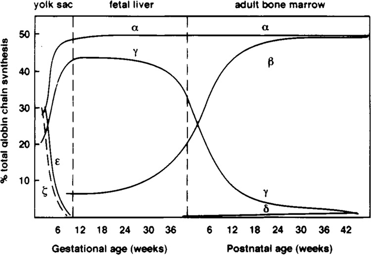
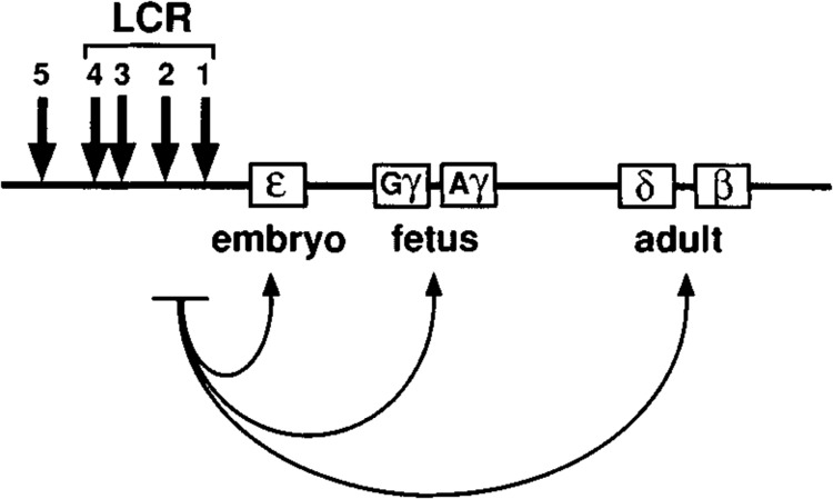
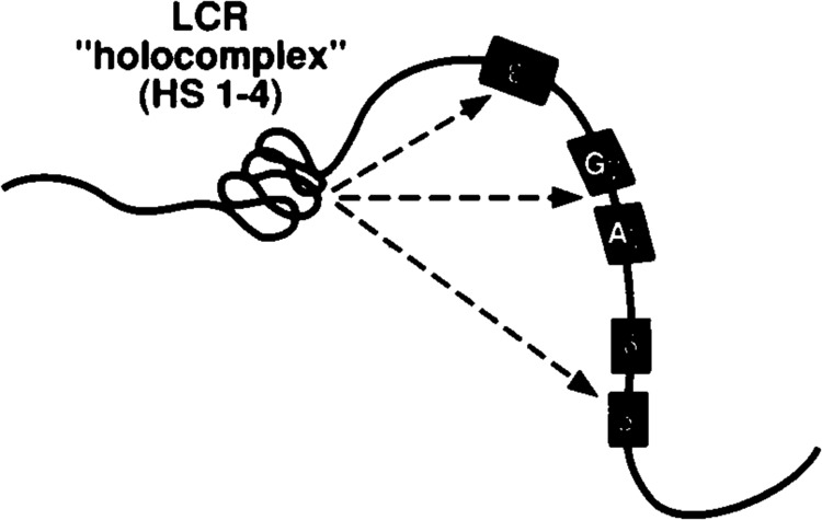
"Hemoglobin switching," or the sequential expression of globin genes in erythroid cells during development, has provided an important paradigm for tissue- and stage-specific gene regulation. Over the past decade, regulatory DNA sequences and transcription factors involved in controlling the expression of individual globin genes in erythroid cells have been identified. The picture that has emerged indicates that gene proximal control elements collaborate with a "locus control region" located far upstream - probably via a DNA looping mechanism - to ensure that each gene is turned on only in erythroid cells and at the appropriate time during development. Interactions among the various regulatory sequences are thought to be mediated and stabilized by an array of tissue-specific and ubiquitous proteins. Chromatin structure plays a critical but still poorly understood role in this process.


Figures

FIG. 1
FIG. 1
Stage-specific expression of α- and β-like globin genes. Changes in globin gene expression during development are associated with changes in the site of production (yolk sac, fetal liver, adult bone marrow) and morphology (size, presence or absence of nucleus) of erythroid cells. The α-like globin genes are ζ (embryonic) and α (fetal, adult); the β-like globin genes are ϵ (embryonic), γ (fetal), and β, δ (adult). Their protein products assemble into heterotetramers to form functional hemoglobin protein.
FIG. 2
FIG. 2
Structure of the human β-globin locus. The β-LCR extends from 6 to 18 kb upstream of the ϵ-globin gene and contains four strong erythroid-specific DNAse I-hypersensitive sites (represented by arrows) that are present at all stages of development (HS1-4). HS5 is constitutively sensitive to nuclease digestion and may function as an insulator or boundary element. Interactions between the LCR and the promoters of individual globin genes (indicated by curved arrows) are thought to be mediated by stage-specific regulatory proteins that stabilize the formation of DNA loops. The figure is not drawn to scale.
FIG. 3
FIG. 3
Cartoon of the LCR “holocomplex.” The hypersensitive regions of the LCR are thought to interact as a unit or “holocomplex” (see text). This figure was adapted from Wij-gerdeetal. (66).

References

    1. Armstrong J. A.; Emerson B. M. NF-E2 disrupts chromatin structure at the human β-globin locus control region HS2 in vitro. Mol. Cell. Biol. (in press). - PMC - PubMed
    1. Baron M. H.; Maniatis T. Rapid reprogramming of globin gene expression in transient heterokaryons. Cell 46:599–602; 1986. - PubMed
    1. Baron M. H.; Maniatis T. Regulated expression of human α- and β-globin genes in transient heterokaryons. Mol. Cell. Biol. 11:1239–1247; 1991. - PMC - PubMed
    1. Baron M. H. Reversibility of the differentiated state in somatic cells. Curr. Opin. Cell Biol. 5:1050–1056; 1993. - PubMed
    1. Baron M. H. Transcriptional control of globin gene switching during vertebrate development. Biochim. Biophys. Acta (in press). - PubMed

Publication types

LinkOut - more resources