. 1996;6(3):139-49.

Characterization of mouse fibronectin alternative mRNAs reveals an unusual isoform present transiently during liver development

Affiliations

Characterization of mouse fibronectin alternative mRNAs reveals an unusual isoform present transiently during liver development

G K Górski et al. Gene Expr. 1996.

Abstract

Fibronectins are found in many extracellular matrices as well as being abundant plasma proteins. The plasma isoforms of fibronectin, which are synthesized in the adult by liver hepatocytes, differ from those derived from most other cells and tissues due to alternative mRNA splicing. Studies in several vertebrates have indicated that FN alternative splicing is regulated spatially and temporally during development. The mouse represents an attractive organism in which to study the regulation of fibronectin splicing during development, but the patterns of fibronectin alternative splicing were not known for this species. Mouse fibronectin cDNA clones were isolated and sequenced, revealing > 95% identity with rat fibronectin at the amino acid level; all three segments that undergo alternative splicing are well conserved. RNase protection and RT-PCR were used to determine the patterns of alternative splicing that occur in fibroblasts and adult liver, sources of cellular and plasma fibronectins. Only A-B-mRNAs were detected in liver, and three V region variants were observed, corresponding to the protein isoforms V120, V95, and V0. Fibroblasts produced mRNAs that were heterogeneous for A and B splicing, but all RNAs contained V120. These patterns contrast with the embryonic form (B+A+V120). Characterization of fibronectin mRNAs from livers of fetal and newborn mice revealed that a significant level of B+ mRNA was present throughout late gestation, declining at birth. Little A+ mRNA was present, and the adult liver V region pattern was observed at all stages. Thus, fibronectin splicing changes during liver development are noncoordinate. One consequence of this temporal regulation is the transient synthesis of B+ mRNAs, including a novel isoform, B+A-V0.


Figures

FIG. 1
FIG. 1
Structure of cDNA clones and RNase protection probes. At the top is a schematic diagram indicating the modular organization of the FN monomer based on the sequence of other vertebrate FNs (22). Type I repeats are depicted as narrow rectangles, Type II repeats as ovals, Type III repeats as squares, and untranslated regions of the mRNA are shown as thick lines. The alternatively spliced segments are indicated as A, B, and V; major cell adhesion signals are indicated above. The structures of the RNase protection probes are indicated below the schematic (A and B). The portions of the molecule encoded by each cDNA clone are indicated below, and the clones are identified at the left.
FIG. 2
FIG. 2
Deduced partial mouse FN protein sequence. The composite sequence was derived from sequencing mfn3 in entirety, most of mfn2, and small portions of the other clones. The sequence is presented to emphasize the repeating structure of the protein; conserved residues characteristic of FN Type III repeat units are indicated above the aligned sequences. Key features are underlined and described in the text.
FIG. 3
FIG. 3
Establishment of FN splicing pattern in the mouse. (A) RNase protection analysis of fibroblast and liver RNAs with a probe specific for the A exon. Samples were applied to a denaturing 6% polyacrylamide gel; lane 1, probe alone (arrow); lane 2, yeast tRNA control; lane 3, fibroblast RNA; lane 4, liver RNA. Fibroblasts contain both forms; liver contains only A – mRNAs. (B) RNase protection analysis of fibroblast and liver RNA with a probe specific for the B exon. Samples were analyzed and lanes loaded as in (A); arrow indicates position of intact probe. Fibroblasts contain both forms; liver contains only B – mRNAs. (C) RT-PCR analysis of V region splicing. Primers that flank the V region were used in PCR amplification of cDNA prepared from fibroblast (lane 1) or liver RNAs (lanes 2 and 3, independent preparations); after 35 cycles of amplification, samples were analyzed on a 6% nondenaturing polyacrylamide gel. Products of 607, 532, and 247 bp were observed for liver, but only the largest was detected in fibroblasts. The precise boundaries of the smaller forms were confirmed by cloning and sequencing of the amplified products; the resulting protein subdomains are indicated in Fig. 2.
FIG. 4
FIG. 4
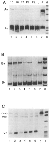
FN isoforms present during liver development. RT-PCR analysis of liver RNAs isolated from E15 (lanes 1), E16 (lanes 2), E17 (lanes 3), postnatal day 1 (lanes 4 and 5), and adult (lane 6) mice and from fibroblasts (lane 7). Lanes M, MspI cut pBR322; from top bands are 622, 527, 404, 309, 242/ 238, 217, 201. Note that not all bands are visible in every panel, but the uppermost band always corresponds to the 622-bp fragment. The light bands present in (A) and (B) between the labeled products are PCR artifacts of unknown origin that are not always evident. Also, the E16 sample seems to consistently give a stronger signal than the other embryonic samples, but changes in cycle number did not change the patterns observed (not shown). (A) Amplification of A+ and A – species (515 and 245 bp); samples were analyzed after 35 cycles on a 6% nondenaturing polyacrylamide gel. (B) Amplification of B+ and B – species (505 and 232); samples were analyzed after 35 cycles on a 6% nondenaturing polyacrylamide gel. (C) Amplification of V120, V95, and VO species (607, 532, and 247 bp); samples were analyzed after 30 cycles on a 2% agarose gel.
FIG. 5
FIG. 5
Detection of a B+ A – V0 FN mRNA isoform. At the top is a diagram of the experimental strategy to determine the V composition of mRNAs that contain B. Below left, RT-PCR analysis of E17 liver RNA using IBF and VR primers; sample was subjected to 35 cycles of amplification and analyzed on a 1% agarose gel. Only two bands were observed, corresponding to V120/V95 (bands of 2512 and 2437 bp did not resolve) and V0 (2152 bp); thus, there was no evidence for either additional variants or incompletely processed RNAs. Another aliquot of this sample was cut with BamHI and the products resolved on a 6% polyacrylamide gel (right). A single large fragment was observed (> 1.0 kbp, not shown) as well as three smaller fragments correponding to the expected sizes of 550, 475, and 190 bp for the V120, V95, and V0 RNA isoforms.
FIG. 6
FIG. 6
Similarity plot of mouse, human, and rat fibronectins. Amino acid sequences translated from human (Genbank K00799, with Ml8177, the human EIIIB sequence, added to it) and rat (XI5906) FN sequences were aligned with the partial mouse sequence shown in Fig. 2. These aligned sequences were then analyzed for the level of identity (scale at left) by the Plotsimilarity program, with a sliding window of 40. The average level of identity is indicated by the dotted horizontal line; identities were assigned a score of 1.0; nonidentities, 0. Regions of the protein that are subjected to exclusion by alternative splicing are indicated by the labeled bars at the top.
FIG. 7
FIG. 7
V region sequence comparison. The sequences of the V regions of seven vertebrate FNs are shown. P, Pleurodeles waltii (12), X, Xenopus laevis (13), C, Gallus domesticus (32); H, Homo sapiens (25); B, Bos tarsus (51); R, Rattus norvegicus (48); M, Mus musculus (this study). Vertical lines indicate identities, and asterisks highlight residues shared by all species. The LDV cell adhesion site is indicated by underlining. A consensus for a second adhesion signal is shown at the bottom (RGD; see text).

Similar articles

Cited by

References

    1. Aota S.-I.; Nagai T.; Yamada K. M. Characterization of regions of fibronectin besides the arginine-glycine-aspartic acid sequence required for adhesive function of the cell-binding domain using site-directed mutagenesis. J. Biol. Chem. 266:15938–15943; 1991. - PubMed
    1. Barkalow F. J. B.; Schwarzbauer J. E. Localization of the major heparin-binding site in fibronectin. J. Biol. Chem. 266:7812–7818; 1991. - PubMed
    1. Bennett V. D.; Pallante K. M.; Adams S. L. The splicing pattern of fibronectin mRNA changes during chondrogenesis resulting in an unusual form of the mRNA in cartilage. J. Biol. Chem. 266:5918–5924; 1991. - PubMed
    1. Blatti S. P.; Foster D. N.; Ranganathan G.; Moses H. L.; Getz M. J. Induction of fibronectin gene transcription and mRNA is a primary response to growth-factor stimulation of AKR-2B cells. Proc. Natl. Acad. Sci. USA 85:1119–1123; 1988. - PMC - PubMed
    1. Borsi L.; Balza E.; Allemanni G.; Zardi L. Differential expression of the fibronectin isoform containing the ED-B oncofetal domain in normal fibroblast cell lines originating from different tissues. Exp. Cell Res. 199:98–105; 1992. - PubMed

Publication types

LinkOut - more resources