. 1997 Mar 15;17(6):1891-7.
doi: 10.1523/JNEUROSCI.17-06-01891.1997.

The neuroprotective activity of group-II metabotropic glutamate receptors requires new protein synthesis and involves a glial-neuronal signaling

Affiliations

The neuroprotective activity of group-II metabotropic glutamate receptors requires new protein synthesis and involves a glial-neuronal signaling

V Bruno et al. J Neurosci. .

Abstract

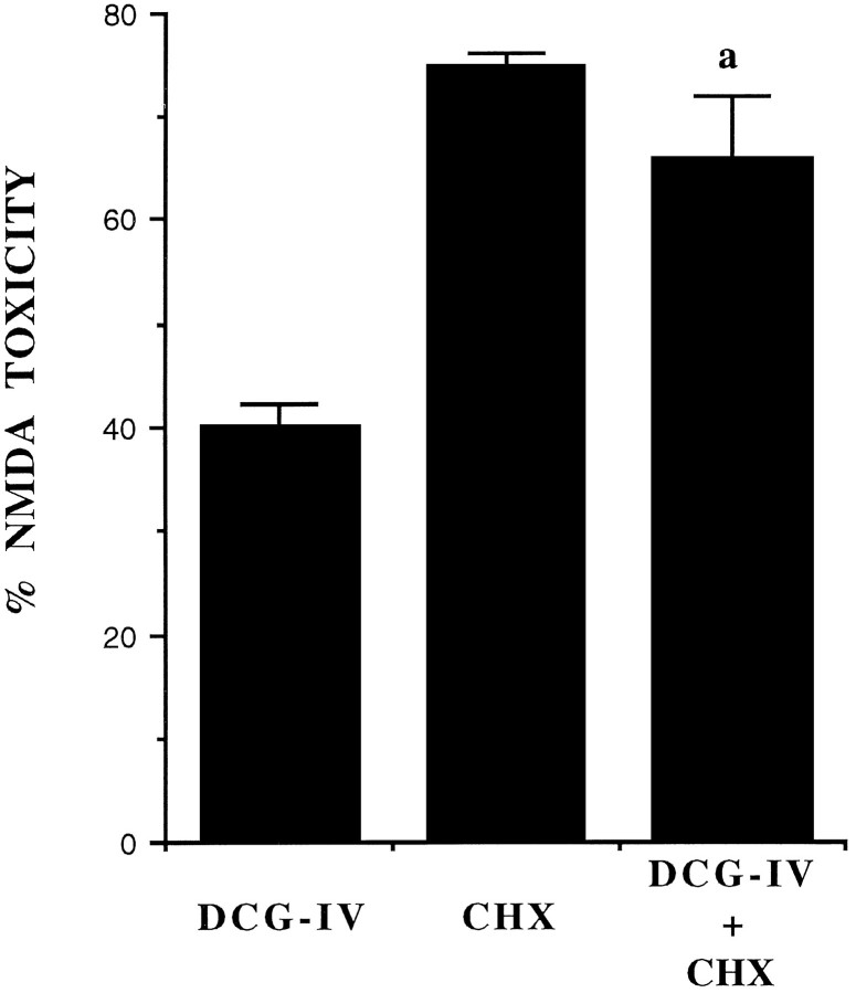
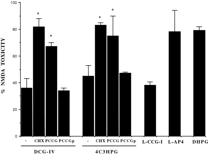
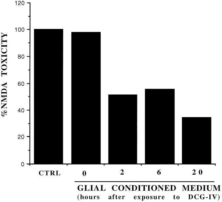
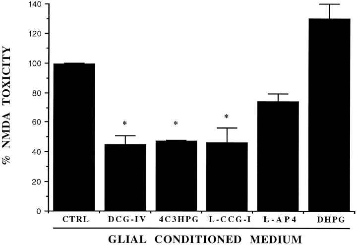
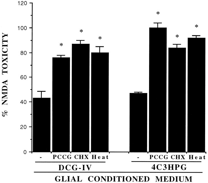
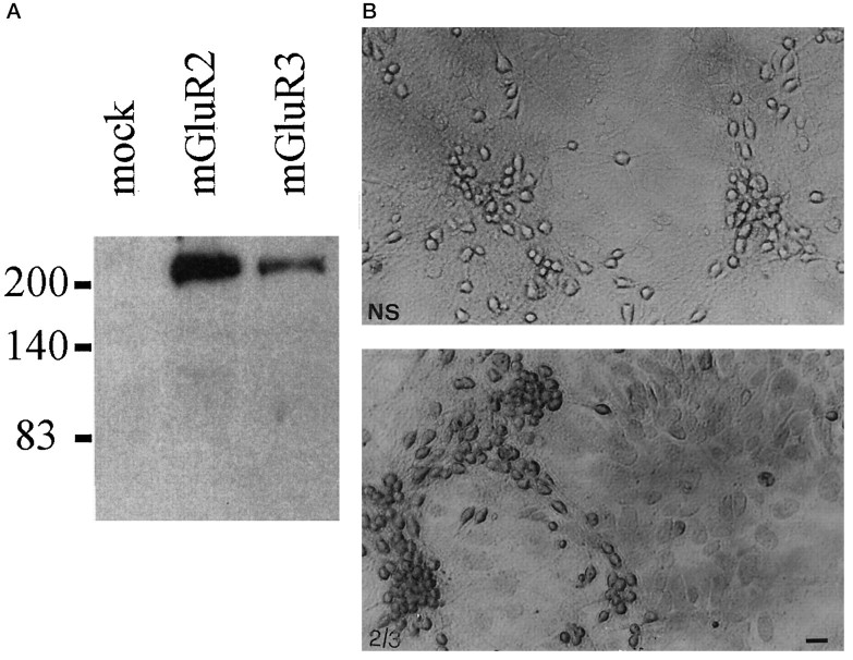
The group-II metabotropic glutamate (mGlu) receptor agonists (2S,1'R, 2'R,3'R)-2-(2,3-dicarboxycyclopropyl)glycine (DCG-IV), S-4-carboxy-3-hydroxyphenylglycine (4C3HPG), and (2S,1'S, 2'S)-2-(carboxycyclopropyl)glycine (L-CCG-I) protected mouse cortical neurons grown in mixed cultures against excitotoxic degeneration induced by a 10 min pulse with NMDA. Protection was observed not only when agonists were added in combination with NMDA but also when they were transiently applied to cultures 6-20 hr before the NMDA pulse. In both cases, neuroprotection was reduced by the group-II mGlu receptor antagonist (2S,1'S,2'S,3'R)-2-(2'-carboxy-3'-phenylcyclopropyl)glycine (PCCG-IV), as well as by the protein synthesis inhibitor cycloheximide (CHX). Both neurons and astrocytes in mixed cultures were immunostained with an antibody that recognized mGlu2 and mGlu3 receptors in recombinant cells. To determine whether astrocytes played any role in the neuroprotection mediated by group-II mGlu receptors, we exposed pure cultures of cortical astrocytes to DCG-IV, 4C3HPG, or L-CCG-I for 10 min. The astrocyte medium collected 2-20 hr after the exposure to any of these drugs was highly neuroprotective when transferred to mixed cultures treated with NMDA. This protective activity was reduced when CHX was applied to astrocyte cultures immediately after the transient exposure to group-II mGlu receptor agonists. We conclude that neuroprotection mediated by group-II mGlu receptors in cultured cortical cells requires new protein synthesis and involves an interaction between neurons and astrocytes.


Figures

Fig. 1.
Fig. 1.
The protein synthesis inhibitor cycloheximide (CHX) attenuates the neuroprotective activity of DCG-IV in mixed cultures of cortical cells. Cultures were exposed to a toxic pulse with 100 μm NMDA in the absence or presence of DCG-IV. CHX (500 ng/ml) was applied to the cultures immediately after the NMDA pulse. Addition of 100 μm NMDA resulted in the necrotic death of 70 ± 8% of the neuronal population (n = 32), without any apparent damage to the underlying monolayer of astrocytes. This value was set at 100%. In untreated cultures, the number of trypan-blue-positive dead neurons was always <3% of the total population. Values indicate the mean ± SEM and were calculated from four independent experiments (n = 4 individual wells for each experiment). In each experiment, SD was <10% of the mean value.
Fig. 2.
Fig. 2.
Neuroprotective activity of group-II mGlu receptor agonists applied to mixed cultures for 10 min, 6 hr before the toxic pulse with NMDA. DCG-IV, 1 μm; 4C3HPG, 50 μm; L-CCG-I, 10 μm; l-AP4, 10 μm; DHPG, 50 μm; PCCG-IV, 20 μm; CHX, 500 ng/ml. CHX was applied immediately after the transient exposure to DCG-IV or 4C3HPG and was maintained during the 6 hr interval preceding the NMDA pulse. PCCG-IV was applied either in combination with DCG-IV or 4C3HPG (labeled as PCCG) or immediately after the exposure to DCG-IV or 4C3HPG (labeled asPCCGp). In the latter case, PCCG-IV was maintained during the 6 hr interval preceding the NMDA pulse (labeled asPCCGp). Values (mean ± SEM) were calculated from three to six independent experiments (n = 4 individual wells in each experiment). *p < 0.01 [one-way ANOVA + Fisher protected least significant difference (PLSD)] when compared with values obtained with DCG-IV or 4C3HPG without addition of CHX or PCCG-IV. Neuroprotection by DCG-IV or 4C3HPG was also significant when both drugs were transiently applied 12 or 20 hr before the NMDA pulse (not shown).
Fig. 3.
Fig. 3.
Neuroprotective activity of the glial-conditioned medium collected from pure cultures of astrocytes, at different times after a 10 min exposure to DCG-IV (1 μm). Values were obtained from a representative experiment and calculated from the mean of four individual determinations. Values relative to the glial-conditioned medium collected at 2, 6, or 20 hr after exposure to DCG-IV were significantly different as compared with values obtained with control (CTRL) astrocyte medium (i.e., with medium from astrocytes treated with the buffer alone) or with the medium collected from astrocyte cultures immediately after washing out DCG-IV (labeled as 0) (p < 0.01 by one-way ANOVA + Fisher PLSD test). This experiment was repeated two times with similar results.
Fig. 4.
Fig. 4.
Neuroprotective activity of the conditioned medium collected from pure cultures of astrocytes 20 hr after a 10 min exposure to the indicated mGlu receptor agonists. CTRL, Control medium (i.e., medium collected from astrocyte cultures treated with the buffer alone). DCG-IV, 1 μm;4C3HPG, 50 μm; L-CCG-I, 10 μm; l-AP4, 10 μm; DHPG, 50 μm. Values (mean ± SEM) were calculated from three to five independent experiments (n = 4 individual wells in each experiment). *p < 0.01 (one-way ANOVA + Fisher PLSD) when compared with CTRL.
Fig. 5.
Fig. 5.
The neuroprotective activity of the glial-conditioned medium collected from pure cultures of astrocytes transiently exposed to DCG-IV (1 μm) or 4C3HPG (50 μm) is attenuated under the following conditions: (1) when PCCG-IV (PCCG, 20 μm) is present during the exposure to DCG-IV or 4C3HPG; (2) when CHX (500 ng/ml) is applied to cultured astrocytes immediately after the exposure to DCG-IV or 4C3HPG and maintained afterward; or (3) when the medium of astrocytes treated with DCG-IV or 4C3HPG is exposed to 100°C for 20 min (Heat) just before being transferred to mixed cortical cultures. Values are mean ± SEM and were calculated from three to five independent experiments (n = 4 individual wells in each experiment). *p < 0.01 when compared with the glial-conditioned medium collected from astrocytes treated with DCG-IV or 4C3HPG alone.
Fig. 6.
Fig. 6.
A, Immunoblotting with purified mGluR2/3 antibodies on lysates from HEK 293 cells transfected with a plasmid encoding the mGlu2 or -3 receptor. Note that the antibody reacts mostly with a high molecular weight band, which may correspond to receptor aggregates (Hayashi et al., 1993). A 100 kDa band corresponding to the deduced molecular weight of mGlu2 and -3 receptors was detected only after long-term exposure. Untransfected HEK 293 cells (mock) or HEK 293 cells transfected with mGlu1a, -5a, and -4 receptor cDNA (not shown) were not stained by the mGlu2/3 receptor antibody. B, Immunostaining of mixed cultures of cortical cells with the mGlu2/3 receptor antibody. Note that both neurons and astrocytes are stained by the antibody. 2/3, Specific staining; NS, nonspecific staining. Scale bar, 20 μm. We also performed Western blot analysis on protein extracts from membranes prepared from (1) pure cultures of mouse astrocytes, (2) pure cultures of rat astrocytes, (3) mouse cerebral cortex, and (4) rat cerebral cortex (60 μg of protein loaded for each lane). In extracts from mouse or rat cerebral cortex, the mGlu2/3 receptor antibody labeled exclusively a 100 kDa band and an additional band of higher molecular weight, which may correspond to receptor aggregates (not shown). No immunolabeling was observed in extracts from either mouse or rat cultured astrocytes. This suggests that the antibody that we have used is specific for mGlu2/3 but the amount of receptor(s) expressed by cultured astrocytes is possibly too low to be detected by Western blot analysis.

References

    1. Altemus KL, Colwell CS, Levine MS. Modulation of excitotoxic damage in the neostriatum by metabotropic glutamate receptors. Soc Neurosci Abstr. 1995;21:217.6.
    1. Balazs R, Miller S, Romano C, de Vries A, Cotman CW. Metabotropic glutamate receptors in cerebral cortical astrocytes: pharmacological properties of mGluR5 and interaction between PLC- and adenylate cyclase (AC)-coupled transduction systems. 2nd International Meeting on Metabotropic Glutamate Receptors. Neuropharmacology. 1996;35:A2.
    1. Bruno V, Copani A, Battaglia G, Raffaele R, Shinozaki H, Nicoletti F. Protective effect of the metabotropic glutamate receptor agonist, DCG-IV, against excitotoxic neuronal death. Eur J Pharmacol. 1994;256:109–112. - PubMed
    1. Bruno V, Battaglia G, Copani A, Giffard RG, Raciti G, Raffaele R, Shinozaki H, Nicoletti F. Activation of class II or III metabotropic glutamate receptors protects cultured cortical neurons against excitotoxic degeneration. Eur J Neurosci. 1995;7:1906–1913. - PubMed
    1. Buisson A, Choi DW. The inhibitory mGluR agonist, S-4-carboxy-3-hydroxy-phenylglycine selectively attenuates NMDA neurotoxicity and oxygen-glucose deprivation-induced neuronal death. Neuropharmacology. 1995;34:1081–1087. - PubMed

MeSH terms

LinkOut - more resources