. 1997 Mar 15;17(6):1993-2005.
doi: 10.1523/JNEUROSCI.17-06-01993.1997.

Functional expression of the heteromeric "olfactory" cyclic nucleotide-gated channel in the hippocampus: a potential effector of synaptic plasticity in brain neurons

Affiliations

Functional expression of the heteromeric "olfactory" cyclic nucleotide-gated channel in the hippocampus: a potential effector of synaptic plasticity in brain neurons

J Bradley et al. J Neurosci. .

Abstract

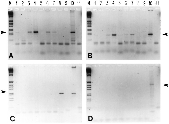
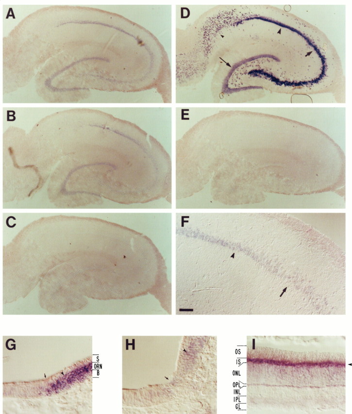
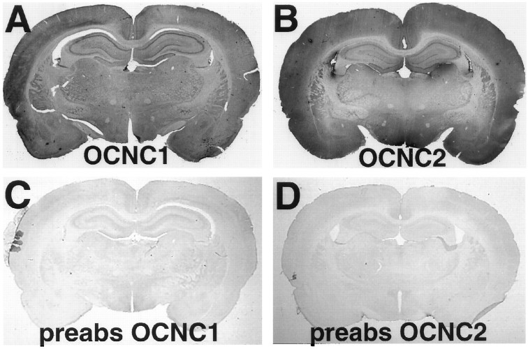
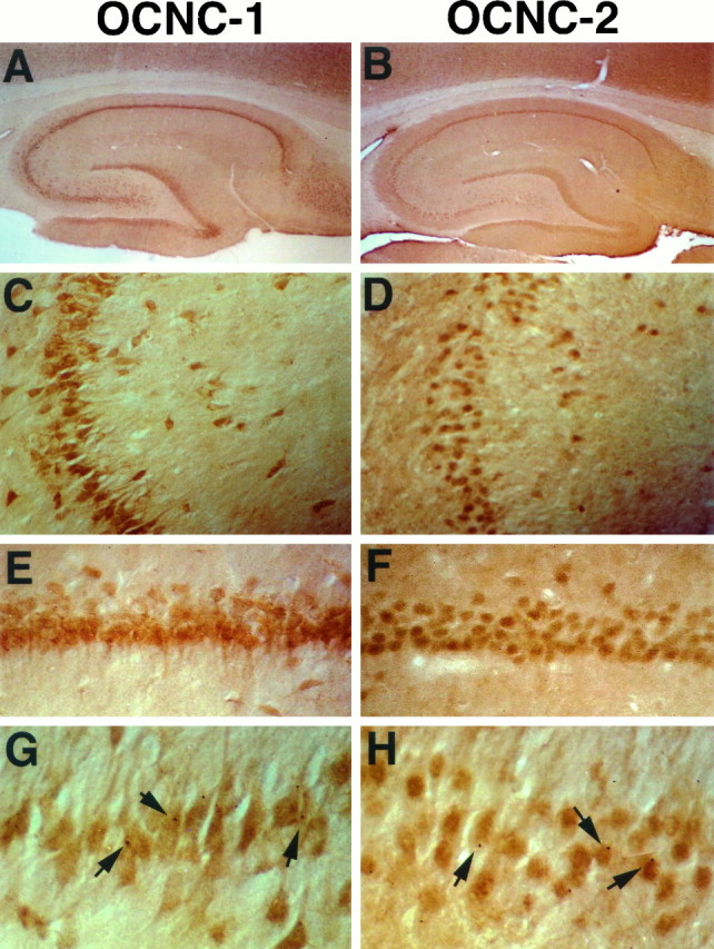
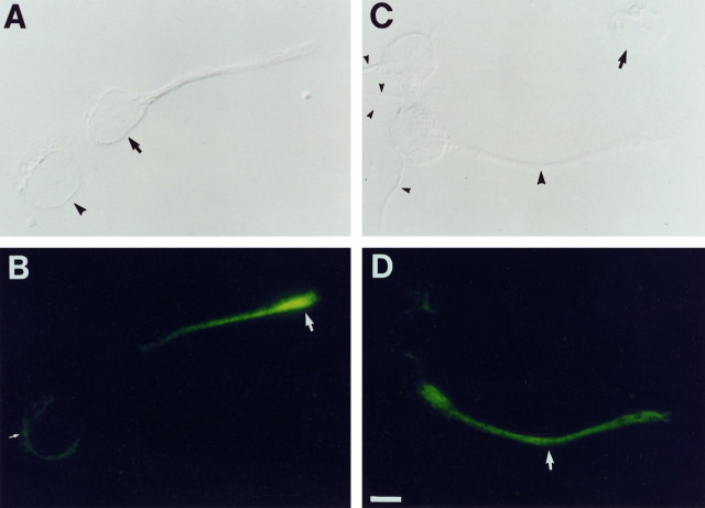
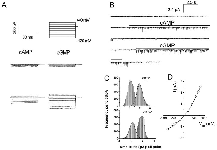
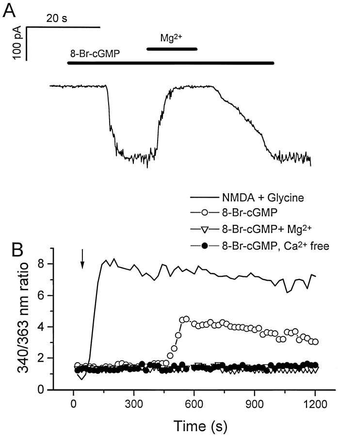
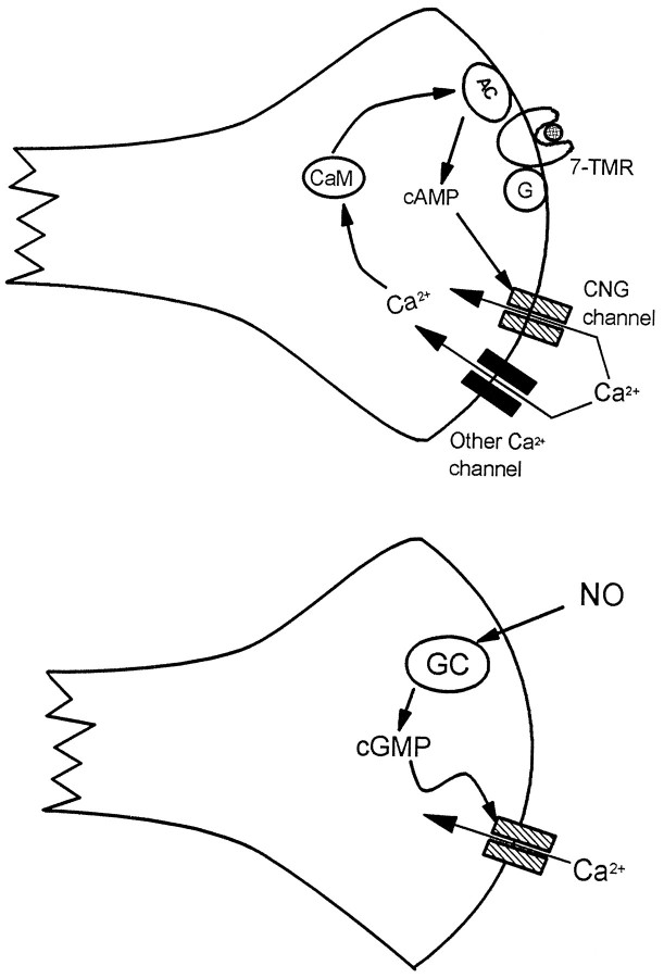
Cyclic nucleotide-gated (cng) channels are important components of signaling systems mediating sensory transduction. In vertebrate photoreceptors, light activates a signaling cascade that causes a decrease in intracellular cGMP concentrations, closing retinal cng channels. Signal transduction in olfactory receptor neurons is believed to proceed via G-protein-mediated elevation of intracellular cAMP in response to odorant binding by 7-helix receptors. cAMP opens the olfactory cng channel, which is highly permeable to Ca2+. Here we demonstrate by in situ hybridization and immunohistochemistry with subunit-specific antibodies that both subunits of the heteromeric rat olfactory cng channel are also widely expressed in the brain. Expression of the retinal rod cng channel, however, can be detected only in the eye. In the adult hippocampus, the olfactory cng channel is expressed on cell bodies and processes of CA1 and CA3 neurons. In cultured embryonic hippocampal neurons, the channel is localized to a subset of growth cones and processes. We recorded conductances with the electrophysiological characteristics of the heteromeric olfactory cng channel in excised inside-out patches from these cultured neurons. We also show that Ca2+ influx into hippocampal neurons in response to cyclic nucleotide elevation can be detected using fura-2 imaging. Cyclic nucleotide elevation has been implicated in several mechanisms of synaptic plasticity in the hippocampus, and these mechanisms also require elevation of intracellular Ca2+. Our results suggest that the "olfactory" cng channel could regulate synaptic efficacy in brain neurons by modulating Ca2+ levels in response to changes in cyclic nucleotide concentrations.


Figures

Fig. 1.
Fig. 1.
Olfactory cng channel mRNAs are expressed in the brain. Semiquantitative RT-PCR assays were used to examine expression of mRNAs encoding rOCNC1 (A), rOCNC2 (B), and rRCNC1 (C). Primers specific for the 3′ untranslated regions of the three mRNAs were used to amplify oligo-dT-primed cDNA made from RNA from various adult rat tissues (35 cycles of amplification). In D, primers spanning an intron/exon boundary from the rOCNC2 gene were used for amplification, to control for the possible presence of genomic DNA in the mRNA preparations. The cDNA concentrations were normalized by amplification (15 and 20 cycles) with β-actin primers, and ∼1 ng of cDNA was used per amplification. Lanes: M, DNA markers;1–9, cDNAs from 1, cortex;2, liver; 3, olfactory bulb;4, nasal epithelium; 5, brainstem;6, cerebellum; 7, hippocampus;8, eye; 9, heart; 10, genomic rat DNA; 11, no DNA. The positions of the appropriate amplified bands are indicated by arrows. The rOCNC1 PCR product is 159 bp (A); the rOCNC2 product is 122 bp (B); the rRCNC1 product is 202 bp (C); and the genomic intron product is ∼363 bp (D). The figure is a negative image of an ethidium bromide-stained agarose gel.
Fig. 2.
Fig. 2.
In situ hybridization analysis of olfactory cng channel mRNA expression in the adult hippocampus. mRNAs were detected by high-stringency in situhybridization to 20 μm sections using digoxigenin-labeled antisense RNA probes derived from nonconserved regions of the channel mRNAs (primarily 3′ untranslated regions), followed by detection with AP-conjugated anti-digoxigenin antibody. A positive signal is indicated by the presence of a purple AP reaction product. A–F, Coronal sections of adult hippocampus: A, rOCNC1;B, rOCNC2; C, rRCNC1; D, SCG10 (positive control); E, I7 olfactory receptor (negative control); F, high-power view of rOCNC1 mRNA expression; G, rOCNC1 probe control (horizontal section of olfactory/respiratory epithelium border); H, rOCNC2 probe control (same as G); I, rRCNC1 probe control (horizontal section of retina). In A and B, but not inC, purple reaction product is visualized in the dentate gyrus, CA3, and CA1 cell body layers [compare withD, in which these regions are labeled dentate gyrus (thin arrow), CA3 (thick arrow), CA1 (large arrowhead)]. The level of nonspecific background hybridization is shown in E; I7 olfactory receptor mRNA is not expressed in the brain. F, Higher-magnification view showing the border between the CA3 (thick arrow) and CA1 (arrowhead) regions. In the probe control panels (G–I), note that hybridization of the rOCNC1 and rOCNC2 probes (arrowheads) is restricted to the olfactory receptor neuron (ORN) layer of the olfactory epithelium [the border between respiratory (left) and olfactory (right) epithelium is at the arrow in G andH; also see Bradley et al., 1994]. S, Sustentacular cell layer; B, basal cell layer. InI, hybridization of the rRCNC1 probe is largely restricted to the inner segments (IS) of the photoreceptors (arrowhead); the other layers are outer segments (OS), outer nuclear layer (ONL), outer plexiform layer (OPL), inner nuclear layer (INL), inner plexiform layer (IPL), and ganglion cell layer (GL). rOCNC1 and rOCNC2 probes do not hybridize to retina. The AP development reaction was incubated for 20 hr for the sections inA–C and E–H and for 4 hr for the sections in D and I. Scale bars:A–E, 290 μm; F, 72 μm; G–I, 36 μm.
Fig. 3.
Fig. 3.
Immunoblot analysis of rOCNC1 and rOCNC2 in the hippocampus. Homogenates were prepared from whole adult hippocampus, and 200 μg of protein per gel lane were electrophoretically separated on a 7–15% SDS-PAGE and transferred onto a nitrocellulose membrane. After blocking, lanes were separated and probed with either anti-rOCNC1 antiserum or anti-rOCNC2 mAb. rOCNC1 is 78 kDa, and rOCNC2 is 67 kDa (− lanes). In each case, immunoreactivity was blocked when the antibodies were preabsorbed with a 10-fold excess of the appropriate purified fusion protein antigen (+ lanes).
Fig. 4.
Fig. 4.
Immunolocalization of rOCNC1 and rOCNC2 in whole brain sections. Coronal sections of adult rat brains were immunostained, using horseradish peroxidase immunohistochemistry, with affinity-purified antibody to rOCNC1 (A, C) or anti-rOCNC2 mAb (B, D). In C andD, the antibodies were preabsorbed with a 10-fold excess of the appropriate fusion protein. Widespread immunoreactivity is seen with both antibodies. Staining is particularly strong in the hippocampal pyramidal cell layers and in the dentate gyrus. A high level of staining is also observed in cortex.
Fig. 5.
Fig. 5.
Immunolocalization of rOCNC1 and rOCNC2 in hippocampal sections. A, C, E, and G show staining with affinity-purified antibody to rOCNC1; B, D, F, and H show staining with anti-rOCNC2 mAb. Immunoreactivity to rOCNC1 and rOCNC2 is essentially colocalized; it is seen in hippocampal CA1 (C, D) and CA3 (E–H) regions, the subiculum, and the dentate gyrus. Staining for both is most prominent in cell bodies and proximal dendrites. Note the punctate staining in G andH (arrows). Photomicrographs were taken at 25× magnification (A, B), 100× magnification (C, D), 200× magnification (E, F), and 400× magnification (G, H).
Fig. 6.
Fig. 6.
Expression of rOCNC2 on processes and growth cones of cultured embryonic hippocampal neurons. Two-day-old dissociated hippocampal cultures from E17 embryos were fixed with methanol and incubated with anti-rOCNC2 mAb followed by FITC-labeled secondary antibody. In A, C, cells are visualized using DIC optics; in B, D, rOCNC2 expression is visualized using fluorescence. One cell in A (arrowhead) has not yet extended a process, and rOCNC2 is localized to internal membranes within this cell (small white arrow inB). The other cell (arrow inA) has extended a short process. rOCNC2 is no longer localized to the cell body but is present at high levels on the growth cone (white arrow in B) and at lower levels on the process. In C, two of the cells have extended short processes (small arrowheads), and one has a longer process (large arrowhead). rOCNC2 is selectively expressed on the longer process (white arrowin D). The arrow in Cindicates a cell that does not seem to express rOCNC2 at detectable levels. Scale bar, 7.2 μm.
Fig. 7.
Fig. 7.
Whole-cell and excised inside-out patch recording of cng channels from cultured embryonic hippocampal neurons.A, Whole-cell recording. Currents activated in a cell by diffusion from patch pipettes backfilled with 100 μm cAMP (left) or with 100 μm cGMP in a different cell (right). The top traces show the voltage commands for each episode in a trial, with the time and current scales indicated at the left. The middle traces are currents recorded immediately after patch rupture, and the bottom traces are of currents recorded 4 min after patch rupture. B, Inside-out patch recording. Continuous recordings from an inside-out patch excised from a cell body of a hippocampal neuron are shown. The patch was held at −60 mV. Cyclic nucleotides were perfused onto the cytoplasmic side of the patch for 10 sec intervals, at the times indicated by the horizontal bars. The pipette and perfusion solutions were symmetrical and free of divalent cations. C, D, Amplitude histograms and current–voltage relations (I–V). The amplitude histograms were constructed from a patch perfused with 10 μm cAMP. The I–V relation was plotted for the same patch.
Fig. 8.
Fig. 8.
Effect of membrane-permeable cyclic nucleotide on cultured hippocampal neurons. A, Effects of 8-bromo-cGMP on a hippocampal neuron from a 7-d-old culture. The recording was performed in a whole-cell patch-clamp configuration. The recording chamber was perfused constantly with bath solution. The holding potential was −80mV. 8-bromo-cGMP was diluted in the bath solution or in a bath solution containing 10 mm MgCl2 in a final concentration of 1 mm. Duration of drug delivery is indicated by the horizontal bars. B, Fura-2 calcium measurement of cultured hippocampal neurons. Measurements were made from separate coverslips of the same neuronal culture. The measurements were the averages of responses of multiple cells (8-bromo-cGMP, 6 cells; NMDA, 16 cells; 8-bromo-cGMP with extracellular Mg2+, 13 cells; 8-bromo-cGMP without extracellular Ca2+, 11 cells). The arrowindicates the starting point for drug application. The long delay before the onset of the drug response in this system is attributable to the slow diffusion of 8-bromo-cGMP into the space between the immersion objective and the slide (an upright microscope was used in these experiments). Drugs were present at all times, and no perfusion was used during the measurements. The final concentrations of the drugs were NMDA, 100 μm; glycine, 10 μm; 8-bromo-cGMP, 1 mm; and Mg2+, 10 mm.
Fig. 9.
Fig. 9.
Potential functions of “olfactory” cng channels in nerve terminals in the brain. On the left, a hypothetical positive feedback loop is depicted in which agonist (hatched circle) binding to a 7-helix receptor (7-TMR) coupled to AC activation via a G-protein causes production of cAMP, opening the cng channel and admitting Ca2+. This Ca2+, together with Ca2+ entering through other channels, binds to CaM, which further stimulates AC activity. Note also that this loop might be “self-damping,” because Ca2+/CaM binds to the olfactory channel and reduces its affinity for cAMP (for review, see Finn et al., 1996). On the right, NO activates GC, producing cGMP, which causes opening of the cng channel and an influx of Ca2+.

References

    1. Ahmad I, Leinders-Zufall T, Kocsis JD, Shepherd GM, Zufall F, Barnstable CJ. Retinal ganglion cells express a cGMP-gated cation conductance activatable by nitric oxide donors. Neuron. 1994;12:155–165. - PubMed
    1. Barnstable CJ, Wei JY. Isolation and characterization of the alpha-subunit of rod photoreceptor cGMP-gated cation channel. J Mol Neurobiol. 1995;6:289–302. - PubMed
    1. Bradley J, Li J, Davidson N, Lester HA, Zinn K. Heteromeric olfactory cyclic nucleotide-gated channels: a subunit that confers increased sensitivity to cAMP. Proc Natl Acad Sci USA. 1994;91:8890–8894. - PMC - PubMed
    1. Broillet MC, Firestein S. Direct activation of the cyclic nucleotide-gated channel through modification of sulfhydryl-groups by NO compounds. Neuron. 1996;16:377–385. - PubMed
    1. Buck L, Axel R. A novel multigene family may encode odorant receptors: a molecular basis for odor recognition. Cell. 1991;65:175–187. - PubMed

Publication types

Associated data