. 1997 Mar 15;17(6):2018-29.
doi: 10.1523/JNEUROSCI.17-06-02018.1997.

Cell fate specification and symmetrical/asymmetrical divisions in the developing cerebral cortex

Affiliations

Cell fate specification and symmetrical/asymmetrical divisions in the developing cerebral cortex

M C Mione et al. J Neurosci. .

Abstract

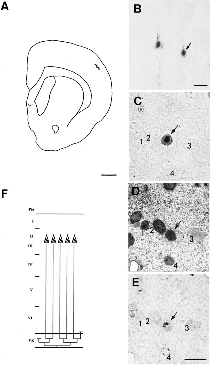
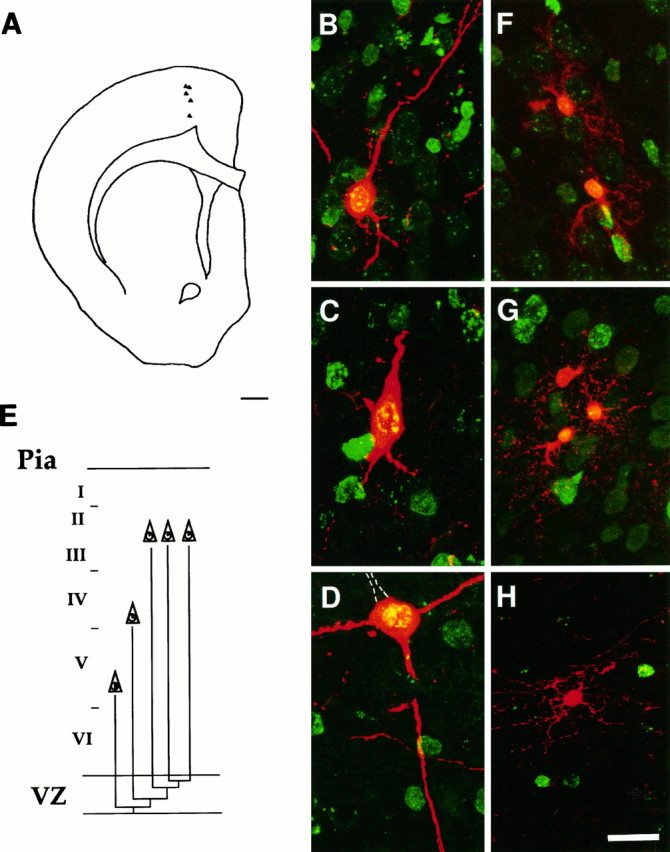
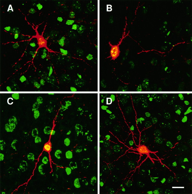
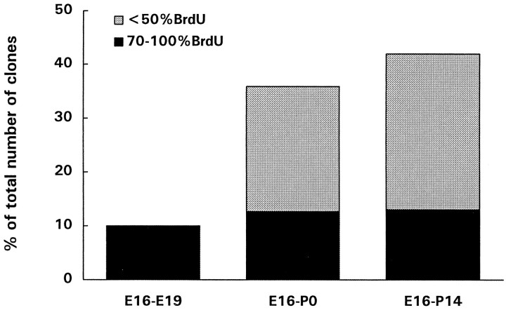
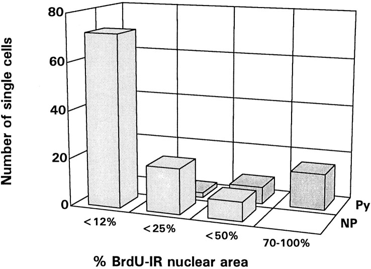
Two different modes of cell division are adopted by progenitor cells to generate the neurons and glia of the cerebral cortex: they either divide symmetrically to generate other progenitors or a pair of postmitotic cells or divide asymmetrically to generate both a progenitor and a postmitotic cell. In this study we used a lineage marker, the BAG retrovirus, in embryonic day 16 rats in combination with bromodeoxyuridine (BrdU) to identify patterns of cell generation in the cerebral cortex, and investigated the relationship between the phenotype of cells and the history of their lineages. The location, phenotype and birth order of clonally related cells were studied in the subsequent 3 weeks. Only pyramidal neurons and/or astrocytes formed discrete clusters in which several generations of family members were present, whereas nonpyramidal neurons were found exclusively in pairs or as single cells. Analysis of BrdU levels in these cells showed that nonpyramidal neurons were originally part of larger clones and were found dispersed in the neocortex because of tangential migration of their progenitors, dispersion of postmitotic cells, or death of clonal relatives. These results suggest that both symmetrical and asymmetrical division can be adopted by progenitor cells to generate cortical neurons and glial cells and that cell extrinsic events contribute to the isolation of nonpyramidal neurons.


Figures

Fig. 1.
Fig. 1.
Horizontal clusters. A, Camera lucida drawing showing the location of β-gal+cells of cluster 18 (Table 1). These cells were all located in layers II and III and distributed within two consecutive 100-μm-thick Vibratome sections. Glutamate immunoreactivity was used as a marker of pyramidal neurons. Two of the cells of this cluster are displayed inB, and one (arrow) is shown in adjacent semithin sections (C–E): unstained (C) or after immunostaining for glutamate (D) or BrdU (E). The other cells of this cluster were all immunoreactive for glutamate and displayed similar low levels of BrdU immunoreactivity. None of the cells was immunoreactive for GABA, a marker of nonpyramidal neurons. Four neighboring neurons (1–4) are shown in all three sections as landmarks. Asterisks mark the same blood vessel.F, A possible family tree for this cluster. To obtain five neurons with <12% BrdU-immunoreactive nuclear area, the progenitor cells must have divided symmetrically at least three times (corresponding clonal size, 8 cells) after incorporation of retrovirus and BrdU. Only five cells were found, which suggests that at least one postmitotic [line ending above the ventricular zone (VZ)] and one progenitor cell (line ending within the VZ) were lost (through death or migration) from this cluster. Scale bars: A, 1 mm;BE, 20 μm.
Fig. 2.
Fig. 2.
Radial clusters. A, Camera lucida drawing showing the location of β-gal+ cells of cluster 18 (see Table 2). Cells were located in different layers, from layers V to II, but displayed very little lateral displacement.B–D, Laser scanning confocal micrograph of cells located in layer II (B), layer IV (C), or layer V (D). β-gal+ cells were stained (red) with a polyclonal β-gal antiserum, followed by streptavidin–Texas Red, whereas BrdU immunoreactivity (green) was detected with a monoclonal antiserum followed by FITC. Coexistence appears yellow. All cells displayed morphological features of pyramidal neurons, with visible basal and/or apical dendrites. The cell depicted in Dhad a prominent apical dendrite (dotted lines) in the adjacent section. The levels of BrdU immunoreactivity were higher in the cell located in layer V (D, immunoreactive nuclear area, ∼50%) and progressively lower for the cell located in layer IV (C, immunoreactive nuclear area, 12–25%) and II (B, immunoreactive nuclear area, <12%).E, A possible family tree of this cluster. To obtain five neurons with these levels of BrdU, the progenitors must have undergone three asymmetrical and one symmetrical division after the incorporation of retrovirus and BrdU to give rise to postmitotic daughters during the first three divisions and two terminal postmitotic cells from the last symmetrical division. Glial clusters usually showed a clear radial alignment; the cells were grouped in several subunits and distributed along different layers. F–H, Examples of confocal micrographs of clonally related glial cells stained with β-gal+ antiserum and Texas Red. In F andG, small clusters of β-gal+ astrocytes can be recognized by their thin, spider-like processes. InH, an oligodendrocyte, part of cluster 7 (see Table 3), shows the characteristic parallel processes. The levels of BrdU immunoreactivity (here visualized with FITC) were very low in all glial cells, indicating that their progenitors must have divided several times after retroviral and BrdU incorporation before these cells were born. Scale bars: A, 1 mm; B–H, 20 μm.
Fig. 3.
Fig. 3.
Two cell clusters and isolated cells. An example of a two-cell cluster, composed of nonpyramidal (A) and pyramidal (B) neurons (cluster 21 in Table 4). Confocal micrographs showing β-gal immunoreactivity in red(Texas Red) and BrdU immunoreactivity in green (FITC). Coexistence is shown in yellow. The pyramidal cell was characterized by the presence of dendritic spines along the apical dendrite, whereas the nonpyramidal neuron showed intense staining of its many dendrites. BrdU levels were low in both cells, suggesting that they may have been generated by the same division or the same order of divisions after the incorporation of retrovirus and BrdU by their ancestor. C, D, Examples of isolated β-gal+ cells, double-immunostained for β-gal (Texas Red) and BrdU (FITC) in the cortex of 2-week-old rats given injections of retrovirus and BrdU at E16. Only isolated pyramidal neurons, like the cell depicted in C, were characterized by high levels of BrdU immunoreactivity. The majority of isolated β-gal+ cells, however, was nonpyramidal neurons (D), which displayed low levels of BrdU immunoreactivity. Scale bars, 20 μm.
Fig. 4.
Fig. 4.
An isolated bipolar β-gal+ cell from the cortex of a 2-week-old rat is shown in A (Vibratome section) and in B–D in adjacent semithin sections (arrows), either unstained (B) or after immunostaining for GABA (C) or BrdU (D). This cell was GABA immunoreactive (a marker of nonpyramidal neurons), as were many other isolated β-gal+ cells at this age, and displayed 12–25% BrdU-immunoreactive nuclear area. Three neighboring neurons (1–3) and blood vessels (asterisks) are shown as landmarks. Scale bars, 20 μm.
Fig. 5.
Fig. 5.
Histogram showing the percentages of isolated β-gal+ cells over the total number of clusters and pairs at different ages, after the injection of retrovirus and BrdU at E16, and the proportion of these cells that displayed high levels of BrdU immunoreactivity (dark bars). Isolated β-gal+ cells were studied in E19 rat embryos (3 d after injections), in newborn rats, and in 2-week-old rats. Less than 10% of the clones found at E19 were composed of a single cell, all of which displayed high levels of BrdU immunoreactivity. At later times after the injections, isolated β-gal+ cells increased in number, possibly as a result of tangential migration and/or death of clonal relatives. However, the number of single-cell clones (isolated cells with high levels of BrdU immunoreactivity) remained constant and represented ∼10% of all clusters at any age examined. The results obtained are pooled from at least five animals for each age group.
Fig. 6.
Fig. 6.
The phenotype and BrdU levels of 130 isolated β-gal+ cells found in the cortex of 18 two-week-old rats given injections of retrovirus and BrdU at E16 are plotted here. A vast majority of cells were nonpyramidal neurons with low levels of BrdU immunoreactivity (light gray bars). Isolated nonpyramidal neurons with intermediate levels of BrdU immunoreactivity were also found. However, all isolated β-gal+ cells with 70–100% BrdU-immunoreactive nuclear area, indicative of single-cell clones, were pyramidal cells (dark bars).

References

    1. Acklin SE, van der Kooy D. Clonal heterogeneity in the germinal zone of the developing rat telencephalon. Development. 1993;118:175–192. - PubMed
    1. Anderson DJ. The neural crest lineage problem: neuropoiesis? Neuron. 1989;3:1–12. - PubMed
    1. Artavanis-Tsakonas S, Matsuno K, Fortini ME. Notch signaling. Science. 1995;268:225–232. - PubMed
    1. Austin CP, Cepko CL. Cellular migration patterns in the developing mouse cerebral cortex. Development. 1990;110:713–732. - PubMed
    1. Bayer SA, Altman J. Neocortical development. Raven; New York: 1991.

Publication types

LinkOut - more resources