. 1997 Feb 24;136(4):907-18.
doi: 10.1083/jcb.136.4.907.

Induction of neurite outgrowth through contactin and Nr-CAM by extracellular regions of glial receptor tyrosine phosphatase beta

Affiliations

Induction of neurite outgrowth through contactin and Nr-CAM by extracellular regions of glial receptor tyrosine phosphatase beta

T Sakurai et al. J Cell Biol. .

Abstract

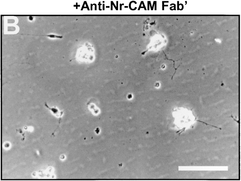
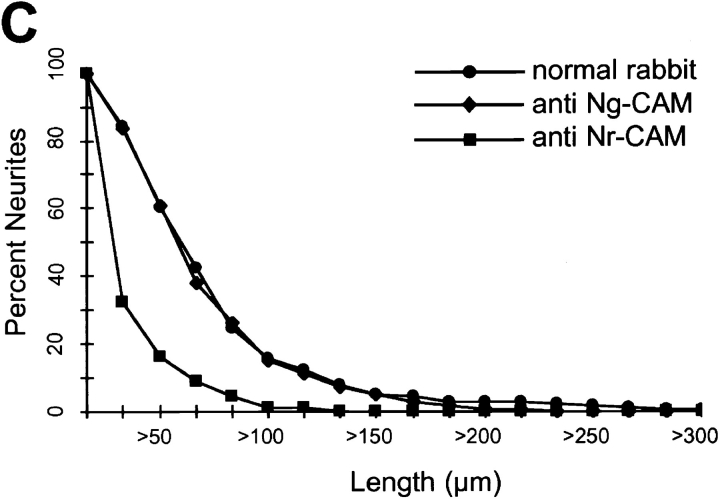
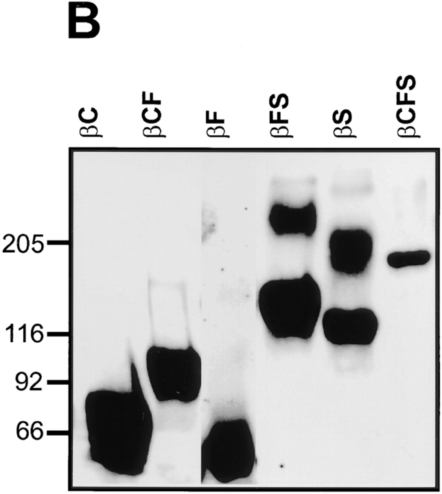
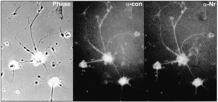
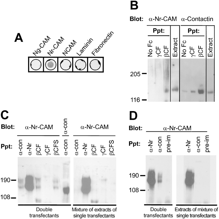
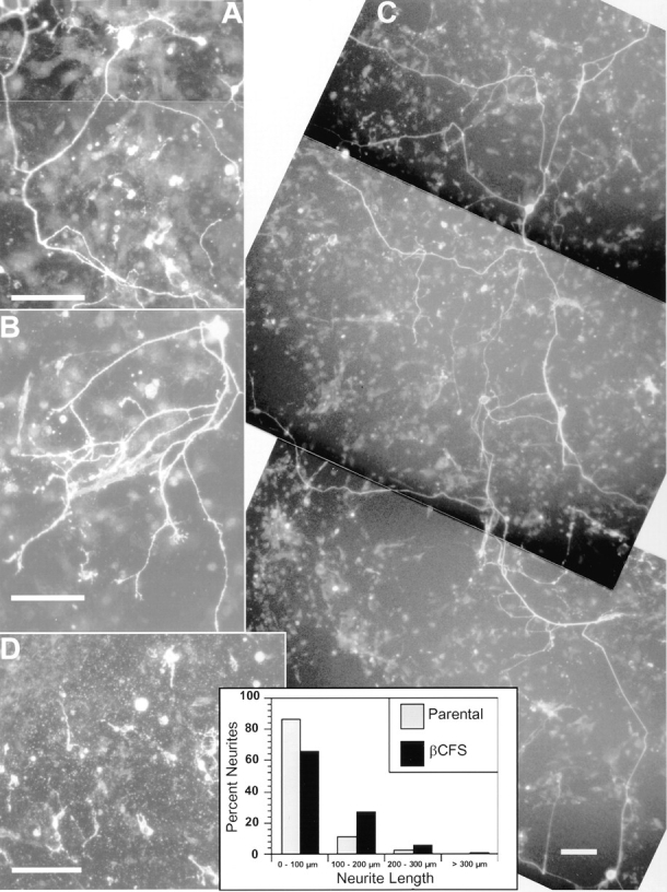
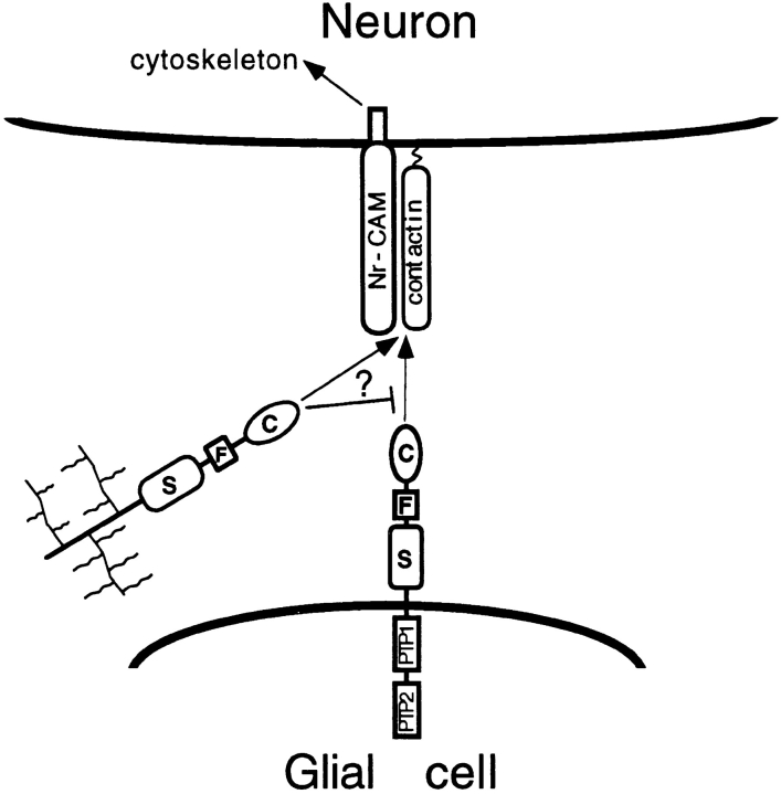
Receptor protein tyrosine phosphatase beta (RPTPbeta) is expressed as soluble and receptor forms with common extracellular regions consisting of a carbonic anhydrase domain (C), a fibronectin type III repeat (F), and a unique region called S. We showed previously that a recombinant Fc fusion protein with the C domain (beta C) binds to contactin and supports neuronal adhesion and neurite growth. As a substrate, betaCFS was less effective in supporting cell adhesion, but it was a more effective promoter of neurite outgrowth than betaCF. betaS had no effect by itself, but it potentiated neurite growth when mixed with betaCF. Neurite outgrowth induced by betaCFS was inhibited by antibodies against Nr-CAM and contactin, and these cell adhesion molecules formed a complex that bound betaCFS. NIH-3T3 cells transfected to express betaCFS on their surfaces induced neuronal differentiation in culture. These results suggest that binding of glial RPTPbeta to the contactin/Nr-CAM complex is important for neurite growth and neuronal differentiation.


Figures

Figure 3
Figure 3
Inhibition by anti-contactin antibodies of neurite outgrowth induced by βCFS. Primary chick tectal cells were plated on dishes coated with βCFS fusion protein (80 μg/ml) and cultured for 48 h in the presence of Fab′ fragments (500 μg/ml) of nonimmune (A) or anti-contactin polyclonal antibody (B). The plates were fixed and photographed. Quantification of lengths of tectal cell neurites on βCFS fusion protein (C) in the presence of 100 μg/ml Fab′ fragments of nonimmune and anti-contactin polyclonal antibodies, or no added Fab′. The percentage of neurons with neurites longer than a given length in μm was determined as described in Materials and Methods. The average neurite lengths without Fab′ were 67 ± 8 μm (n = 100), with normal rabbit 73 ± 7 μm (n = 57), and with anti-contactin 16 ± 2 μm (n = 82). This level of anti-contactin produced maximal inhibition insofar as higher levels of anti-contactin did not yield greater levels of inhibition; the average lengths at 300 and 500 μg/ml were 22 ± 2 (n = 99) and 25 ± 3 μm (n = 119), respectively. Bar, 100 μm.
Figure 3
Figure 3
Inhibition by anti-contactin antibodies of neurite outgrowth induced by βCFS. Primary chick tectal cells were plated on dishes coated with βCFS fusion protein (80 μg/ml) and cultured for 48 h in the presence of Fab′ fragments (500 μg/ml) of nonimmune (A) or anti-contactin polyclonal antibody (B). The plates were fixed and photographed. Quantification of lengths of tectal cell neurites on βCFS fusion protein (C) in the presence of 100 μg/ml Fab′ fragments of nonimmune and anti-contactin polyclonal antibodies, or no added Fab′. The percentage of neurons with neurites longer than a given length in μm was determined as described in Materials and Methods. The average neurite lengths without Fab′ were 67 ± 8 μm (n = 100), with normal rabbit 73 ± 7 μm (n = 57), and with anti-contactin 16 ± 2 μm (n = 82). This level of anti-contactin produced maximal inhibition insofar as higher levels of anti-contactin did not yield greater levels of inhibition; the average lengths at 300 and 500 μg/ml were 22 ± 2 (n = 99) and 25 ± 3 μm (n = 119), respectively. Bar, 100 μm.
Figure 4
Figure 4
Inhibition by anti– Nr-CAM antibodies of neurite extension induced by βCFS. Primary chick tectal cells were plated on dishes coated with βCFS fusion protein (80 μg/ml) and cultured for 48 h in the presence of Fab′ fragments (500 μg/ml) of nonimmune (A) or anti– Nr-CAM polyclonal antibody (B). The plates were fixed and photographed. Quantification of lengths of tectal cell neurites on βCFS fusion protein (C) and on Ng-CAM (D) in the presence of Fab′ fragments. The average neurite lengths on βCFS (C) treated with Fab′ were 59 ± 4 μm (n = 193) for normal rabbit, 55 ± 2 μm (n = 302) for anti–Ng-CAM, and 16 ± 3 μm (n = 93) for anti–NrCAM. The average neurite lengths on Ng-CAM (D) treated with Fab′ were 83 ± 5 μm (n = 198) for normal rabbit, 34 ± 2 μm (n = 118) for anti–Ng-CAM, and 80 ± 5 μm (n = 187) for anti–Nr-CAM. The percentage of neurons with neurites longer than a given length in micrometers was determined as described in Materials and Methods. The results of one representative experiment are shown, and similar results were obtained in five different experiments. Anti–Nr-CAM polyclonal antibody, inhibited neurite outgrowth on Nr-CAM substrate completely at the concentration of 500 μg/ml (data not shown). Bar, 100 μm.
Figure 4
Figure 4
Inhibition by anti– Nr-CAM antibodies of neurite extension induced by βCFS. Primary chick tectal cells were plated on dishes coated with βCFS fusion protein (80 μg/ml) and cultured for 48 h in the presence of Fab′ fragments (500 μg/ml) of nonimmune (A) or anti– Nr-CAM polyclonal antibody (B). The plates were fixed and photographed. Quantification of lengths of tectal cell neurites on βCFS fusion protein (C) and on Ng-CAM (D) in the presence of Fab′ fragments. The average neurite lengths on βCFS (C) treated with Fab′ were 59 ± 4 μm (n = 193) for normal rabbit, 55 ± 2 μm (n = 302) for anti–Ng-CAM, and 16 ± 3 μm (n = 93) for anti–NrCAM. The average neurite lengths on Ng-CAM (D) treated with Fab′ were 83 ± 5 μm (n = 198) for normal rabbit, 34 ± 2 μm (n = 118) for anti–Ng-CAM, and 80 ± 5 μm (n = 187) for anti–Nr-CAM. The percentage of neurons with neurites longer than a given length in micrometers was determined as described in Materials and Methods. The results of one representative experiment are shown, and similar results were obtained in five different experiments. Anti–Nr-CAM polyclonal antibody, inhibited neurite outgrowth on Nr-CAM substrate completely at the concentration of 500 μg/ml (data not shown). Bar, 100 μm.
Figure 8
Figure 8
βS induces neurite growth when combined with βCF. Primary chick tectal cells (5 × 10⁴ cells) were plated on purified βCFS, βCF, βS (40 μg/ml), or on combinations (40 μg/ml final concentration) of the latter two as noted in the right panels of the figure. After 48 h, cultures were fixed and photographed. The graph shows quantification of the lengths of neurites on βCF and βCFS and on mixtures of βCF + βS at different ratios. Neurite lengths were measured and analyzed as described in Materials and Methods. The average neurite lengths were: βCFS, 73 ± 7 μm (n = 109); βCF, 26 ± 2 μm (n = 148); 1X[CF] + 3X[S], 67 ± 6 μm (n = 161); 2X[CF] + 2X[S], 39 ± 3 μm (n = 153); and 3X[CF] + 1X[S], 27 ± 2 μm (n = 192). Representative data are shown, and similar results were obtained in three experiments. Bar, 100 μm.
Figure 8
Figure 8
βS induces neurite growth when combined with βCF. Primary chick tectal cells (5 × 10⁴ cells) were plated on purified βCFS, βCF, βS (40 μg/ml), or on combinations (40 μg/ml final concentration) of the latter two as noted in the right panels of the figure. After 48 h, cultures were fixed and photographed. The graph shows quantification of the lengths of neurites on βCF and βCFS and on mixtures of βCF + βS at different ratios. Neurite lengths were measured and analyzed as described in Materials and Methods. The average neurite lengths were: βCFS, 73 ± 7 μm (n = 109); βCF, 26 ± 2 μm (n = 148); 1X[CF] + 3X[S], 67 ± 6 μm (n = 161); 2X[CF] + 2X[S], 39 ± 3 μm (n = 153); and 3X[CF] + 1X[S], 27 ± 2 μm (n = 192). Representative data are shown, and similar results were obtained in three experiments. Bar, 100 μm.
Figure 1
Figure 1
Fusion proteins representing extracellular region of RPTPβ. (A) Schematic representations of the short form of RPTPβ and different subdomains used to construct fusion proteins with human IgG-Fc. The short receptor form (the deletion variant) consists of an NH2-terminal carbonic anhydrase domain (C), fibronectin type III repeat (F), a spacer region (S), and cytoplasmic protein tyrosine phosphatase domains (PTP). Vertical bar represents the transmembrane region (TM). (B) Expression of the chimeric IgG molecules in COS cells. Various RPTPβ fusion proteins containing different combinations of C, F, and S regions as illustrated in A, were purified, separated on an SDS gel, and immunoblotted with antibodies against human IgG. The diffuse higher molecular weight components in βFS and βS probably represent proteoglycans insofar as they were not observed after chondroitinase treatment (Sakurai et al., 1996), indicating that the short form of RPTPβ is a “part time” proteoglycan. Molecular weight makers are shown in kD.
Figure 1
Figure 1
Fusion proteins representing extracellular region of RPTPβ. (A) Schematic representations of the short form of RPTPβ and different subdomains used to construct fusion proteins with human IgG-Fc. The short receptor form (the deletion variant) consists of an NH2-terminal carbonic anhydrase domain (C), fibronectin type III repeat (F), a spacer region (S), and cytoplasmic protein tyrosine phosphatase domains (PTP). Vertical bar represents the transmembrane region (TM). (B) Expression of the chimeric IgG molecules in COS cells. Various RPTPβ fusion proteins containing different combinations of C, F, and S regions as illustrated in A, were purified, separated on an SDS gel, and immunoblotted with antibodies against human IgG. The diffuse higher molecular weight components in βFS and βS probably represent proteoglycans insofar as they were not observed after chondroitinase treatment (Sakurai et al., 1996), indicating that the short form of RPTPβ is a “part time” proteoglycan. Molecular weight makers are shown in kD.
Figure 2
Figure 2
Extracellular regions of RPTPβ promote neuronal adhesion and neurite outgrowth. Primary chick tectal cells were plated on purified βCFS or βCF fusion proteins (80 μg/ml), Ng-CAM (20 μg/ml), and anti-Ng-CAM monoclonal antibody 4B9 (100 μg/ml of IgG) and incubated for 48 h. After fixation with paraformaldehyde, photographs were taken. In each case the average length of neurites was measured (see text). Bar, 100 μm.
Figure 5
Figure 5
Coexpression of Nr-CAM and contactin on tectal neurons. Primary chick tectal cells were isolated on dishes coated with βCFS fusion protein, as described in the legend to Fig. 3, and stained by immunofluorescence with monoclonal antiF11 (α-con) and polyclonal anti–Nr-CAM antibodies (αNr) as described in Materials and Methods. Note that the majority of cells that bound to βCFS were stained by both antibodies to contactin and Nr-CAM.
Figure 6
Figure 6
Binding of domains in RPTPβ to various adhesion molecules. Proteins (10 μg/ml) were coated on dishes and incubated with different RPTPβ fusion proteins (0.5–1 μg/ml). Binding of the Fc fusion proteins was detected by biotinylated anti–human Fc antibody, followed by streptavidin-alkaline phosphatase, and visualized by NBT/BCIP alkaline phosphatase substrates. Only fusion proteins containing the S domain bound to Ng-CAM (Ng), Nr-CAM (Nr), and N-CAM (N), while fusion proteins containing the C domain bound to contactin (Con). None of the fusion proteins tested bound to laminin (Lm) or fibronectin (Fn). Similar results were obtained in three independent experiments.
Figure 7
Figure 7
Interactions among contactin, Nr-CAM, and RPTPβ. (A) Binding of human contactin–Fc fusion protein to adhesion molecules was detected as described in Fig. 6. Contactin–Fc fusion protein bound most strongly to Nr-CAM but not to other purified CAMs, including rat L1 (data not shown). (B) Coprecipitation of Nr-CAM with contactin. Proteins were precipitated (Ppt) from detergent extracts of E14 chick embryo brain membranes (extract) with Pansorbin that had been incubated with culture medium (no Fc) as a control, or with conditioned medium containing βCF or γCF Fc fusion proteins. Precipitated proteins were resolved on 7.5% SDS-PAGE and immunoblotted with polyclonal antibodies against chick Nr-CAM and monoclonal antibodies against chick F11/contactin. The specificity of the anti– Nr-CAM and anti-contactin antibodies was demonstrated in immunoblots of the unfractionated brain extracts which revealed species of ∼140 and 130 kD, respectively. Note that strong signals for Nr-CAM and contactin were only seen in association with βCF. By comparison with the βCF precipitates, much lower levels of contactin were consistently found in the controls with no Fc and with γCF, indicating the nonspecific level of binding of contactin to Pansorbin. (C) COS7 cells were transiently transfected with cDNAs of human contactin and chick Nr-CAM alone (single transfectants) or in combination (double transfectants). After 72 h, the cells were lysed and extracts were precipitated with protein A beads coated with anti-contactin (α-con) or anti– Nr-CAM (α-Nr) antibodies, or with Fc fusion proteins with βCF, γCF, and βCFS, as indicated. The eluted proteins were resolved on SDS-PAGE and immunoblotted (Blot) with polyclonal antibodies against chick Nr-CAM and human contactin. Contactin was detected as a 130-kD protein, and Nr-CAM appeared as two species of ∼190 and 140 kD representing the full length protein and its major cleavage product (Kayyem et al., 1992). (D) COS7 cells transfected as described in C, with plasmids encoding either Nr-CAM or contactin, were released from the dishes with trypsin and cocultured on tissue culture dishes for 48 h, and then extracted. The extracts were immunoprecipitated using antibodies against Nr-CAM or contactin or with preimmune (pre-im) sera and immunoblotted with antibodies against Nr-CAM. Note the presence of Nr-CAM in the anti-contactin precipitates only in the double transfectants. Molecular weight markers are shown in kD.
Figure 9
Figure 9
βCFS transfectants promote neurite outgrowth and differentiation of chick primary neurons. Chick tectal cells (1,000 cells/ well) were cultured for 24 h on confluent monolayers of 2.2 fibroblasts stably transfected with the βCFS/EK chimera (A–C) or on the parental cells (D). After fixation, neurons were stained with anti–Ng-CAM polyclonal antibodies; staining with anti–Nr-CAM antibody was essentially the same as that observed for anti–NgCAM antibody (data not shown). On monolayer of the βCFS/EK chimera (A–C) but not on the parental cells (D), 5–10 cells/well had long neurites. Note that neurons with long processes have large, oval-shaped cell bodies. Neurite lengths were measured on monolayers of the parental cells and the cells expressing βCFS/EK, as described in Materials and Methods, and plotted as histograms. The average neurite lengths were 46 ± 5 (n = 73) on the parental cells and 82 ± 6 (n = 172) on the βCFS/EK transfectants. Bars, 100 μm.
Figure 10
Figure 10
Model of interaction of RPTPβ with contactin and NrCAM. Our results support the hypothesis that contactin and NrCAM bind to each other laterally in the plasma membrane and interact with RPTPβ which is expressed by glia as receptor forms (only the short form is shown) and as a secreted proteoglycan (wavy branches represent glycosaminoglycan chains). The CFS region of RPTPβ induces neurite outgrowth through interaction with contactin and Nr-CAM. Neurite outgrowth might involve signaling mediated by Nr-CAM or other transmembrane receptors. The cytoplasmic region of Nr-CAM binds to ankyrin (Davis and Bennett, 1994) and contains a COOH-terminal SFV amino acid sequence that has been predicted to bind to PDZ domains (Kornau et al., 1995). The secreted form of RPTPβ may also interact with neuronal receptors or inhibit binding of the glial receptor forms.

References

    1. Alves F, Vogel W, Mossie K, Millauer B, Hofler H, Ullrich A. Distinct structural characteristics of discoidin I subfamily receptor tyrosine kinases and complementary expression in human cancer. Oncogene. 1995;10:609–618. - PubMed
    1. Barnea G, Silvennoinen O, Shannan B, Honegger AM, Canoll PD, D'Estachio P, Levy JL, Laforgia S, Huebner K, Musacchio JM, et al. Identification of a carbonic anhydrase-like domain in the extracellular region of RPTP-γ defines a new subfamily of receptor tyrosine phosphatases. Mol Cell Biol. 1993;13:1497–1506. - PMC - PubMed
    1. Barnea G, Grumet M, Milev P, Silvennoinen O, Levy JB, Sap J, Schlessinger J. Receptor tyrosine phosphatase β is expressed in the form of a proteoglycan and binds to the extracellular matrix protein tenascin. J Biol Chem. 1994a;269:14349–14352. - PubMed
    1. Barnea G, Grumet M, Sap J, Margolis RU, Schlessinger J. Close similarity between a receptor-linked tyrosine phosphatase and a rat brain proteoglycan. Cell. 1994b;76:205. - PubMed
    1. Bixby JL, Lilien J, Reichardt LF. Identification of the major proteins that promote neuronal process outgrowth on Schwann cells in vitro. J Cell Biol. 1988;107:353–361. - PMC - PubMed

Publication types

MeSH terms