Comparative Study
. 1997 Mar;78 ( Pt 3)(Pt 3):547-51.
doi: 10.1099/0022-1317-78-3-547.

The Lassa fever virus L gene: nucleotide sequence, comparison, and precipitation of a predicted 250 kDa protein with monospecific antiserum

Affiliations
Comparative Study

The Lassa fever virus L gene: nucleotide sequence, comparison, and precipitation of a predicted 250 kDa protein with monospecific antiserum

I S Lukashevich et al. J Gen Virol. 1997 Mar.

Abstract

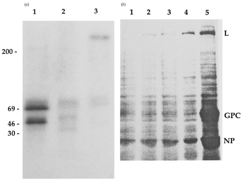
The large (L) RNA segment of Lassa fever virus (LAS) encodes a putative RNA-dependent RNA polymerase (RdRp or L protein). Similar to other arenaviruses, the LAS L protein is encoded on the genome-complementary strand and is predicted to be 2218 amino acids in length (253 kDa). It has an unusually large non-coding region adjacent to its translation start site. The LAS L protein contains six motifs of conserved amino acids that have been found among arenavirus L proteins and core RdRp of other segmented negative-stranded (SNS) viruses (Arena-, Bunya- and Orthomyxoviridae). Phylogenetic analyses of the RdRp of 20 SNS viruses reveals that arenavirus L proteins represent a distinct cluster divided into LAS-lymphocytic choriomeningitis and Tacaribe-Pichinde virus lineages. Monospecific serum against a synthetic peptide corresponding to the most conserved central domain precipitates a 250 kDa product from LAS and lymphocytic choriomeningitis virus-infected cells.


Figures

Fig. 1
Fig. 1
Conserved regions in LAS and LCM L proteins. Pairwise sequence comparisons were performed using GAP software (UWGCG), with a gap size of 3.0. Lines signify amino acid identity and one or two dots signify increasing amino acid similarity. (a) Amino-terminal conserved sequences. Numbers indicate the position of the amino acid. Amino acid residues conserved for Arena- and Bunyaviridae are shown in bold. (b) RdRp core motifs of LAS and LCM viruses, in accord with motifs originally designated by Poch et al. (1990). Amino acid residues strictly conserved for other SNS viruses are underlined and in bold.
Fig. 2
Fig. 2
Genetic comparison of SNS viruses based on the analysis of RdRp sequences. Phylogenetic analysis of amino acid differences between RdRp was carried out by maximum parsimony (PAUP 3.1.1) using a heuristic search option and a weight matrix based on the minimum number of nucleotide substitutions required to convert one amino acid to another. Bootstrap confidence limits were calculated from 500 replicates and are indicated below the lines. Only branches present in >50% of the trees are shown. Horizontal branch lengths are indicated above the lines and are proportional to the number of substitution steps. (a) Phylogenetic tree built on the basis of the RdRp core sequences of SNS viruses. (b) Phylo-genetic analysis of arena- and bunyavirus RdRp sequences. Amino-terminal conserved motifs (Muller et al., 1994) and core sequences were included in a 678 residue data set. All RdRp sequences were taken from the latest release of the GenBank and EMBL databases: LCM (J04331); TAC (J04340); PIC (D. G. Harnish, S. Zheng & S. Polyak, unpublished data); BUN, Bunyamwera (X14383); LAC, La Crosse (U12396); RVF, rift valley fever (X56464); TSW, tomato spotted wilt (D10066); HTN, Hantaan 76–118 (X55901); SEO, Seoul 80–39F (X56492):PUU, Puumala 18–20 (M63194); SN, Sin Nombre NMH10 (X56464); UUK, Uukuniemi (D10759); TOS, Toscana (X68414); DUG, Dugbe (U15018); InfA, influenza A/PR/8/34 (J02151); InfB, influenza B/AnAr/1/66 (M20479); InfC, influenza C/JJ/50 (M28060); DHO, Dhori/India/1313/61 (M65866); RSV, rice stripe virus (D31879). The core sequence of HRS (human respiratory syncytial virus) RdRp was used as an outgroup example (M75730).
Fig. 3
Fig. 3
(a) Detection of 250 kDa L protein in LCM- or LAS-infected cells. Immunoprecipitation of 35S-labelled proteins from LAS-(lane 1) and LCM- (lanes 2 and 3) infected cells in EDTA-containing (lanes 1 and 2) and magnesium-containing (lane 3) lysis buffers. Positions of marker proteins are indicated. (b) Immunoprecipitation of labelled proteins from LAS-infected Vero cells in a magnesium-containing lysis buffer. Lane 1, precipitation with non-immune rabbit serum; lanes 2, 3, and 4, precipitation with L peptide-specific rabbit serum after the second, third and fourth immunizations, respectively; lane 5, virus-specific proteins precipitated with monkey serum produced against purified LAS. Positions of virus-specific L, GP-C and NP proteins are indicated.

References

    1. Auperin DD, Compans RW, Bishop DHL. Nucleotide sequence conservation at the 3′ termini of the virion RNA species of new world and old world arenaviruses. Virology. 1982;121:200–203. - PubMed
    1. Auperin DD, Sasso DR, McCormick JB. Nucleotide sequence of the glycoprotein gene and intergenic region of the Lassa virus S genome RNA. Virology. 1986;154:155–167. - PubMed
    1. Bowen MD, Peters CJ, Nichol ST. The phylogeny of New World (Tacaribe complex) arenaviruses. Virology. 1996;219:285–290. - PubMed
    1. Chizhikov VE, Spiropoulou DF, Morzunov SP, Monroe MC, Peters CJ, Nichol ST. Complete genetic characterization and analysis of isolation of Sin Nombre virus. Journal of Virology. 1995;69:8132–8136. - PMC - PubMed
    1. Clegg JCS. Molecular phylogeny of the arenaviruses and guide to published sequence data. In: Salvato MS, editor. The Arenaviridae. New York: Plenum Press; 1993. pp. 175–187.

Publication types