. 1997 Mar 4;94(5):1795-9.
doi: 10.1073/pnas.94.5.1795.

Inhibition by uridine but not thymidine of p53-dependent intestinal apoptosis initiated by 5-fluorouracil: evidence for the involvement of RNA perturbation

Affiliations

Inhibition by uridine but not thymidine of p53-dependent intestinal apoptosis initiated by 5-fluorouracil: evidence for the involvement of RNA perturbation

D M Pritchard et al. Proc Natl Acad Sci U S A. .

Abstract

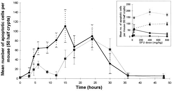
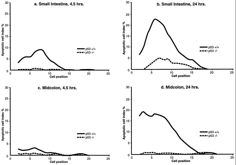
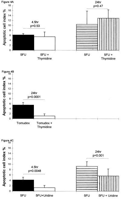
The epithelia from the crypts of the intestine are exquisitely sensitive to metabolic perturbation and undergo cell death with the classical morphology of apoptosis. Administration of 40 mg/kg 5-fluorouracil (5-FU) to BDF-1 p53+/+ mice resulted in an increase in p53 protein at cell positions in the crypts that were also those subjected to an apoptotic cell death. In p53-/- mice apoptosis was almost completely absent, even after 24 hr. 5-FU is a pyrimidine antimetabolite cytotoxin with multiple mechanisms of action, including inhibition of thymidylate synthase (TS), which gives rise to DNA damage, and incorporation into RNA. The inhibition of TS can be increased by coadministration of folinic acid and can be abrogated by administration of thymidine. The incorporation of 5-FU into RNA is inhibited by administration of uridine. p53-Dependent cell death induced by 5-FU was only inhibited by administration of uridine. Uridine had no effect on the apoptosis initiated by 1 Gy of gamma-radiation. Although thymidine abrogated apoptosis induced by the pure TS inhibitor Tomudex, it had no effect on 5-FU-induced apoptosis, and coadministration of folinic acid did not increase apoptosis. The data show that 5-FU-induced cell death of intestinal epithelial cells is p53-dependent and suggests that changes in RNA metabolism initiate events culminating in the expression of p53.


Figures

Figure 1
Figure 1
Time course showing total number of apoptotic events counted in 50 half-crypts (4 experimental animals at each time point) of small intestine (⧫) and midcolon (▪) of BDF1 mice, following 40 mg/kg i.p. injection of 5-FU. (Inset) Levels of apoptosis induced in BDF1 intestine with 5-FU doses ranging from 4 to 800 mg/kg. The total number of apoptotic events occurring in 50 half-crypts (4 experimental animals at each point) are shown. ⧫ and ▴, Small intestine at 4.5 and 24 hr, respectively; ▪ and ×, midcolon at 4.5 and 24 hr, respectively, after drug administration. (Error bars = SD.)
Figure 2
Figure 2
Examples of apoptotic figures (arrows) in (A) small intestine and (B) midcolon of BDF1 mice following 40 mg/kg 5-FU (staining of 3-μm paraffin sections; hematoxylin/eosin, ×1000). p53 protein expression identified by the affinity-purified CM5 polyclonal antibody in BDF1 mouse small intestine (C and D) and midcolon (E and F), 24 hr following the i.p. injection of 40 mg/kg 5-FU. p53-Positive cells are darkly stained; arrows, apoptotic cells. (×1000).
Figure 3
Figure 3
Cell positional distribution of apoptosis in the intestinal crypts of p53 wild-type (+/+) and homozygously null (−/−) mice (four animals in each experimental group, with data smoothed over three cell positions; ref. 13), following the i.p. administration of 40 mg/kg 5-FU. (a) Small intestine, after 4.5 hr. (b) Small intestine, after 24 hr. (c) Midcolon, after 4.5 hr. (d) Midcolon, after 24 hr.
Figure 4
Figure 4
Effects on apoptotic cell index percentage in the small intestinal crypts of BDF1 mice. (Error bars represent SD; statistical analysis was by a two-tailed Student’s t test.) (A) ▪, 4.5 hr after 40 mg/kg 5-FU (n = 3); □, 4.5 hr after 40 mg/kg 5-FU and contemporaneous 500 mg/kg thymidine (n = 4); ▤, 24 hr after 40 mg/kg 5-FU (n = 4); ▥, 24 hr after 40 mg/kg 5-FU and contemporaneous 500 mg/kg thymidine with further 500 mg/kg thymidine at 8 and 16 hr (n = 4). (B) ▪, 24 hr after 10 mg/kg Tomudex (n = 4); □, 24 hr after 10 mg/kg Tomudex and contemporaneous 500 mg/kg thymidine with further 500 mg/kg thymidine at 8 and 16 hr (n = 4). (C) ▪, 4.5 hr after 40 mg/kg 5-FU (n = 4); □, 4.5 hr after 40 mg/kg 5-FU with 3500 mg/kg uridine given 2 hr after 5-FU (n = 4); ▤, 24 hr after 40 mg/kg 5-FU (n = 11); ▥, 24 hr after 40 mg/kg 5-FU with 3500 mg/kg uridine given 2 hr after 5-FU (n = 12).

References

    1. Lane D P. Nature (London) 1992;358:15–16. - PubMed
    1. Yonish-Rouach E, Resnitzky D, Lotem J, Sachs L, Kimchi A, Oren M. Nature (London) 1991;353:345–347. - PubMed
    1. Wu L, Bayle J H, Elenbaas B, Pavletich N P, Levine A J. Mol Cell Biol. 1995;15:497–504. - PMC - PubMed
    1. Potten C S. Cancer Metastasis Rev. 1992;11:179–195. - PubMed
    1. Potten C S. In: Radiation and Gut. Potten C S, Hendry J H, editors. Amsterdam: Elsevier Science; 1995. pp. 1–31.

Publication types

MeSH terms