

DEK, an autoantigen involved in a chromosomal translocation in acute myelogenous leukemia, binds to the HIV-2 enhancer
- PMID: 9050861
- PMCID: PMC19999
- DOI: 10.1073/pnas.94.5.1811
DEK, an autoantigen involved in a chromosomal translocation in acute myelogenous leukemia, binds to the HIV-2 enhancer
Abstract
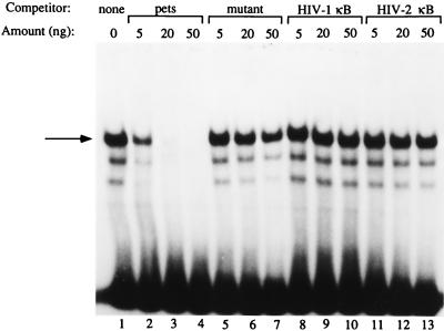
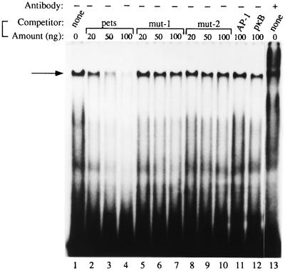
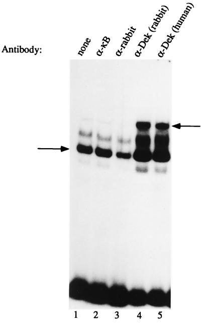
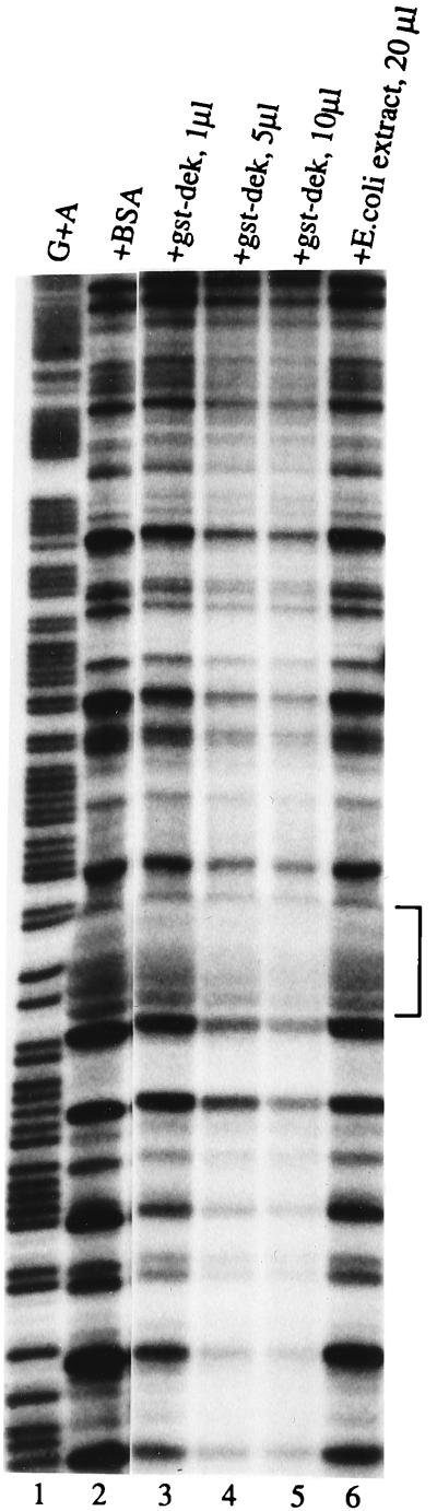
The product of the dek oncogene is the 43-kDa DEK nuclear protein. DEK was first identified in a fusion with the CAN nucleoporin protein in a specific subtype of acute myelogenous leukemia. DEK has also been shown to be an autoantigen in patients with pauciarticular onset juvenile rheumatoid arthritis. Further, the last 65 amino acids of DEK can partially reverse the mutation-prone phenotype of cells from patients with ataxia-telangiectasia. However, in spite of these significant disease associations, the function of DEK has remained unclear. The HIV-2 peri-ets (pets) site is a TG-rich element found between the two Elf-1 binding sites in the HIV-2 enhancer. The pets element mediates transcriptional activation whether the enhancer is stimulated by phorbol 12-myristate 13-acetate (PMA) alone, phytohemagluttinin (PHA) alone, PMA plus PHA, soluble antibodies to the T cell receptor, immobilized antibodies to the T cell receptor, or by antigen. Previously, we purified and characterized the pets factor, demonstrating that it is a 43-kDa nuclear protein. We now describe the identification of DEK as this 43-kDa pets factor. Using a modified Southwestern screening procedure, we find that DEK can recognize the pets element. We demonstrate the ability of recombinant DEK to bind specifically to the pets site using the electrophoretic mobility shift assay (EMSA) and DNase I footprinting. "Supershift" EMSA further confirms that DEK is the dominant protein binding to the pets site in T cell extracts. Our findings show that DEK is a site-specific DNA binding protein that is likely involved in transcriptional regulation and signal transduction. This has implications for multiple pathogenic processes, including hematologic malignancies, arthritis, ataxia-telangiectasia, and AIDS caused by HIV-2.
Figures
References
-
- von Lindern M, Breems D, van Baal S, Adriaansen H, Grosveld G. Genes Chromosomes Cancer. 1992;5:227–234. - PubMed
-
- Soekarman D, von Lindern M, van der Plas D C, Selleri L, Bartram C R I, Martiat P, Culligan D, Padua R A, Hasper-Voogt K P, Hagemeijer A, Grosveld G. Leukemia. 1992;6:489–494. - PubMed
-
- Fornerod M, Boer J, van Baal S, Jaegle M, von Lindern M, Murti K G, Davis D, Bonten J, Buijs A, Grosveld G. Oncogene. 1995;10:1739–1748. - PubMed
Publication types
MeSH terms
Substances
Grants and funding
LinkOut - more resources
Full Text Sources
Other Literature Sources
Molecular Biology Databases
Miscellaneous
