. 1997 Mar 4;94(5):1822-7.
doi: 10.1073/pnas.94.5.1822.

Hepatitis B virus (HBV) envelope glycoproteins vary drastically in their sensitivity to glycan processing: evidence that alteration of a single N-linked glycosylation site can regulate HBV secretion

Affiliations

Hepatitis B virus (HBV) envelope glycoproteins vary drastically in their sensitivity to glycan processing: evidence that alteration of a single N-linked glycosylation site can regulate HBV secretion

A Mehta et al. Proc Natl Acad Sci U S A. .

Abstract

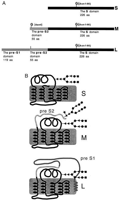
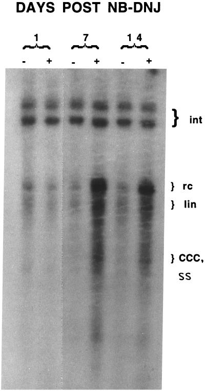
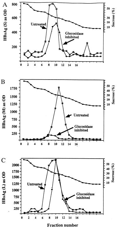
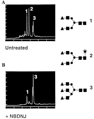
The role of N-linked glycosylation and glycan trimming in the function of glycoproteins remains a central question in biology. Hepatitis B virus specifies three glycoproteins (L, M, and S) that are derived from alternate translation of the same ORF. All three glycoproteins contain a common N-glycosylation site in the S domain while M possesses an additional N-glycosylation site at its amino terminus. In the presence of N-butyl-deoxnojirimycin (an inhibitor of alpha-glucosidase) virions and the M protein are surprisingly retained. Preliminary evidence suggests that the retained M protein is hyperglucosylated and localized to lysosomal vesicles. In contrast, the S and L proteins are secreted, and their glycosylation state is unaffected by the presence of the inhibitor. Site-directed mutagenesis provides evidence that virion secretion requires the glycosylation sequon in the pre-S2 domain of M. This highlights the potential role of the M protein oligosaccharide as a therapeutic target.
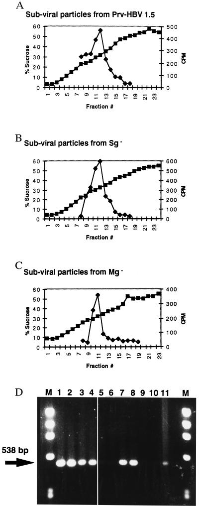

Figures

Figure 1
Figure 1
(A) The HBV envelope proteins. All three proteins have a common N-linked glycosylation site at position 146 of the S domain (marked with a G). The M protein contains an additional glycan site at amino acid 4 of the pre-S2 domain (marked with a G). The L protein, while containing the pre-S2 glycosylation sequon, only utilizes the shared S glycan site (1). (B) Structural diagram of the three HBV envelope proteins with attached N-linked glycans. The pre-S1 and pre-S2 domains are indicated. B is based upon ref. . The glycan structures are as follows: ♦, mannose; ▴, N-acetylglucosamine; •, galactose; ▪, sialic acid; ★, fucose.
Figure 2
Figure 2
Southern blot of accumulated intracellular HBV DNA from untreated and α-glucosidase-inhibited HepG2.215 cells. HepG2.2.15 cells were either left untreated or treated with 1000 μg/ml of the α-glucosidase inhibitor NB-DNJ for the indicated times and the intracellular HBV DNA detected as described. The location of the HBV integrated (Int), relaxed circular (rc), linear (lin), covalently closed circular (CCC), and single-stranded (SS) DNA are indicated. Increases in the rc and lin form of HBV DNA are clearly evident. As the HBV SS DNA comigrates with the CCC DNA it is difficult to determine which of these increases.
Figure 3
Figure 3
Detection of the HBV surface antigens in subviral particles secreted from untreated or α-glucosidase-inhibited cells by sucrose density gradient. Medium from untreated cells and cells treated with 1000 μg/ml of the α-glucosidase inhibitor NB-DNJ for 10 days was collected and resolved through sucrose density gradients as described. HBV-specific envelope proteins were detected by ELISA using antibodies and methods described previously (10). (AC) Detection of individual envelope antigens using antibodies for the HBV S, M, and L proteins, respectively. Markers are untreated (▪) or 1000 μg/ml (•) of NB-DNJ.
Figure 4
Figure 4
Analysis of the major glycan structures on subviral particles purified from untreated (A) and α-glucosidase-inhibited (B) cells. Glycans were removed from subviral particles by hydrazinolysis and fluorescently labeled at their reducing end as described. The determined glycan structures are shown along the right. The glycan structures are as follows: ▴, galactose; ▪, N-acetylglucosamine; •, mannose; ★, fucose.
Figure 5
Figure 5
Analysis of the subviral and viral particles released from the glycosylation-incompetent expression vectors. Indicated expression vectors were transfected into HepG2 cells and analyzed for the ability to support the secretion of subviral and viral particles as described in materials and methods. Secretion of subviral particles from the (A) PRV-HBV 1.5 vector, (B) Sg vector, and (C) Mg vector, as resolved through a sucrose density gradient. (D) Secretion of virus as detected by PCR. The arrow indicates the location of the 538-bp PCR product. First and last lanes marked “M” are DNA molecular weight standards. Lanes: 1 and 2, HepG2.2.15 cells; 3 and 4, from the control vector PRV-HBV 1.5; 5 and 6, from the Sg Mg vector which secretes no virus; 7 and 8, from the Sg vector; 9 and 10, the Mg vector which also secretes no virus; 11, a control for the PCR.

Similar articles

Cited by

References

    1. Heermann K H, Gerlich W H. In: Molecular Biology of HBV. Maclachlan A, editor. Boca Raton, FL: CRC; 1992. pp. 109–143.
    1. Heermann K H, Goldman I, Schwartz W, Seyffarth T, Baumgarten H, Gerlich W H. J Virol. 1984;52:396–402. - PMC - PubMed
    1. Bruss V, Ganem D. Proc Natl Acad Sci USA. 1991;88:1059–1063. - PMC - PubMed
    1. Ueda K, Tsurimoto T, Matsubara K. J Virol. 1991;65:3521–3529. - PMC - PubMed
    1. Ganem D. Curr Top Microbiol Immunol. 1991;168:61–84. - PubMed

Publication types

MeSH terms

LinkOut - more resources