. 1997 Mar 4;94(5):1840-5.
doi: 10.1073/pnas.94.5.1840.

The ataxia-telangiectasia gene product, a constitutively expressed nuclear protein that is not up-regulated following genome damage

Affiliations

The ataxia-telangiectasia gene product, a constitutively expressed nuclear protein that is not up-regulated following genome damage

K D Brown et al. Proc Natl Acad Sci U S A. .

Abstract

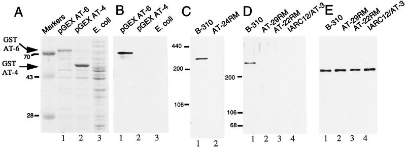
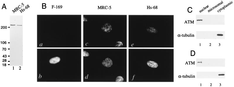
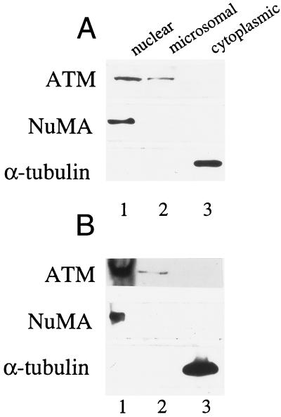
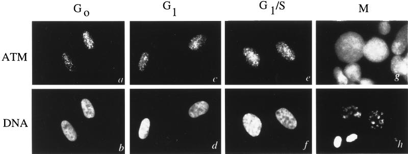
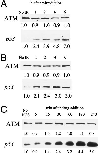
The product of the ataxia-telangiectasia gene (ATM) was identified by using an antiserum developed to a peptide corresponding to the deduced amino acid sequence. The ATM protein is a single, high-molecular weight protein predominantly confined to the nucleus of human fibroblasts, but is present in both nuclear and microsomal fractions from human lymphoblast cells and peripheral blood lymphocytes. ATM protein levels and localization remain constant throughout all stages of the cell cycle. Truncated ATM protein was not detected in lymphoblasts from ataxia-telangiectasia patients homozygous for mutations leading to premature protein termination. Exposure of normal human cells to gamma-irradiation and the radiomimetic drug neocarzinostatin had no effect on ATM protein levels, in contrast to a noted rise in p53 levels over the same time interval. These findings are consistent with a role for the ATM protein in ensuring the fidelity of DNA repair and cell cycle regulation following genome damage.


Figures

Figure 1
Figure 1
Characterization of ATM protein antiserum pAb 132. (A) Lysates from E. coli transformed with the plasmids pGEX AT-6 (lane 1) and pGEX AT-4 (lane 2) following fusion protein induction and untransformed E. coli (lane 3) were subjected to SDS/PAGE and Coomassie blue staining. Migration of the specific GST–AT fusion proteins are indicated by arrowheads. (B) Extracts shown in A were immunoblotted with pAb 132. (C) A total of 50 μg of B-310 (lane 1, wild type for ATM) and AT24RM (lane 2, homozygous mutant for ATM) cell extracts were immunoblotted with pAb 132. (D) A total of 50 μg of SDS-lysate from B-310 lymphoblasts (lane 1) and A-T patient lines AT29RM (lane 2), AT22RM (lane 3), and IARC12/AT3 (lane 4) were immunoblotted with pAb 132. (E) Blot displayed in D was stripped and reprobed with an antisera to NuMA.
Figure 2
Figure 2
Localization of ATM protein in normal human fibroblasts. (A) A total of 50 μg of SDS lysate from the normal human fibroblast lines MRC-5 (lane 1) and Hs-68 (lane 2) was immunoblotted with affinity purified pAb 132. (B) F-169 (a and b), MRC-5 (c and d), and Hs-68 (e and f) cells were stained with affinity purified pAb 132 (a, c, and e), and Hoechst 22358 (b, d, and f). (C) MRC-5 fibroblasts (1 × 10⁷) were fractionated and 10% of the nuclear (lane 1), microsomal (lane 2), and cytoplasmic (lane 3) fractions were immunoblotted with pAb 132 (Upper) or 1% of each fraction were immunoblotted with antitubulin (Lower). (D) Hs-68 fibroblasts (1 × 10⁷) were fractionated and analyzed as in C.
Figure 3
Figure 3
Analysis of ATM protein in fractionated human lymphoblasts and peripheral blood lymphocytes. (A) B-310 lymphoblasts (1 × 10⁷) were fractionated. Ten percent of the resultant nuclear (lane 1), microsomal (lane 2), and cytoplasmic (lane 3) fractions were immunoblotted with pAb 132 (Top). To evaluate purity of the preparations, 2% of each fraction was analyzed with NuMA antisera (Middle), or 1% of each fraction was analyzed with anti-tubulin (Bottom). (B) Normal peripheral blood lymphocytes (2 × 10⁸) were fractionated and analyzed as in A.
Figure 4
Figure 4
Localization of ATM protein at various cell cycle phases. Cells were synchronized at various cell cycle phases and processed for immunofluorescence with affinity-purified pAb 132 (a, c, e, and g) and Hoechst 22358 (b, d, f, and h). (a and b) Cells synchronized at G0. (c and d) Cells synchronized at G1. (e and f) Cells synchronized at G1/S. (g and h) Cells synchronized in mitosis.
Figure 5
Figure 5
Effects of genome damaging agents on cellular ATM levels. (A) Hs-68 fibroblasts were exposed to 5 Gy of ionizing radiation. Unirradiated cells (No IR) and cells 1, 2, 4, and 6 h after exposure were analyzed for ATM protein (Upper) and p53 (Lower) levels by immunoblotting. (B) B-310 lymphoblasts were exposed to 5 Gy of IR and analyzed as in A. (C) The normal human fibroblast cell line F-2054 was treated with NCS and extracts from treated cells at 5, 15, 30, 60, 120, and 240 min following drug addition, and untreated cells (No NCS) were analyzed for ATM protein (Upper) and p53 (Lower). Relative immunoblot signal intensity (determined by densitometric scanning of exposed films) is displayed below each band. Minor differences in recorded ATM protein levels are likely due to slight inconsistencies in protein loading and/or protein transfer.

References

    1. Sedgwick R P, Boder E. In: Handbook of Clinical Neurology. Vinken P J, Bruyn G W, editors. Amsterdam: North-Holland; 1972. pp. 267–339.
    1. Shiloh Y. Eur J Hum Genet. 1995;3:116–138. - PubMed
    1. Swift M, Morrell D, Massey R B, Chase C L. New Eng J Med. 1991;325:1831–1836. - PubMed
    1. Easton D F. Int J Radiat Biol. 1994;66:S187–S182. - PubMed
    1. Painter R B, Young B R. Proc Natl Acad Sci USA. 1980;77:7315–7317. - PMC - PubMed

Publication types

MeSH terms