. 1997 Mar 4;94(5):1852-6.
doi: 10.1073/pnas.94.5.1852.

Type III collagen is crucial for collagen I fibrillogenesis and for normal cardiovascular development

Affiliations

Type III collagen is crucial for collagen I fibrillogenesis and for normal cardiovascular development

X Liu et al. Proc Natl Acad Sci U S A. .

Abstract

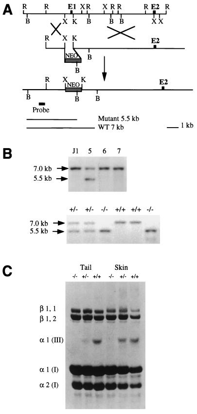
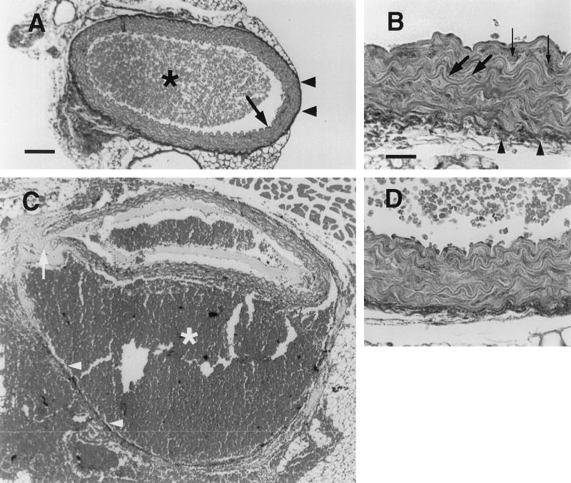
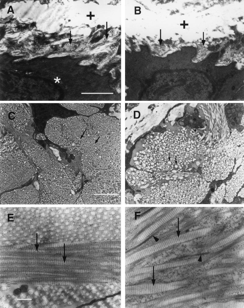
Type III collagen is a fibrillar forming collagen comprising three alpha1(III) chains and is expressed in early embryos and throughout embryogenesis. In the adult, type III collagen is a major component of the extracellular matrix in a variety of internal organs and skin. Mutations in the COL3A1 gene have been implicated as a cause of type IV Ehlers-Danlos syndrome, a disease leading to aortic rupture in early adult life. To directly study the role of Col3a1 in development and disease, we have inactivated the Col3a1 gene in embryonic stem cells by homologous recombination. The mutated allele was transmitted through the mouse germ line and homozygous mutant animals were derived from heterozygous intercrosses. About 10% of the homozygous mutant animals survived to adulthood but have a much shorter life span compared with wild-type mice. The major cause of death of mutant mice was rupture of the major blood vessels, similar to patients with type IV Ehlers-Danlos syndrome. Ultrastructural analysis of tissues from mutant mice revealed that type III collagen is essential for normal collagen I fibrillogenesis in the cardiovascular system and other organs.


Figures

Figure 1
Figure 1
Targeted deletion of the Col3a1 gene in mouse ES cells and generation of mutant mice. (A) Schematic diagram of the strategy used to target the Col3a1 gene. (Top) Restriction map of the Col3a1 gene genomic DNA fragment which covers the promoter, the first two exons, and the first intron of the Col3a1 gene. (Middle) The 18-kb replacement-type targeting vector carrying a neo gene driven by a phosphoglycerate kinase promoter, which replaced the XbaI and KpnI DNA fragment of the Col3a1 gene. (Bottom) The targeted allele in which the promoter region and the first exon that codes for the signal peptide of type III procollagen were deleted by homologous recombination; the genomic probe external to the 5′ homologous arm is indicated, which hybridizes to a 5.5-kb and a 7-kb DNA fragment from mutant and wild-type alleles, respectively. B, BamHI; K, KpnI; R, EcoRI; X, XbaI. (B Upper) Southern blot of ES cell clones. DNA from parental J1 ES cells and independently cloned G418-resistant ES cell clones number 5, 6, and 7 was digested with BamHI and hybridized to the external probe shown in A. Clones containing the expected 5.5-kb BamHI fragment diagnostic for homologous recombinant were obtained at a frequency of 1 in 30. The blot was rehybridized to a neo probe to verify a single integration. Three independent recombinant ES cell clones contributed to the germ line of recipient embryos after blastocyst injection. (Lower) Southern blot of offspring from a Col3a1+/− × Col3a1+/− cross. Tail DNA was extracted and digested with BamHI and analyzed as described in ref. . (C) Collagens from tails and skin of wild-type mice and type III collagen mutants. Collagens were resolved by SDS/PAGE and stained with Coomassie blue. The type III collagen molecule consists of three α1 (III) chains. The β1,1 and β1,2 dimers and the α1(I) and α2(I) chains are all from type I collagen and serve as internal controls for the loading. Skin collagens from wild-type mice (far right lane) were underloaded.
Figure 2
Figure 2
Masson’s trichrome staining of cross sections of aorta of wild-type and Col3a1 mutant mice. (A) Wild-type aorta composed of intima and media (arrow) and adventitia (arrowheads). There are blood cells (star) in the lumen. (B) High magnification of wild-type aorta. Elastic fiber (large arrows) and smooth muscle cells (small arrows) of the intima can be seen. (C) A dissecting aneurysm of mutant aorta. The intima and media (arrow) were ruptured and blood (star) filled in between media and adventitia (arrowheads). (D) High magnification of mutant aorta. [Bar = 100 μm (A and C) and 25 μm (B and D).]
Figure 3
Figure 3
A type III collagen-deficient mouse with a skin wound on its left shoulder.
Figure 4
Figure 4
Transmission electron microscopic analyses of aorta and skin from wild-type and mutant mice. (A) Collagen fibrils (arrows) are around the smooth muscle cell (star) in the media of wild-type aorta. White areas marked with crosses are elastic fibers. (B) Collagen fibrils are missing around the smooth muscle cell (arrows) in the media of mutant aorta. (C) Cross section of the collagen fibrils in the adventitia of wild-type aorta. Arrows point to individual fibrils. The diameter of the collagen fibrils is smaller and relatively uniform compared with the fibrils (arrows) of mutant aorta in D. (E) Skin section of wild-type mouse. The collagen fibrils (arrows) are uniform in diameter. (F) Skin section of mutant mouse. The collagen fibrils are often thicker (arrows) or thinner (arrowheads) than control fibrils and are not uniform in diameter. [Bar = 1 μm (AD) and 0.2 μm (E and F).]
Figure 5
Figure 5
Comparison of the diameters of collagen fibrils in the adventitia of aorta of wild-type and mutant mice. A 2 μm × 2 μm area in the adventitia of either wild-type or mutant aorta was randomly chosen, and all the fibrils in this area were measured for their diameters and counted.

References

    1. Prockop D J, Kivirikko K I. N Engl J Med. 1984;311:376–386. - PubMed
    1. Kontusaari S, Tromp G, Kuivaniemi H, Romanic A M, Prockop D J. J Clin Invest. 1990;86:1465–1473. - PMC - PubMed
    1. Kuivaniemi H, Tromp G, Bergfeld W F, Kay M, Helm T N. J Invest Dermatol. 1995;105:352–356. - PubMed
    1. Olsen B R. Experientia. 1995;51:194–195. - PubMed
    1. Wong R S, Follis F M, Shively B K, Wernly J A. Ann Thorac Surg. 1995;60:1439–1443. - PubMed

Publication types

MeSH terms