

The antigen-binding domain of a human IgG-anti-F(ab')2 autoantibody
- PMID: 9050877
- PMCID: PMC20015
- DOI: 10.1073/pnas.94.5.1902
The antigen-binding domain of a human IgG-anti-F(ab')2 autoantibody
Abstract
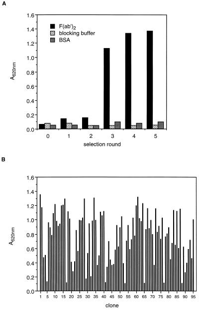
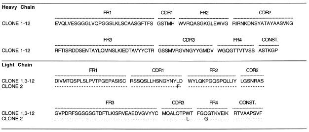
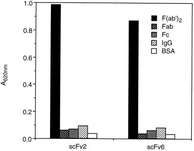
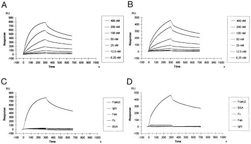
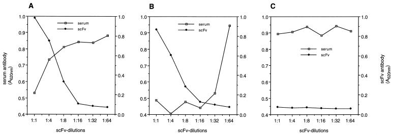
Recent studies revealed an immunoregulatory role of natural IgG-anti-F(ab')2 antibodies in both healthy individuals and patients with certain diseases. The implication of anti-F(ab')2 antibodies in the pathogenesis of diseases prompted us to study the gene segment structure of their antigen-binding domains and their binding characteristics. cDNA was prepared from the lymphocytes of a patient with a high IgG-anti-F(ab')2 serum titer. Variable heavy and light gene segments were amplified by PCR and inserted into a phagemid surface expression vector. Single-chain antibodies displayed on the phage surface were screened for binding to F(ab')2 fragments. The subsequent analysis of 95 single clones demonstrated that they all bound specifically to F(ab')2. Sequence analyses of 12 clones showed that 11 were identical and 1 contained a silent point mutation in the heavy chain and three amino acid exchanges in the light chain. The heavy chains belonged to the V(H)3 and the light chains to the V(kappa)2 gene family. The 11 identical light-chain genes were completely homologous to a germ-line sequence (DPK-15). Binding assays showed that the single-chain antibodies bind to F(ab')2, but not to Fab, Fc, or intact IgG. This binding pattern was confirmed by surface plasmon resonance studies, which revealed a relatively high affinity (Ka = 2.8 x 10(7) M(-1)). The strong binding capacity was further demonstrated by competitive inhibition of the serum anti-IgG antibody's interaction with antigen. The present study defines for the first time to our knowledge the gene segment structure of the antigen-binding domain of two human IgG-anti-F(ab')2 autoantibody clones and describes the binding kinetics of the purified monomeric fragments.
Figures
References
-
- Bona C, Cazenave P A. Lymphocyte Regulation by Antibodies. New York: Wiley; 1981. pp. 205–213.
-
- Bona C, Siminovitch K, Zanetti M, Theofilopoulos A. The Molecular Pathology of Autoimmune Diseases. London: Harwood; 1993.
-
- Terness P, Kirschfink M, Navolan D, Dufter C, Kohl I, Opelz G, Roelcke D. Blood. 1995;85:548–551. - PubMed
Publication types
MeSH terms
Substances
Associated data
- Actions
- Actions
- Actions
- Actions
LinkOut - more resources
Full Text Sources
Other Literature Sources
