. 1997 Mar 4;94(5):1919-24.
doi: 10.1073/pnas.94.5.1919.

syk kinase activation by a src kinase-initiated activation loop phosphorylation chain reaction

Affiliations

syk kinase activation by a src kinase-initiated activation loop phosphorylation chain reaction

O El-Hillal et al. Proc Natl Acad Sci U S A. .

Abstract

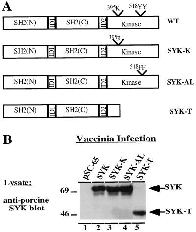
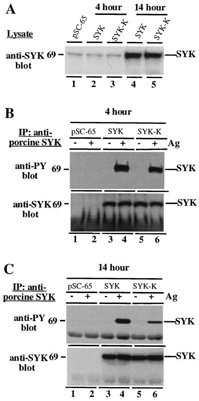
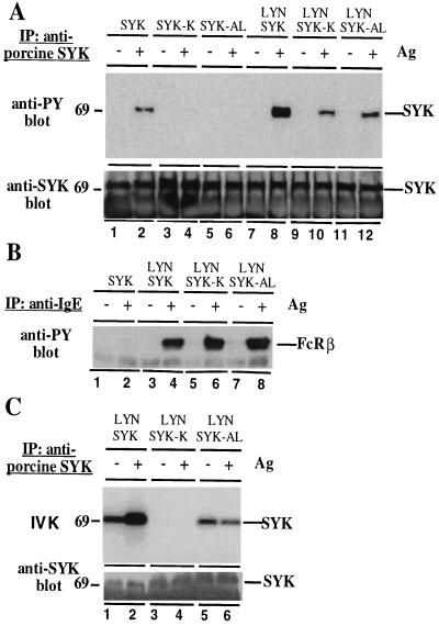
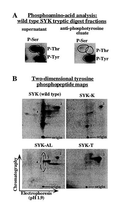
Activation of the syk tyrosine kinase occurs almost immediately following engagement of many types of antigen receptors, including Fc receptors, but the mechanism through which syk is activated is currently unclear. Here we demonstrate that Fc receptor-induced syk activation occurs as the result of phosphorylation of the syk activation loop by both src family kinases and other molecules of activated syk, suggesting that syk activation occurs as the result of a src kinase-initiated activation loop phosphorylation chain reaction. This type of activation mechanism predicts that syk activation would exhibit exponential kinetics, providing a potential explanation for its rapid and robust activation by even weak antigen receptor stimuli. We propose that a similar mechanism may be responsible for generating rapid activation of other cytoplasmic tyrosine kinases, such as those of the Bruton tyrosine kinase/tec family, as well.


Figures

Figure 1
Figure 1
Structure and expression of porcine syk mutants. (A) Schematic representation of syk cDNA constructs used for construction of recombinant vaccinia viruses. SH2(N), NH2 terminal Src homology 2 domain; SH2(C), COOH terminal Src homology 2 domain; ID1, interdomain region between the tandem SH2 domains; ID2, interdomain region between SH2(C) and the tyrosine kinase domain. The amino acid residues of wild-type syk that are mutated in the syk-K (Lys-395 to arginine) and syk-AL mutants (Tyr-518 and Tyr-519 to phenylalanine) are indicated. (B) Expression of various syk mutants in RBL-2H3 cells. RBL-2H3 cells were infected with the indicated viruses for 14 h. Cells were then lysed and analyzed by immunoblotting with an anti-porcine syk-specific antibody.
Figure 2
Figure 2
syk tyrosine phosphorylation is generated primarily by syk transphosphorylation and phosphorylation by other kinases. (A) Relative expression of syk with 4-h or 14-h vaccinia infections. RBL-2H3 cells were infected with the indicated viruses for either 4 h (lanes 1–3) or 14 h (lanes 4 and 5). Cells were then lysed and analyzed by immunoblotting with an anti-syk antibody capable of recognizing both rat and porcine syk. This experiment was done as a pilot experiment to determine the expression time course of syk after infection. The experiments below were done separately using the conditions defined in this experiment. (B) Tracer expression of wild-type syk or syk-K results in similar levels of Fc receptor-induced tyrosine phosphorylation. RBL-2H3 cells were loaded with IgE, infected for 4 h with the indicated recombinant vaccinia viruses, stimulated or not with antigen, and lysed. (Upper) Lysates were subjected to precipitation with anti-porcine syk antibody and analyzed by anti-phosphotyrosine immunoblotting. (Lower) The blot was then stripped and reprobed by anti-syk immunoblotting to verify equivalent amounts of syk protein in each lane. (C) High level overexpression of wild-type syk or syk-K results in substantially reduced tyrosine phosphorylation of syk-K relative to wild-type syk. Identical experiment to above except that RBL-2H3 cells were infected for 14 h with the indicated recombinant vaccinia viruses. (Upper) Lysates were subjected to precipitation with anti-porcine syk antibody and analyzed by anti-phosphotyrosine immunoblotting. (Lower) The blot was then stripped and reprobed by anti-syk immunoblotting to verify equivalent amounts of syk protein in each lane.
Figure 3
Figure 3
Analysis of syk tyrosine phosphorylation and activation in 3T3E cells. (A) syk-K and syk-AL have decreased Fc receptor-induced tyrosine phosphorylation relative to wild-type syk. 3T3E cells were loaded with IgE, infected with the indicated combinations of viruses, triggered (+) or not (−) with antigen, and lysed. (Upper) Lysates were immunoprecipitated with rabbit polyclonal anti-porcine syk antibody and analyzed by immunoblotting with anti-phosphotyrosine antibody. (Lower) The blot was then stripped and reprobed by anti-syk immunoblotting to verify equivalent amounts of syk protein in each lane. Note that longer exposures of the anti-phosphotyrosine blot reveal very low levels of phosphorylation of syk-K and syk-AL in lanes 4 and 6, which is likely attributable to endogenous SRC. (B) lyn activity is not differentially affected by syk, syk-K, or syk-AL. 3T3E cells were loaded with IgE, infected with the indicated combinations of viruses, triggered (+) or not (−) with antigen, and lysed. Lysates were immunoprecipitated with rabbit polyclonal anti-IgE antibody to isolate phosphorylated Fc receptor complexes and analyzed by immunoblotting with anti-phosphotyrosine antibody. The band that runs just under FcRβ in all lanes is the light chain of IgE, which is detected by the peroxidase-conjugated anti-mouse secondary antibody used to develop the blot. (C) Fc receptor-induced tyrosine phosphorylation activates the tyrosine kinase activity of wild-type syk, but not the syk-AL mutant. Similar experiment to above, in that cells were infected with the indicated viruses, and then triggered, lysed, and immunoprecipitated as in A. However, in this experiment, each anti-porcine syk immunoprecipitate was split into two samples. One sample was analyzed by in vitro kinase assay (Upper), while the second sample was analyzed by anti-phosphotyrosine immunoblotting to confirm the expected pattern of syk tyrosine phosphorylation (this produced results identical to those in A; data not shown), and was then stripped and subjected to anti-syk immunoblotting to determine the relative amount of syk protein present in each assay (Lower).
Figure 4
Figure 4
Phosphopeptide analysis of syk tyrosine phosphorylation sites. Tryptic peptides derived from 32P-labeled wild-type syk, syk-K, syk-AL, and syk-T were generated as described, and then immunoprecipitated with anti-phosphotyrosine to generate a population of bound peptides (eluate fraction) and unbound peptides (supernatant fraction). (A) Anti-phosphotyrosine immunoprecipitation of tryptic peptides effectively eliminates serine/threonine phosphorylated peptides from the bound peptides (eluate fraction). Phosphoamino acid analysis was performed on aliquots of the supernatant (Left) or eluate (Right) fractions from the anti-phosphotyrosine immunoprecipitation of wild-type syk tryptic peptides. (B) Two-dimensional phosphopeptide analyses of the eluate fractions. For all syk constructs, equal amounts of eluate fractions were subjected to two-dimensional phosphopeptide mapping. The set of spots inside the ellipse in the wild-type syk map are grouped together as “peptide 4” because over several experiments the labeling of the spots above and below the main spot was variable. We believe that these spots are isoforms of a single peptide generated by contaminating protease activity in the trypsin preparation as we have previously described (19). The increased intensity of peptide 2 phosphorylation in the syk-T map relative to the other maps is reproducible, but is of unclear origin.

References

    1. Ravetch J V, Kinet J P. Annu Rev Immunol. 1991;9:457–492. - PubMed
    1. Metzger H. Immunol Rev. 1992;125:37–48. - PubMed
    1. Weiss A, Littman D R. Cell. 1994;76:263–274. - PubMed
    1. Cambier J C. J Immunol. 1995;155:3281–3285. - PubMed
    1. Benhamou M, Ryba N J, Kihara H, Nishikata H, Siraganian R P. J Biol Chem. 1993;268:23318–23324. - PubMed

MeSH terms

LinkOut - more resources