. 1997 Mar 4;94(5):1955-60.
doi: 10.1073/pnas.94.5.1955.

Peptide inhibition of glomerular deposition of an anti-DNA antibody

Affiliations

Peptide inhibition of glomerular deposition of an anti-DNA antibody

B Gaynor et al. Proc Natl Acad Sci U S A. .

Abstract

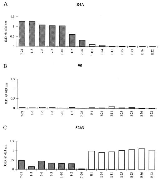
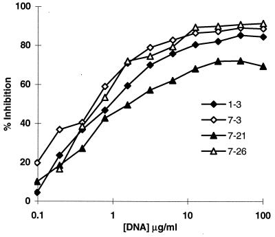
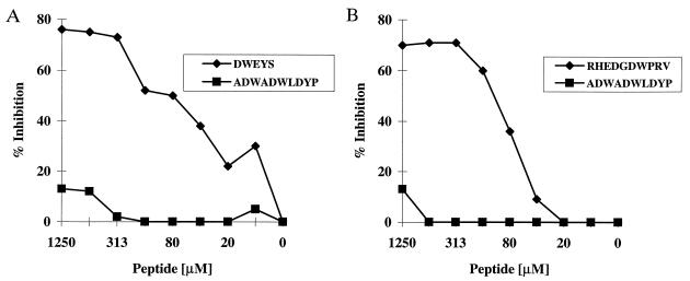
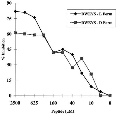
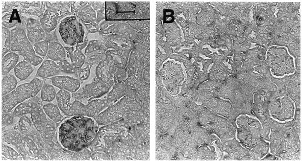
Antibodies to double-stranded DNA are pathognomonic of systemic lupus erythematosus and deposit in the kidneys of lupus patients to cause glomerulonephritis. Recent data suggest that a significant proportion of anti-DNA antibodies may cross-react with renal antigens and be sequestered in the kidney by virtue of this cross-reactivity. If this is true, antigenic competition for pathogenic antibodies might prevent their deposition in kidneys and the ensuing tissue damage. To generate surrogate antigens that could be used for this purpose, we have used peptide display phage libraries to identify peptides that react with R4A, a pathogenic mouse monoclonal anti-DNA antibody that deposits in glomeruli. We have demonstrated that the peptides bind in or near the double-stranded DNA binding site. Furthermore, the peptides are bound preferentially by the R4A antibody as compared with two closely related antibodies derived from it, one of which deposits in renal tubules and one of which displays no renal pathogenicity. Administration of one of these peptides in a soluble form protects mice from renal deposition of the R4A anti-DNA antibody in vivo. This represents a new therapeutic approach in systemic lupus erythematosus that focuses on protecting target organs from antibody mediated injury.


Figures

Figure 1
Figure 1
R4A, 95, and 52b3 binding by ELISA to various phage clones. Microtiter 96-well plates were coated with 1 μg/ml purified antibody, and 2.5 × 10¹⁰ phage were added to each well, followed by biotinylated antibody to M13 phage, and streptavidin-alkaline phosphatase. OD after addition of substrate was monitored at 405 nm. Shaded boxes (left) are R4A selected clones, and open boxes (right) are 52b3 selected clones. (A) R4A reactivity with R4A and 52b3 selected clones. (B) 95 reactivity with R4A and 52b3 selected clones. (C) 52b3 reactivity with R4A and 52b3 selected clones.
Figure 2
Figure 2
Inhibition of R4A binding to selected phage by calf thymus dsDNA. 1–3, 7–3, 7–21, and 7–26 are R4A selected phage clones containing the consensus motif to which R4A showed binding by ELISA (Fig. 1A). Microtiter 96-well plates were coated with 2.5 × 10¹⁰ phage from each R4A selected phage clone. R4A at a concentration of 1 μg/ml was incubated with varying concentrations of dsDNA for 1 hr at 37°C and then transferred to the plates precoated with phage. Following a 2-hr incubation at 37°C, the plates were washed, and alkaline phosphatase linked goat anti-mouse IgG2b antibody was added for 1 hr at 37°C. OD after addition of substrate was monitored at 405 nm.
Figure 3
Figure 3
Peptide inhibition of anti-dsDNA antibody binding to dsDNA. Immulon II plates were coated overnight with 100 μg/ml dsDNA. R4A and 52b3 at a final concentration of 5 μg/ml were incubated with varying concentrations of the specific (DWEYS or RHEDGDWPRV, respectively) or irrelevant (ADWADWLDYP) peptide and were transferred to the plate precoated with dsDNA. After a 2-hr incubation at 37°C, the plates were washed and alkaline phosphatase-linked goat anti-mouse IgG2b antibody was added for 1 hr at 37°C. After addition of substrate, OD was monitored at 405 nm. (A) DWEYS vs. ADWADWLDYP inhibition of R4A binding to dsDNA. (B) RHEDGDWPRV vs. ADWADWLDYP inhibition of 52b3 binding to dsDNA.
Figure 4
Figure 4
DWEYS (l and d peptides) inhibition of R4A binding to dsDNA. Immulon II plates were coated overnight with 100 μg/ml salmon sperm dsDNA. R4A at a final concentration of 5 μg/ml was incubated with varying concentrations of l or d DWEYS, and transferred to the plate precoated with dsDNA. After a 2-hr incubation at 37°C, the plates were washed, and alkaline phosphatase-linked goat anti-mouse IgG2b antibody was added for 1 hr at 37°C. OD after addition of substrate was monitored at 405 nm.
Figure 5
Figure 5
Peptide inhibition of R4A deposition in glomeruli. R4A (75 μg) and peptide (150 μg) were injected i.p. into SCID mice. Mice were killed 16 hr later, and the kidneys immunostained for IgG deposition. (A) A representative section from a mouse injected with R4A, and the R4A-specific l peptide DWEYS. (B) A representative section from a mouse injected with R4A, and the R4A-specific d peptide DWEYS. More anti-dsDNA antibody is present in glomeruli from mice injected with the l peptide.

References

    1. Pearson L, Lightfoot R W., Jr J Immunol. 1981;126:16–19. - PubMed
    1. Koffler D. Annu Rev Med. 1974;25:149–164. - PubMed
    1. Tsao B P, Ohnishi K, Cheroutre H, Mitchell B, Teitell M, Mixter P, Kronenberg M, Hahn B H. J Immunol. 1992;149:350–358. - PubMed
    1. Foster M H, Cizman B, Madaio M P. Lab Invest. 1993;69:494–507. - PubMed
    1. Naparstek Y, Ben-Yehuda A, Madaio M P, Bar-Tana R, Schuger L, Pizov G, Neeman Z, Cohen I R. Arthritis Rheum. 1990;33:1554–1559. - PubMed

Publication types

MeSH terms

LinkOut - more resources