. 1997 Feb 3;185(3):579-82.
doi: 10.1084/jem.185.3.579.

Purinergic modulation of interleukin-1 beta release from microglial cells stimulated with bacterial endotoxin

Affiliations

Purinergic modulation of interleukin-1 beta release from microglial cells stimulated with bacterial endotoxin

D Ferrari et al. J Exp Med. .

Abstract

Microglial cells express a peculiar plasma membrane receptor for extracellular ATP, named P2Z/P2X7 purinergic receptor, that triggers massive transmembrane ion fluxes and a reversible permeabilization of the plasma membrane to hydrophylic molecules of up to 900 dalton molecule weight and eventual cell death (Di Virgilio, F. 1995. Immunol. Today, 16:524-528). The physiological role of this newly cloned (Surprenant, A., F. Rassendren, E. Kawashima, R. A. North and G. Buell, 1996. Science (Wash. DC). 272:735-737) cytolytic receptor is unknown. In vitro and in vivo activation of the macrophage and microglial cell P2Z/P2X7 receptor by exogenous ATP causes a large and rapid release of mature IL-1 beta. In the present report we investigated the role of microglial P2Z/P2X7 receptor in IL-1 beta release triggered by LPS. Our data suggest that LPS-dependent IL-1 beta release involves activation of this purinergic receptor as it is inhibited by the selective P2Z/P2X7 blocker oxidized ATP and modulated by ATP-hydrolyzing enzymes such as apyrase or hexokinase. Furthermore, microglial cells release ATP when stimulated with LPS. LPS-dependent release of ATP is also observed in monocyte-derived human macrophages. It is suggested that bacterial endotoxin activates an autocrine/paracrine loop that drives ATP-dependent IL-1 beta secretion.


Figures
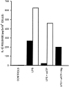
Figure 1
Figure 1
Oxidized ATP inhibits LPS-dependent release of IL-1β. N13 microglial cells were incubated in 24-well plates in RPMI medium supplemented with 10% FCS at a concentration of 2 × 10⁶ and incubated 24 h in the presence or absence (controls) of 10 μg/ml LPS. In the experiments with oATP, cells were treated with this inhibitor (300 μM) for 2 h and then rinsed before addition of LPS. Stimulation with nigericin (20 μM) was performed for 30 min after removal of oATP. Closed bars, IL-1β; open bars, IL-6. Data are averages of duplicate determinations from a single experiment repeated on three separate occasions. Similar results were obtained with the N9 cell line.
Figure 2
Figure 2
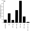
Modulation of IL-1β release by apyrase and hexokinase in N13 microglial cells. (A) Where indicated, cells were incubated in the presence of apyrase (apy, 0.4 U/ml) or hexokinase (hex, 100 μg/ml) throughout LPS treatment (10 μg for 24 h). As a control, the enzymes were boiled for 30 min (b apy and b hex) before being added to the cell monolayers. Pretreatment with oATP (300 μM) was peformed for 2 h; then the monolayers were rinsed and challenged with the different stimuli. (B) Cells were first stimulated for 2 h with LPS (10 μg/ml), and then stimulated with either 1 mM ATP or ADP for 30 min. Other experimental conditions were as shown in Fig. 1.
Figure 2
Figure 2
Modulation of IL-1β release by apyrase and hexokinase in N13 microglial cells. (A) Where indicated, cells were incubated in the presence of apyrase (apy, 0.4 U/ml) or hexokinase (hex, 100 μg/ml) throughout LPS treatment (10 μg for 24 h). As a control, the enzymes were boiled for 30 min (b apy and b hex) before being added to the cell monolayers. Pretreatment with oATP (300 μM) was peformed for 2 h; then the monolayers were rinsed and challenged with the different stimuli. (B) Cells were first stimulated for 2 h with LPS (10 μg/ml), and then stimulated with either 1 mM ATP or ADP for 30 min. Other experimental conditions were as shown in Fig. 1.
Figure 3
Figure 3
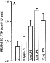
LPS dose response for IL-1β and ATP release. Microglial cells were plated in 24-well plates as described in Fig. 1 for IL-1β secretion or microtiter plastic wells as described in Materials and Methods for ATP release and stimulated with LPS for 24 h in a CO2 incubator at 37°C. For measurement of ATP, release samples were processed as follows: monolayers were rinsed and 100 μl of diluent buffer (Firezyme) were added (see Materials and Methods). Accumulation of extracellular ATP was measured by the luciferin/luciferase assay. Data for IL-1β release are duplicates from a single experiment repeated with similar results with three different batches of microglial cells. Data for ATP release are means of quadruplicate determinations ± SD from a single experiment repeated in three different occasions.
Figure 3
Figure 3
LPS dose response for IL-1β and ATP release. Microglial cells were plated in 24-well plates as described in Fig. 1 for IL-1β secretion or microtiter plastic wells as described in Materials and Methods for ATP release and stimulated with LPS for 24 h in a CO2 incubator at 37°C. For measurement of ATP, release samples were processed as follows: monolayers were rinsed and 100 μl of diluent buffer (Firezyme) were added (see Materials and Methods). Accumulation of extracellular ATP was measured by the luciferin/luciferase assay. Data for IL-1β release are duplicates from a single experiment repeated with similar results with three different batches of microglial cells. Data for ATP release are means of quadruplicate determinations ± SD from a single experiment repeated in three different occasions.
Figure 4
Figure 4
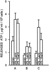
Stimulation with LPS triggers ATP release from human macrophages. Macrophages were isolated from three different donors (A–C ) as described in Materials and Methods and plated in microtiter plastic wells at a concentration of 50 × 103/well. After plating, cells were stimulated for 24 h with LPS and ATP release measured as detailed in Fig. 3. Data are mean ± SD from quadruplicate determinations.

References

    1. Surprenant A, Rassendren F, Kawashima E, North RA, Buell G. The cytolytic P2Z receptor for extracellular ATP identified as a P2X receptor (P2X7) Science (Wash DC) 1996;272:735–738. - PubMed
    1. Ferrari D, Villalba M, Chiozzi P, Falzoni S, RicciardiCastagnoli P, Di Virgilio F. Mouse microglial cells express a plasma membrane pore gated by extracellular ATP. J Immunol. 1996;156:1531–1539. - PubMed
    1. Zimmermann H. Signalling via ATP in the nervous system. Trends Neurosci. 1994;17:420–426. - PubMed
    1. Walev I, Reske K, Palmer M, Bhakdi S. Potassium-inhibited processing of IL-1β in human monocytes. EMBO (Eur Mol Biol Organ) J. 1995;14:1607–1614. - PMC - PubMed
    1. Dinarello CA. Interleukin-1. Rev Infect Dis. 1984;6:51–95. - PubMed

Publication types

MeSH terms