. 1997 Mar;73(1-2):15-27.
doi: 10.1016/s0165-5728(96)00159-2.

Nitric oxide synthase type II expression by different cell types in MHV-JHM encephalitis suggests distinct roles for nitric oxide in acute versus persistent virus infection

Affiliations

Nitric oxide synthase type II expression by different cell types in MHV-JHM encephalitis suggests distinct roles for nitric oxide in acute versus persistent virus infection

D M Grzybicki et al. J Neuroimmunol. 1997 Mar.

Abstract

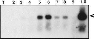
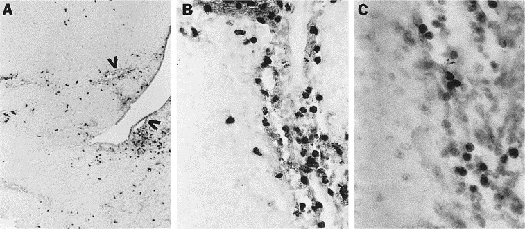
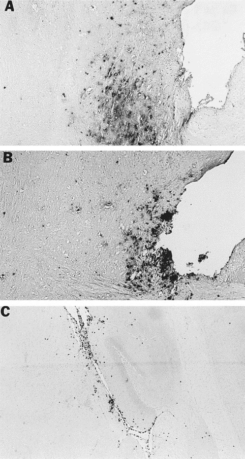
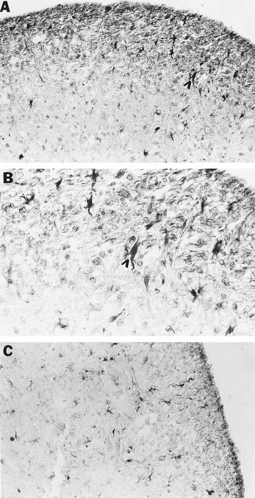
Intranasal inoculation with mouse hepatitis virus strain JHM (MHV-JHM) results in acute meningoencephalitis. We found NOS II mRNA expression in brains of acutely infected animals on days 5 through 7 after infection. In situ hybridization and immunohistochemistry demonstrated NOS II message and protein in infiltrating macrophages. Persistent infection with MHV-JHM results in chronic demyelinating encephalomyelitis. NOS II mRNA was detected in persistently infected spinal cords. In situ hybridization and immunohistochemistry showed expression of NOS II in astrocytes in and around demyelinated lesions. These results suggest the role of NO release in acute versus persistent infection with this virus, and its contribution to the resulting pathology, may be different.


Figures

Fig. 1
Fig. 1
Expression of NOS II mRNA in the CNS of mice with acute or persistent MHV-JHM infection. For acute infection, unimmunized mice were sacrificed at different time points after intranasal inoculation with 50,000 PFU of MHV-JHM and whole brain was harvested. For persistent infection, mice suckled from immunized dams were intranasally inoculated with 50,000 PFU of MHV-JHM. Animals exhibiting hindlimb paralysis weeks later were sacrificed and spinal cord was harvested. RNA extraction followed by RT-PCR/Southern blotting was performed. The expected size cDNA product is 441 bp, indicated by the arrowhead. Lane 1, `no target' control; 2–6, acutely infected mice three, four, five, six, and seven days post-inoculation; 7 and 8, persistent infection with hindlimb paralysis; 9, uninfected control; 10, LPS+IFN-γ induced RAW cells (positive control).
Fig. 2
Fig. 2
Cellular localization of NOS II mRNA in brains of mice acutely infected with MHV-JHM. In situ hybridization utilizing a 35S-labeled NOS II antisense (A) or sense (B) cRNA probe was performed on brain sections from acutely infected mice 7 days after inoculation. Numerous cells exhibit the presence of NOS II mRNA in A (examples indicated by arrows). Magnification, 400×.
Fig. 3
Fig. 3
Demyelinated lesion in ventral spinal cord of mouse with persistent MHV-JHM infection and hindlimb paralysis. White matter (W) chronic active lesion in lower right is hypercellular due to infiltrating chronic inflammatory cells and has multifocal areas of myelin loss evident by LFB staining, seen as rarified areas in the tissue by H and E staining. Magnification, 100×.
Fig. 4
Fig. 4
Localization of NOS II mRNA in spinal cords of mice persistently infected with MHV-JHM. In situ hybridization utilizing a 35S-labeled NOS II antisense (A) or sense (B) cRNA was performed on cord sections from mice exhibiting hindlimb paralysis. Clustering of silver grains is present in the white matter and peripheral gray matter of the cervical spinal cord at the edge of a demyelinated lesion (E) and in the lesion (L) which is histologically similar to that shown in Fig. 3. Magnification, 25×.
Fig. 5
Fig. 5
Cellular localization of NOS II protein in brains of acutely infected mice. Immunohistochemical staining of brain sections utilizing a polyclonal NOS II antibody followed by counterstaining with methyl green was performed on brain sections from mice sacrificed 3–7 days after infection. (A, B) NOS II positive cells in a brain 5 days after infection. Positive cells are seen surrounding blood vessels and migrating into the parenchyma. Vessels are indicated by arrowheads in (A). (C) F4/80 positive cells have a similar pattern of staining to that seen in B. Magnifications: (A) 25×, (B) 250×, (C) 400×.
Fig. 6
Fig. 6
Cellular localization of MHV-JHM viral antigen (A) and NOS II protein (B) in VTA of an acutely infected mouse at 5 days post-infection after immunohistochemical staining utilizing a polyclonal NOS II antibody followed by counterstaining with methyl green. Viral antigen occurs in parenchymal patches, while NOS II positive cells have a conspicuous meningeal and perivascular distribution. NOS II positive cells in the parenchyma often occur singly. (C) NOS II positive cells in the meninges of the hippocampus. Magnification, 25×.
Fig. 7
Fig. 7
Cellular localization of NOS II protein in spinal cords of mice persistently infected with MHV-JHM. Immunohistochemical staining of brain sections utilizing a polyclonal NOS II antibody was performed on spinal cord sections of mice with hindlimb paralysis and chronic-active demyelinating lesions in the spinal cord. (A, B) NOS II positive cells were present in the lesion and around the lesion edge (B is a higher magnification of A). Arrowheads indicate one of many positive cells morphologically resembling astrocytes. (C) GFAP staining reveals a similar pattern to that seen in (A). Magnifications: (A, C) 100×, (B) 250×.

References

    1. Adams, L.B., Hibbs, J.B., Taintor, R.R. and Krahenbuhl, J.L. (1990) Microbiostatic effect of murine-activated macrophages for Toxoplasma gondii. Role for synthesis of inorganic nitrogen oxides from L-arginine. J. Immunol. 144, 2725–2729. - PubMed
    1. Benveniste, E.N. (1992) Inflammatory cytokines within the central nervous system:sources, function, and mechanism of action. Am. J. Physiol. 263, C1–C16. - PubMed
    1. Bailey, O.T., Pappenheimer, A.M., Sargent, F., Cheever, M.D. and Daniels, J.B. (1949) A murine virus (JHM) causing disseminated encephalomyelitis with extensive destruction of myelin II. Pathology. J. Exp. Med. 90, 195–211. - PMC - PubMed
    1. Bauer, J., Berkenbosch, F., Van Dam, A. and Dijkstra, C.D. (1993) Demonstration of interleukin-1b in Lewis rat brain during experimental allergic encephalomyelitis by immunocytochemistry at the light and ultrastructural level. J. Neuroimmunol. 48, 13–22. - PubMed
    1. Bi, Z., Barna, M., Komatsu, T. and Reiss, C.S. (1995) Vesicular stomatitis virus infection of the central nervous system activates both innate and acquired immunity. J. Virol. 69, 6466–6472. - PMC - PubMed

Publication types

LinkOut - more resources