. 1997 Feb;81(2):154-62.
doi: 10.1136/bjo.81.2.154.

Increased expression of angiogenic growth factors in age-related maculopathy

Affiliations

Increased expression of angiogenic growth factors in age-related maculopathy

M Kliffen et al. Br J Ophthalmol. 1997 Feb.

Abstract

Aims/background: The late stages of age-related maculopathy (ARM), especially neovascular macular degeneration (ARMD), can severely affect central vision and are the main cause of blindness in the elderly in the Western world. It has been shown that angiogenic growth factors are present in neovascular membranes in ARMD. However, it is not known if angiogenic growth factors play a role in the onset of neovascularisation.

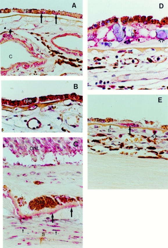
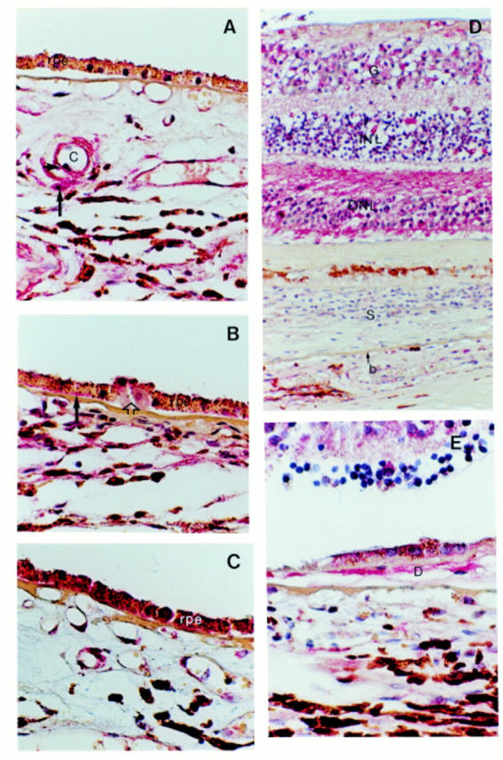
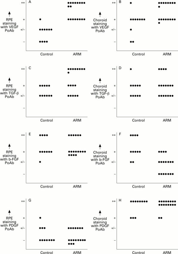
Methods: In order to elucidate the involvement of angiogenic growth factors in the initiation of neovascularisation in early stages of ARM, the expression patterns of VEGF, TGF-beta, b-FGF, and PDGF-AA on 18 human maculae with ARM, and on 11 control specimens were investigated immunohistochemically.

Results: A significantly increased expression of VEGF (p = 0.00001) and TGF-beta (p = 0.019) was found in the retinal pigment epithelium (RPE) of maculae with ARM compared with control maculae. Furthermore, an increased expression of VEGF and PDGF was found in the outer nuclear layer of maculae with ARM.

Conclusion: These results demonstrate an increased expression of VEGF in the RPE, and in the outer nuclear layer in maculae with ARM, that could be involved in the pathogenesis of neovascular macular degeneration. Furthermore, enhanced TGF-beta expression in the RPE cells of maculae with early stages of ARM was shown.


Figures

Figure 1
Figure 1
Light microscopic images of immunohistochemistry with anti-VEGF antibodies. (A) Typical example of VEGF expression in a control macula; VEGF is present in smooth muscle cells (short arrow) of choroidal blood vessels (C) and in some (long arrows) of the retinal pigment epithelial cells (rpe). (B) The expression of VEGF in the RPE is increased in ARM maculae in comparison with control maculae. (C) The outer nuclear layer (ONL) and basal laminar deposit (long arrows) show expression of VEGF in this macula with neovascular macular degeneration. The myofibroblast cells (small arrows) in the scar tissue (S) contain VEGF as well. (D) Expression of VEGF in a large drusen (D) with calcified bodies (open arrow) in an ARM macula. (E) Macrophages (long arrow) that have infiltrated Bruch's membrane (b) in a ARM macula show intense staining for VEGF. A monocyte (open arrow) shows VEGF expression as well. Original magnification × 400.
Figure 2
Figure 2
Light microscopic images of immunohistochemistry with anti-TGF-β (A and B), b-FGF (C), and PDGF-AA (D and E) antibodies. (A) Control maculae show TGF-β expression in smooth muscle cells (long arrow) and endothelial cells (short arrow) of choroidal blood vessels (C). The RPE shows only faint staining in most of the control maculae. (B) TGF-β expression is increased in the RPE of this typical ARM macula. Basal laminar deposit (long arrow) contains TGF-β as well. Note that the most prominent RPE staining is present on top of a drusen (open arrow) that itself shows no staining. (C) A typical example of an ARM macula that shows b-FGF expression in the RPE. (D) PDGF-AA expression in a macula with neovascular macular degeneration. The retina demonstrates PDGF-AA expression, whereas the retina in control maculae shows no staining at all. Note that the expression in the outer nuclear layer (ONL) is more prominent than in the inner nuclear layer (INL) and ganglion cell layer (G). (E) PDGF expression in a large drusen (D) in a macula with neovascular macular degeneration. S = scar tissue, b = Bruch's membrane. Original magnification × 400.
Figure 3
Figure 3
Comparison of the immunohistochemical expression of VEGF (A and B), TGF-β (C and D), b-FGF (E and F), and PDGF-AA (G and H) in RPE (A, C, E, G) and choroid (B, D, F, H) between control maculae and ARM maculae. PoAb = Polyclonal antibody.

References

    1. Br J Ophthalmol. 1976 May;60(5):324-41 - PubMed
    1. Ophthalmology. 1995 Sep;102(9):1261-2 - PubMed
    1. Ophthalmology. 1985 May;92(5):615-27 - PubMed
    1. Arch Ophthalmol. 1985 Dec;103(12):1870-5 - PubMed
    1. Proc Natl Acad Sci U S A. 1987 Aug;84(16):5788-92 - PubMed

MeSH terms

Substances