. 1997 Mar 10;136(5):1109-21.
doi: 10.1083/jcb.136.5.1109.

LI-cadherin-mediated cell-cell adhesion does not require cytoplasmic interactions

Affiliations

LI-cadherin-mediated cell-cell adhesion does not require cytoplasmic interactions

B Kreft et al. J Cell Biol. .

Abstract

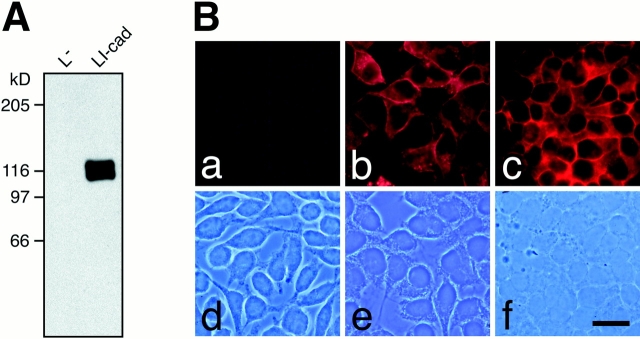
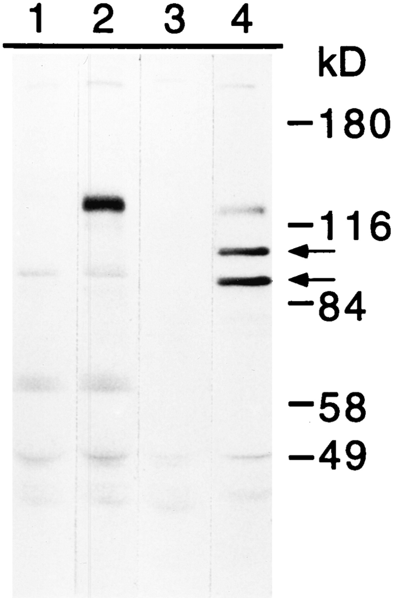
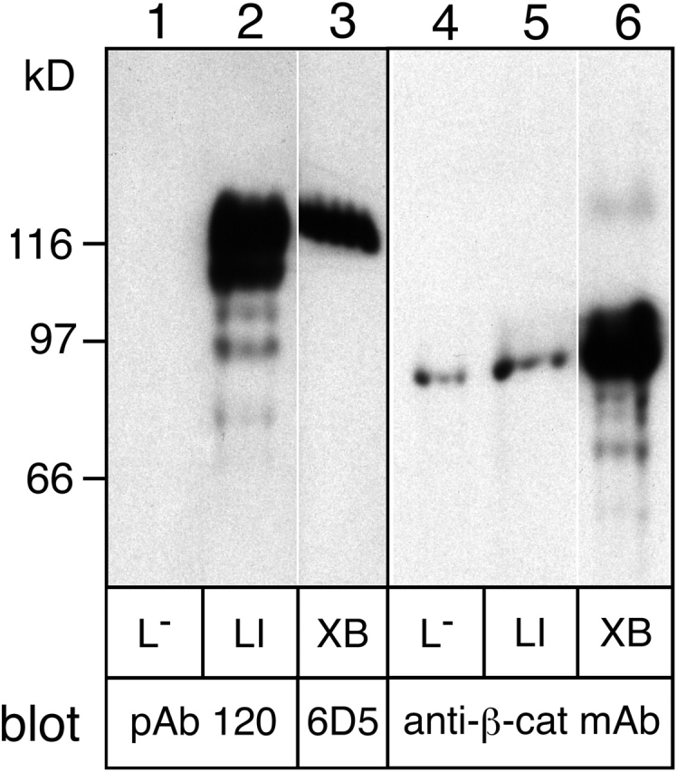
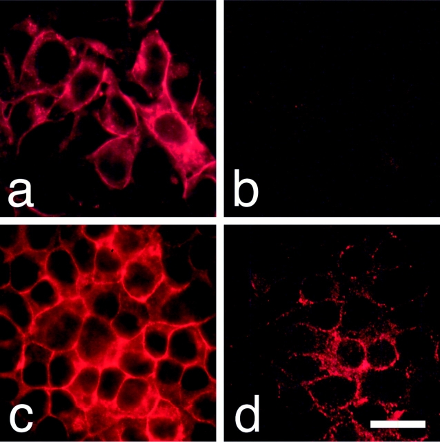
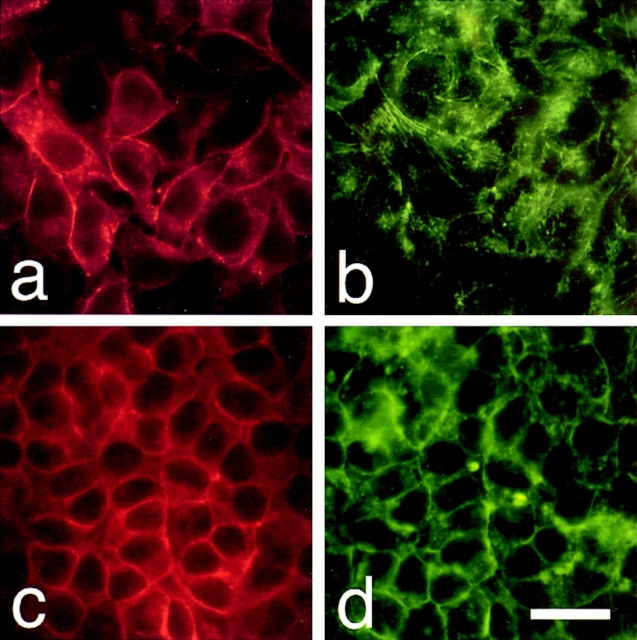
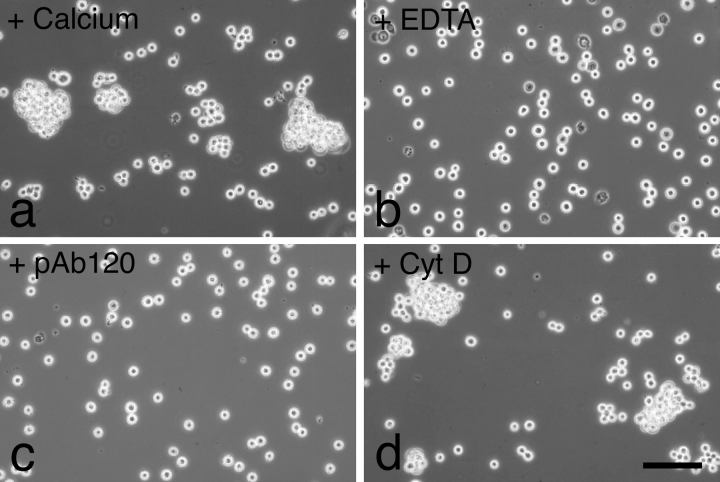
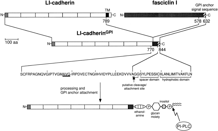
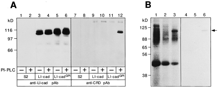
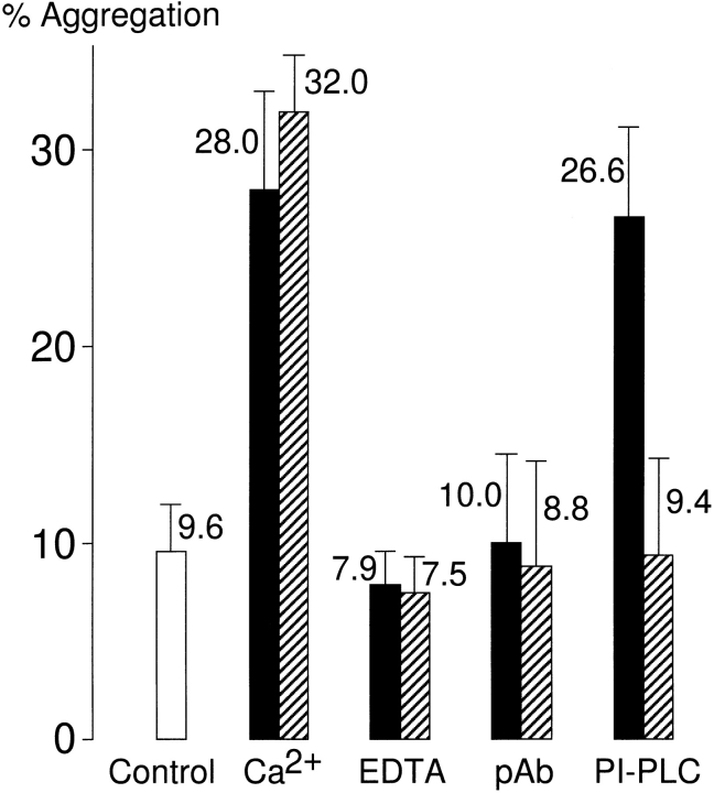
The adhesive function of classical cadherins depends on the association with cytoplasmic proteins, termed catenins, which serve as a link between cadherins and the actin cytoskeleton. LI-cadherin, a structurally different member of the cadherin family, mediates Ca2+-dependent cell-cell adhesion, although its markedly short cytoplasmic domain exhibits no homology to this highly conserved region of classical cadherins. We now examined whether the adhesive function of LI-cadherin depends on the interaction with catenins, the actin cytoskeleton or other cytoplasmic components. In contrast to classical cadherins, LI-cadherin, when expressed in mouse L cells, was neither associated with catenins nor did it induce an upregulation of beta-catenin. Consistent with these findings, LI-cadherin was not resistant to detergent extraction and did not induce a reorganization of the actin cytoskeleton. However, LI-cadherin was still able to mediate Ca2+-dependent cell-cell adhesion. To analyze whether this function requires any interaction with proteins other than catenins, a glycosyl phosphatidylinositol-anchored form of LI-cadherin (LI-cadherin(GPI)) was constructed and expressed in Drosophila S2 cells. The mutant protein was able to induce Ca2+-dependent, homophilic cell-cell adhesion, and its adhesive properties were indistinguishable from those of wild type LI-cadherin. These findings indicate that the adhesive function of LI-cadherin is independent of any interaction with cytoplasmic components, and consequently should not be sensitive to regulatory mechanisms affecting the binding of classical cadherins to catenins and to the cytoskeleton. Thus, we postulate that the adhesive function of LI-cadherin is complementary to that of coexpressed classical cadherins ensuring cell-cell contacts even under conditions that downregulate the function of classical cadherins.


Figures

Figure 1
Figure 1
Expression of LI-cadherin in L cells. (A) Immunoblotting of LI-cadherin. Equal amounts (100 μg) of proteins from parental (L) and transfected L cells expressing LI-cadherin (LI-cad) were separated by SDS-PAGE, transferred to nitrocellulose membranes, and immunoblotted using anti–LI-cadherin pAb120. LI-cadherin appeared as a broad double band with a molecular mass of ∼120 kD. (B) Immunocytochemical staining of LI-cadherin. Parental (a and d) and transfected L cells expressing either LI-cadherin (b and e) or XB/U-cadherin (c and f) were fixed, incubated with anti–LI-cadherin mAb 47.2 (a and b) or anti–XB/U-cadherin mAb 6D5 (c), stained with TRITC-labeled secondary antibodies. The corresponding phase contrast micrographs are shown in panels d–f. LI-cadherin was expressed on the cell surface of transfected L cells and appeared concentrated at sites of cell–cell contact (b). In contrast to classical XB/Ucadherin (c and f), LI-cadherin did not induce an epithelial phenotype, although cell–cell contact regions were enlarged compared to nontransfected L cells (d and e). Bar, ( f  ) 20 μm.
Figure 2
Figure 2
Catenins do not coprecipitate with LI-cadherin. Parental L cells (lanes 1 and 3) and transfected cells expressing either LI-cadherin (lane 2) or XB/U-cadherin (lane 4) were metabolically labeled, lysed, and subjected to immunoprecipitation using anti–LI-cadherin pAb120 (lanes 1 and 2) or anti–XB/ U-cadherin mAb 6D5 (lanes 3 and 4). Precipitates were separated by SDS-PAGE and analyzed by autoradiography. While α- and β-catenin copurified with XB/Ucadherin (lane 4), neither catenins nor any other labeled proteins were coprecipitated with LI-cadherin (lane 2).
Figure 3
Figure 3
β-Catenin expression is not upregulated in LI-cadherin–transfected L cells. Equal amounts (100 μg) of proteins from parental (lanes 1 and 4) and transfected L cells expressing either LI-cadherin (lanes 2 and 5) or XB/U-cadherin (lanes 3 and 6) were separated by SDS-PAGE, transferred to nitrocellulose membranes and immunoblotted using anti–LI-cadherin pAb120 (lanes 1 and 2), anti–XB/U-cadherin mAb 6D5 (lane 3) or anti–βcatenin mAb (lanes 4–6. Although LI-cadherin was highly expressed in transfected L cells (lane 2), the β-catenin expression remained the same as in nontransfected cells (lanes 4 and 5). In contrast, expression of XB/U-cadherin (lane 3) induced a significant increase of β-catenin in L cells (lane 6).
Figure 4
Figure 4
LI-cadherin is not resistant to extraction with NP-40. L cells expressing either LI-cadherin (a and b) or XB/U-cadherin (c and d) were fixed before (a and c) or after (b and d) extraction with 0.5% NP-40. Immunofluorescence staining was performed using anti–LI-cadherin mAb 47.2 (a and b) or anti–XB/U-cadherin mAb 6D5 (c and d) followed by an incubation with secondary TRITC-labeled antibodies. As shown in b, LI-cadherin could be easily extracted with NP-40, while XB/U-cadherin was partially resistant and remained clearly visible at cell–cell contacts under these conditions (d). Bar, (d) 20 μm.
Figure 5
Figure 5
Actin cytoskeleton reorganization is not induced by LIcadherin expression. L cells expressing either LI-cadherin (a and b) or XB/U-cadherin (c and d) were fixed, permeabilized, and the actin cytoskeleton was stained with FITC-phalloidin (b and d). For double labeling, the same cells were incubated with anti–LIcadherin mAb 47.2 (a) or anti–XB/U-cadherin mAb 6D5 (c) followed by staining with secondary TRITC-labeled antibodies. In transfected L cells expressing XB/U-cadherin, the actin cytoskeleton was completely redistributed to sites of cell–cell contact (d). In contrast, expression of LI-cadherin did not promote any significant reorganization of the actin cytoskeleton and stress fibers were still visible (b). Bar, (d) 20 μm.
Figure 6
Figure 6
LI-cadherin mediates aggregation of transfected L cells. Aggregation of LI-cadherin expressing L cells was analyzed in the presence of 2 mM CaCl2 (a), 2 mM EDTA (b) or anti–LI-cadherin pAb120 (c). For the disruption of the cytoskeleton (d), cells were preincubated with 1 μM cytochalasin D for 30 min at 37°C. LI-cadherin acted as a Ca2+-dependent cell adhesion molecule when expressed in L cells. Its function was not affected by the disruption of the actin cytoskeleton.
Figure 7
Figure 7
Construction of GPI-anchored LI-cadherinGPI. The first 789 amino acids representing the entire extracellular domain of LIcadherin were linked to the COOH-terminal 55 amino acids of Drosophila fasciclin I. The fasciclin I–derived portion of LI-cadherinGPI contains a typical signal sequence for GPI anchoring (Coyne et al., 1993; Kodukula et al., 1993). This signal sequence comprises a domain with small amino acids at the first position (representing the putative cleavage/GPI anchor attachment site), and at the third position, followed by a 9–amino acid-spacer domain and a hydrophobic region of 16 amino acids. After cleavage of the signal peptide in the ER, the GPI anchor is linked to the new COOH terminus of the protein via an ethanolamine residue (for reviews see Cross, 1990; Englund, 1993). The last LI-cadherin–derived amino acids (AVG) are underlined in the protein sequence of LIcadherinGPI.
Figure 8
Figure 8
LI-cadherinGPI is expressed as a GPI-anchored molecule in S2 cells. (A) Immunoblotting of LI-cadherinGPI. Equivalent amounts (75 μg) of membrane proteins from untransfected S2 cells (S2) and cells expressing either LI-cadherin (LI-cad) or LI-cadherinGPI (LI-cadGPI) were incubated with or without PI-PLC from T. brucei, separated by SDS-PAGE, and analyzed by immunoblotting using anti–LI-cadherin pAb120 (lanes 1–6). When expressed in S2 cells, both LI-cadherin and LI-cadherinGPI exhibited an apparent molecular mass of ∼110 kD, that was not changed detectably by PI-PLC treatment (lanes 4 and 6). The filter was stripped and reprobed with an antibody against the cross-reacting determinant (anti-CRD pAb) which is exposed solely in PI-PLC–cleaved GPI anchors (lanes 7–12). Only in cells expressing LI-cadherinGPI a single 110-kD protein was stained by anti-CRD pAb after digestion with PI-PLC (lane 12). (B) Metabolic labeling of LI-cadherinGPI with [3H]ethanolamine. After metabolic labeling with [3H]ethanolamine, untransfected S2 cells (lanes 1 and 4) and cells expressing either LI-cadherin (lanes 2 and 5) or LI-cadherinGPI (lanes 3 and 6) were lysed and cellular extracts were subjected to immunoprecipitation using anti–LI-cadherin pAb120. Equivalent amounts (75 μg) of solubilized proteins from the three cell types were separated in lanes 1–3. The corresponding immunoprecipitates are shown in lanes 4–6. The arrow indicates the position of immunoprecipitated LI-cadherinGPI in lane 6 that has incorporated [3H]ethanolamine.
Figure 9
Figure 9
LI-cadherinGPI is a functional cell adhesion molecule when expressed in S2 cells. Aggregation of transfected S2 cells expressing either LI-cadherin (black bars) or LI-cadherinGPI (striped bars) was induced and quantified as percent reduction in particle number. Aggregation was carried out in Schneider's medium (containing 5 mM Ca2+), or after addition of either 30 mM EDTA or anti–LI-cadherin pAb120. For PI-PLC treatment, cells were incubated for 2 h with 1 U/ml PI-PLC from B. thuringiensis before aggregation. Untransfected S2 cells (white bar) in medium containing 5 mM Ca2+ served as an aggregation control. The column height corresponds to the mean of five aggregation experiments; the error bars indicate the standard deviation. LI-cadherinGPI induced aggregation of transfected S2 cells in a Ca2+-dependent manner to the same extent as did wild-type LI-cadherin. Pretreatment with PI-PLC, and thus removal of LI-cadherinGPI from the cell surface, caused a complete inhibition of aggregation.
Figure 10
Figure 10
Surface expression pattern of LI-cadherinGPI. The distribution of LI-cadherinGPI within fixed aggregates of transfected S2 cells was determined by immunofluorescence staining with FITC-labeled anti–LI-cadherin pAb120. LI-cadherinGPI was located at sites of cell–cell contact, but could also be found on cell surface regions that were not in direct contact with neighboring cells. Note that LI-cadherinGPI did not appear in clusters on the cell surface. Bar, 50 μm.
Figure 11
Figure 11
LI-cadherinGPI is a homophilic cell adhesion molecule and is able to interact with native LI-cadherin. Parental S2 cells (a and b) or LI-cadherin–expressing S2 cells (c and d) were labeled with the vital fluorescence membrane dye DiI, mixed with unlabeled LI-cadherinGPI-transfected cells and agitated together. Fluorescence micrographs are shown in a and c, the corresponding phase contrast micrographs are shown in b and d. LI-cadherinGPI mediates cell–cell adhesion in a homotypic manner, since aggregates contained no DiI-labeled parental S2 cells (a and b). S2 cells expressing either native or GPI-anchored LI-cadherin form large mixed aggregates, suggesting that the adhesive properties of both proteins are indistinguishable (c and d). Bar, (d) 100 μm.

Similar articles

Cited by

References

    1. Anderson RGW. Caveolae: where incoming and outgoing messengers meet. Proc Natl Acad Sci USA. 1993;90:10909–10913. - PMC - PubMed
    1. Behrens J, Vakaet L, Friis R, Winterhager E, Van RF, Mareel MM, Birchmeier W. Loss of epithelial differentiation and gain of invasiveness correlates with tyrosine phosphorylation of the E-cadherin/β-catenin complex in cells transformed with a temperature-sensitive v-SRC gene. J Cell Biol. 1993;120:757–766. - PMC - PubMed
    1. Berndorff D, Gessner R, Kreft B, Schnoy N, Lajous-Petter A-M, Loch N, Reutter W, Hortsch M, Tauber R. Liver-intestine cadherin: molecular cloning and characterization of a novel Ca(2+)-dependent cell adhesion molecule expressed in liver and intestine. J Cell Biol. 1994;125:1353–1369. - PMC - PubMed
    1. Birchmeier W, Behrens J. Cadherin expression in carcinomas: role in the formation of cell junctions and the prevention of invasiveness. Biochem Biophys Acta. 1994;1198:11–26. - PubMed
    1. Boller K, Vestweber D, Kemler R. Cell-adhesion molecule uvomorulin is localized in the intermediate junctions of adult intestinal epithelial cells. J Cell Biol. 1985;100:327–332. - PMC - PubMed

Publication types

MeSH terms