. 1997 Apr 1;17(7):2295-313.
doi: 10.1523/JNEUROSCI.17-07-02295.1997.

Distribution of brain-derived neurotrophic factor (BDNF) protein and mRNA in the normal adult rat CNS: evidence for anterograde axonal transport

Affiliations

Distribution of brain-derived neurotrophic factor (BDNF) protein and mRNA in the normal adult rat CNS: evidence for anterograde axonal transport

J M Conner et al. J Neurosci. .

Abstract

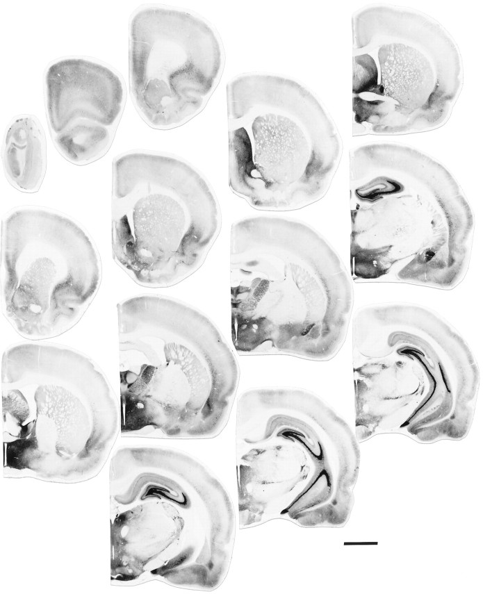
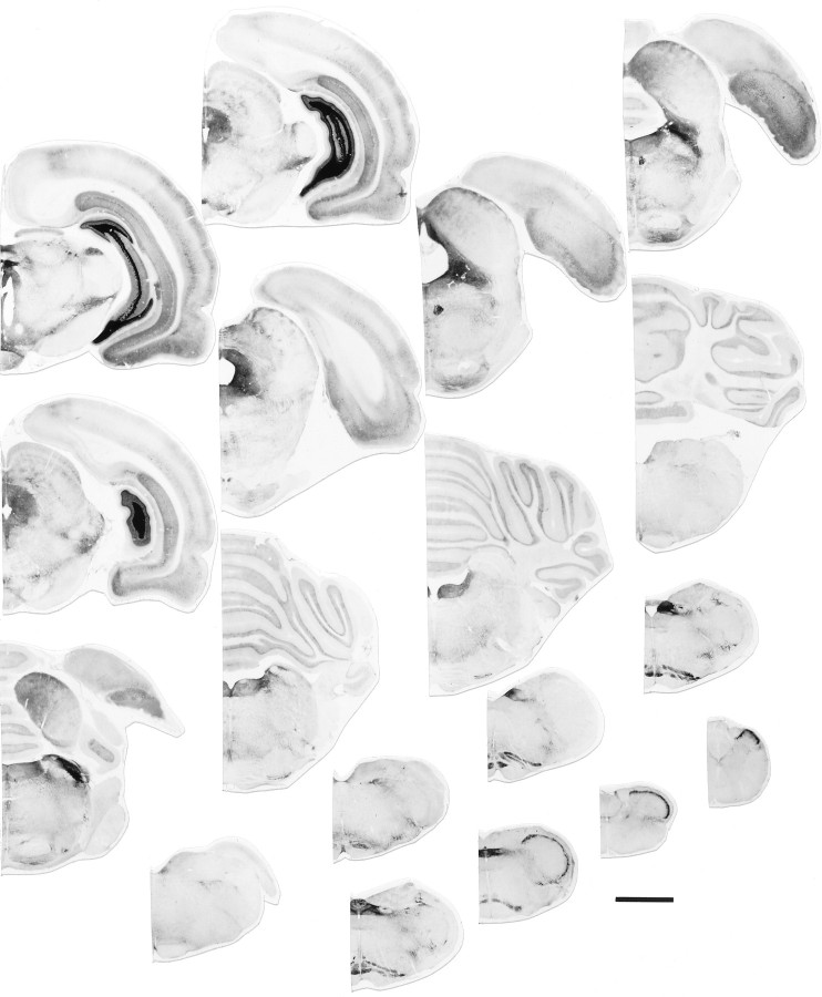
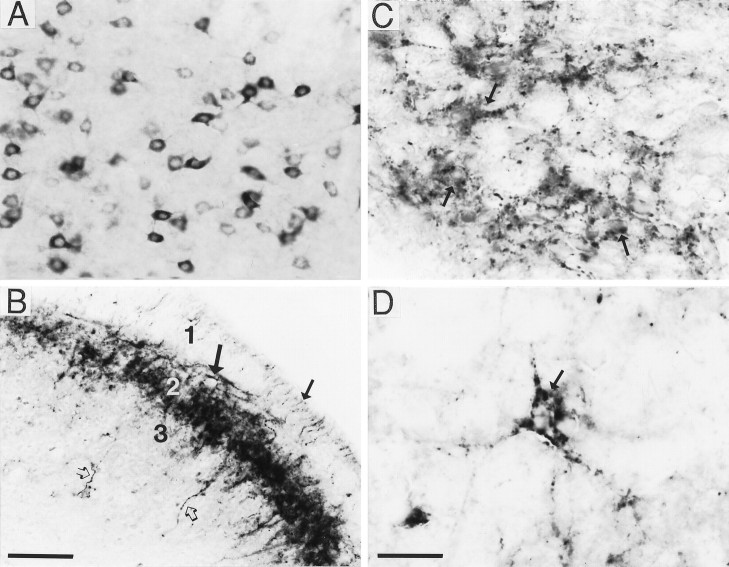
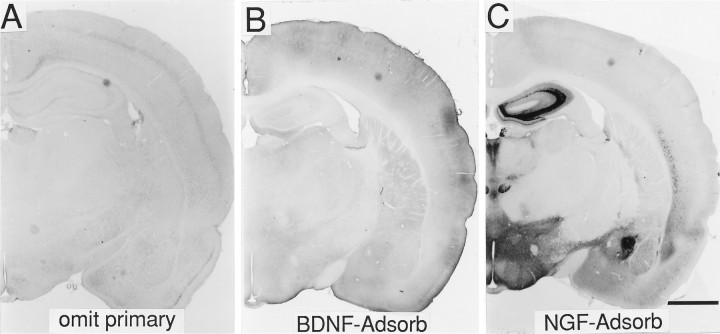
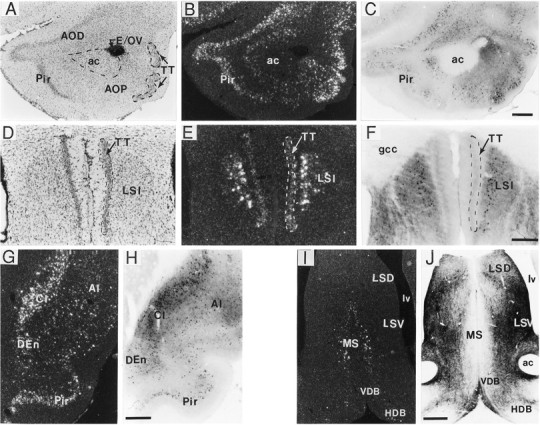
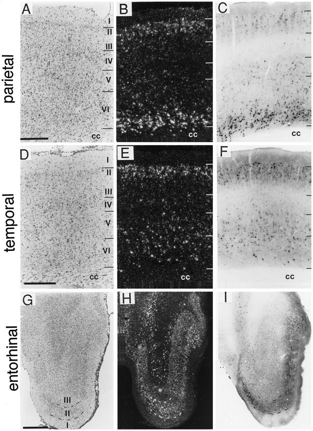
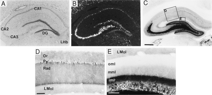
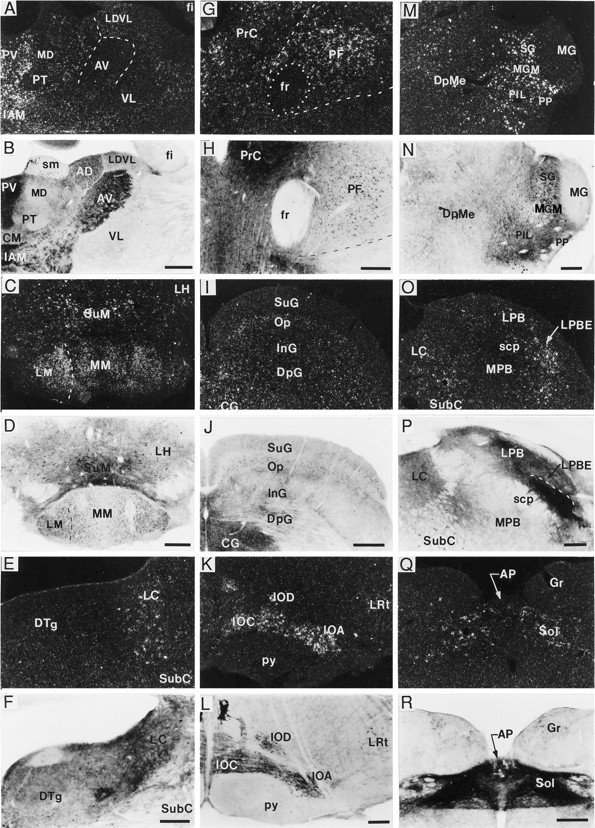
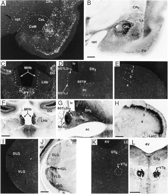
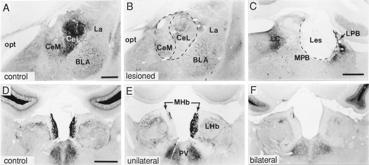
A sensitive immunohistochemical technique was used, along with highly specific affinity-purified antibodies to brain-derived neurotrophic factor (BDNF), to generate a detailed mapping of BDNF immunoreactivity (BDNF-ir) throughout the adult rat CNS. A parallel analysis of sites of BDNF synthesis was performed with in situ hybridization techniques using a cRNA probe to the exon encoding mature rat BDNF protein. These combined data revealed (1) groups of cell bodies containing diffuse BDNF-ir throughout the CNS that were strongly correlated with fields of cells containing BDNF mRNA; (2) varying degrees of BDNF-ir outside of cell bodies, in what appeared to be fibers and/or terminals; and (3) many regions containing extremely heavy BDNF-immunoreactive fiber/terminal labeling that lacked BDNF mRNA (e.g., medial habenula, central nucleus of the amygdala, bed nucleus of stria terminalis, lateral septum, and spinal cord). The latter observation suggested that in these regions BDNF was derived from anterograde axonal transport by afferent systems. In the two cases in which this hypothesis was tested by the elimination of select afferents, BDNF immunostaining was completely eliminated. These data, along with the observation that BDNF-ir was rarely found within dendrites or fibers en passage, suggest that BDNF protein produced in adult CNS neurons is polarized primarily along axonal processes and is preferentially stored in terminals within the innervation target.


Figures

Fig. 1.
Fig. 1.
Distribution of BDNF-ir in the adult rat brain. Both plates show at low magnification a rostrocaudal series of sections (left to right) that were obtained from two female rats immunostained using affinity-purified antibodies specific for BDNF (bright-field illumination). BDNF immunostaining in male rats (not shown) was indistinguishable from that seen in females. Scale bar, 3 mm.
Fig. 1.
Fig. 1.
Distribution of BDNF-ir in the adult rat brain. Both plates show at low magnification a rostrocaudal series of sections (left to right) that were obtained from two female rats immunostained using affinity-purified antibodies specific for BDNF (bright-field illumination). BDNF immunostaining in male rats (not shown) was indistinguishable from that seen in females. Scale bar, 3 mm.
Fig. 2.
Fig. 2.
Characteristic patterns of BDNF immunostaining in the rat CNS. Panels show examples of BDNF immunostaining in basolateral amygdaloid nucleus (A), spinal cord (B), hypothalamus (C), and lateral septum (D) that are representative of the quality of immunolabeling seen throughout the CNS (bright-field illumination). As shown inA, perikaryal labeling was characterized by a diffuse reaction product distributed throughout the cell cytoplasm but excluding the cell nucleus. B–D, Various types of fiber/terminal staining. As seen in spinal cord lamina 1 (B, small arrow) and laminae 3 (B, open arrows), numerous individual fibers were immunolabeled; immunoreactive fibers were also concentrated in lamina 2 (B, large arrow). Other fields contained punctate immunostaining that appeared either diffuse or clustered around unlabeled cell bodies (C, arrows), or well defined, with discrete pericellular baskets around unlabeled cells (D, arrow). Scale bars (shown inB): A, B, 100 μm; (shown inD) C, D, 50 μm.
Fig. 3.
Fig. 3.
Specificity of BDNF immunostaining.A–C, Tissue sections processed for immunohistochemistry with either the BDNF antibody omitted (A), the BDNF antibody solution preadsorbed against purified recombinant human BDNF (B), or the BDNF antibody solution preadsorbed against mouse β-NGF (C) (bright-field illumination). As seen in A and B, when the BDNF antibody was omitted or preadsorbed with BDNF protein, no specific staining was present. In C, preadsorbing against mouse β-NGF revealed a pattern of immunostaining that was identical to sections reacted with the BDNF antibody alone (see Fig. 1). Scale bar, 2 mm.
Fig. 4.
Fig. 4.
BDNF-ir and mRNA in forebrain regions.A–J, Sections through the anterior olfactory nucleus (A–C), caudal portion of the tenia tecta (D–F), claustrum and piriform cortex (G, H), and septal region (I, J) processed for immunohistochemistry (C, F, H, J; bright field) and in situ hybridization (B, E, G, I; dark field). Photomicrographs inA and D are of Nissl-stained tissue. Patterns of BDNF-ir and cRNA labeling were very similar in the anterior olfactory nucleus and rostral tenia tecta (TT) (A–C) and, more caudally, in the claustrum (Cl), endopiriform nucleus (DEn), and piriform cortex (Pir) (G, H). In the caudal portion of the tenia tecta, very light hybridization but no immunostaining were observed (D–F); notably heavy hybridization and immunostaining were present in the adjacent intermediate lateral septum (LSI) (D–F). In septal regions, cRNA-labeled cells were seen only in the medial septum (MS), whereas intense BDNF-ir was localized mostly to fibers/terminals in the lateral septum (I, J). Scale bars (shown inC): A–C, 500 μm; (shown inF): D–F, 200 μm; (shown inH): G, H, 500 μm; (shown inJ): I, J, 500 μm.ac, Anterior commissure; AI, agranular insular cortex; AOD, anterior olfactory nucleus, dorsal part; AOP, anterior olfactory nucleus, posterior part;E/OV, ependymal layer/olfactory ventricle;gcc, genu of the corpus callosum; HDB, horizontal limb of the diagonal band; LSD, lateral septum, dorsal part; LSV, lateral septum, ventral part;lv, lateral ventricle; VDB, vertical limb of the diagonal band.
Fig. 5.
Fig. 5.
Comparison of the distributions of BDNF-ir and mRNA in cortical fields. Sections through parietal (A–C), temporal (D–F), and entorhinal (G–I) cortices were processed forin situ hybridization to localize BDNF mRNA (B, E, H; dark field) or immunohistochemistry to localize BDNF-ir (C, F, I; bright field). Sections through similar planes were Nissl-stained (A, D, G; bright field). In parietal cortex (A–C), the BDNF cRNA heavily labeled neurons in layer VI, moderately in layersII/III, and lightly in layer V; BDNF-ir was very heavy in deep layer VI, lighter in layersII, III, V, and superficial layer VI, and almost undetectable in layer IV. In temporal cortex (D–F), BDNF mRNA was moderately labeled in layers II/III and V/VI, with lighter labeling in layer IV. BDNF-ir in temporal cortex was heaviest in layers II/III, moderate in layersV/VI, and light in layer IV. In entorhinal cortex (G–I), the patterns of BDNF cRNA hybridization and immunoreactivity were similar. Scale bars (shown in A): A–C, 500 μm; (shown inD): D–F, 500 μm; (shown inG): G–I, 1 mm. cc, Corpus callosum.
Fig. 6.
Fig. 6.
Distribution of BDNF-ir and mRNA in hippocampus. Low-magnification (A–C) and high-magnification (D, E) photomicrographs of sections through hippocampus that were either Nissl-stained (A, bright field) or processed for in situ hybridization (B, dark field) or immunohistochemistry (C–E, bright field). In hippocampus, the BDNF cRNA densely labeled pyramidal cells in CA3, CA2, and the hilus, and less densely the CA1 pyramidal cells and the dentate gyrus granule cells (GrDG) (B). BDNF immunostaining densely labeled processes within the mossy fiber system (C). At higher magnification, one could see that in region CA1 BDNF immunostaining was diffuse in strata oriens (Or) and radiatum (Rad) and weaker in stratum lacunosum moleculare (LMol) (D). Scattered neurons in the CA1 pyramidal cell layer (Py) were immunolabeled (D). In the dentate gyrus (DG) molecular layer, a trilaminar pattern of BDNF-ir was seen, with moderately dense labeling in the inner molecular layer (iml), light labeling in the middle molecular layer (mml), and no detectable staining in the outer molecular layer (oml). Scale bars (shown in C):A–C, 500 μm; (shown in D): D, E, 100 μm and 75 μm, respectively. LHb, Lateral habenular nucleus.
Fig. 7.
Fig. 7.
Localization of BDNF-ir and mRNA in thalamic and brainstem regions. Dark-field and bright-field photomicrographs showing BDNF cRNA hybridization (A, C, E, G, I, K, M, O, Q) and BDNF immunolabeling (B, D, F, H, J, L, N, P, R), respectively, in thalamus (A, B), mammillary region (C, D), locus coeruleus (E, F), parafascicular nucleus (PF) (G, H), superior colliculus (I, J), inferior olivary complex (K, L), medial geniculate nucleus (M, N), parabrachial region (O, P), and the nucleus of the solitary tract (Q, R). In many thalamic (A, B, G, H, M, N) and hypothalamic (C, D) areas, the distributions of BDNF mRNA and perikaryal BDNF-ir overlapped; although in some regions [e.g., anteroventral thalamic nucleus (AV)] there was heavy fiber/terminal BDNF-ir in the absence of hybridization (A, B). In other regions, such as the locus coeruleus (LC) (E, F) and nucleus of the solitary tract (Sol) (Q, R), cRNA-labeled cells could be detected, but it was difficult to identify immunostained cells because of the fiber/terminal immunolabeling. Weak labeling with both the cRNA and antibody also overlapped in most layers of superior colliculus, excluding the superficial gray layer, which did not contain cRNA labeling (I, J). In contrast, heavy hybridization and BDNF-ir were detected throughout the inferior olivary complex (K, L). Moderate to heavy BDNF cRNA labeling was found in the external portion of the lateral parabrachial nucleus (LPBE), whereas heavy fiber/terminal BDNF-ir was present in most subregions of the lateral parabrachial subfield (LPB) (O, P); neither cRNA nor antibody labeled the medial portion of the parabrachial nucleus (MPB) (O, P). Scale bars (shown inB, D, J, N): A–D, I, J, M, N, 500 μm, respectively; (shown in F): E, F, 200 μm; (shown in H, P, R): G, H, O, P–R, 250 μm, respectively. AP, Area postrema;CG, central (periaqueductal) gray; DpG, superior colliculus, deep gray layer; DpMe, deep mesencephalic nucleus; DTg, dorsal tegmental nucleus;fr, fasciculus retroflexus; Gr, gracile nucleus; IAM, interanteromedial thalamic nucleus;InG, superior colliculus, intermediate gray layer;IOA, inferior olive, subnucleus A; IOC, inferior olive, subnucleus C; IOD, inferior olive, dorsal nucleus; LDVL, laterodorsal thalamic nucleus, ventrolateral; LH, lateral hypothalamic area;LM, lateral mammillary nucleus; LRt, lateral reticular nucleus; MG, medial geniculate nucleus; MGM, medial geniculate nucleus, medial portion;MM, medial mammillary nucleus; Op, superior colliculus, optic nerve layer; PIL, posterior intralaminar nucleus; PrC, precommissural nucleus; PP, peripeduncular thalamic nucleus;PT, paratenial thalamic nucleus; PV, paraventricular thalamic nucleus; py, pyramidal tract;scp, superior cerebellar peduncle; SG, suprageniculate nucleus; SubC, subcoeruleus nucleus;SuG, superior colliculus, superficial gray layer;SuM, supramammillary nucleus; VL, ventrolateral thalamic nucleus.
Fig. 8.
Fig. 8.
Dense fiber/terminal BDNF-ir was present in many regions lacking BDNF mRNA expression. A–L, Dark-field and bright-field photomicrographs of sections processed for in situ hybridization (A, C–E, I, K) and immunohistochemistry (B, F–H, J, L), respectively, through the amygdala (A, B), habenula (C, F), bed nucleus of stria terminalis (D, G), spinal cord (E, H), lateral geniculate nucleus (I, J), and dorsal/ventral tegmental nucleus (K, L). As seen in Aand B, in the basolateral (BLA) and lateral amygdala (La), many cells were heavily labeled for both mRNA and BDNF-ir, whereas in the central amygdala many immunolabeled fibers/terminals were present but cRNA hybridization was not. Similarly, in the medial habenular nucleus (MHb) (C, F) subregions of the bed nucleus of stria terminalis (D, G), the dorsal (DLG) and ventral regions (VLG) of the lateral geniculate nucleus (I, J), the intergeniculate leaf (IGL) (I, J), and ventral tegmental nucleus (VTg) (K, L), heavy fiber/terminal BDNF-ir was found without detectable mRNA. In spinal cord (cervical level shown), heavily immunostained fibers were present primarily in lamina 2 (H), which lacked BDNF mRNA (E). Scale bars (shown in B): A, B, 500 μm; (shown in F): C, D, F, G, 500 μm; (shown in H): E, H, 150 μm; (shown in J, L):I–L, 500 μm. ac, Anterior commissure; BSTLD, bed nucleus of stria terminalis, lateral dorsal; BSTLJ, bed nucleus of the stria terminalis, juxtacapsular; BSTM, bed nucleus of stria terminalis, medial; CeL, central amygdaloid nucleus, lateral division; CeM, central amygdaloid nucleus, medial division; CPu, caudate putamen (striatum); DEn, dorsal endopiriform nucleus;DG, dentate gyrus; DTg, dorsal tegmental nucleus; HF, hippocampal formation; ic, internal capsule; LatC, lateral cervical nucleus;LC, locus coeruleus; LHb, lateral habenular nucleus; lv, lateral ventricle;opt, optic tract; Pir, piriform cortex;PV, paraventricular thalamic nucleus;SHy, septohypothalamic nucleus; SubG, subgeniculate nucleus; 4V, 4th ventricle.
Fig. 9.
Fig. 9.
Anterograde axonal transport of BDNF by afferent systems. Bright-field photomicrographs showing sections processed for BDNF immunohistochemistry through amygdala (A, B), parabrachial region (C), and habenula (D–F) in control rats (A, D) or rats that received either an electrolytic lesion of the lateral parabrachial nucleus (C) or an aspirative lesion of the septofimbrial and triangular nuclei (E, F; lesion site is not shown). As seen in A–C, BDNF immunolabeling in the central nucleus (Ce) of the amygdala (A) is almost completely eliminated (B) after ablation of cell bodies in the lateral parabrachial nucleus (LPB) (C); in C, the lesion destroyed much of the LPB but spared the medial parabrachial nucleus (MPB). D–F show that BDNF-ir in the medial habenular nucleus (MHb) (D) is lost ipsilateral to an unilateral septofimbrial/triangular septal nuclei lesion (E) or bilaterally after bilateral lesions of these fields (F). Scale bars (shown inA): A, B, 500 μm; C, 500 μm; (shown in D): D–F, 750 μm.BLA, Basolateral amygdala; CeL, central amygdaloid nucleus, lateral division; CeM, central amygdaloid nucleus, medial division; La, lateral amygdala; LC, locus coeruleus; LHb, lateral habenular nucleus; opt, optic tract;PV, paraventricular thalamic nucleus.

References

    1. Alden M, Besson J-M, Bernard J-F. Organization of the efferent projections from the pontine parabrachial area to the bed nucleus of the stria terminalis and neighboring regions: a PHA-L study in the rat. J Comp Neurol. 1994;341:289–314. - PubMed
    1. Berkemeier LR, Winslow JW, Kaplan DR, Nikolics K, Goeddel DV, Rosenthal A. Neurotrophin-5: a novel neurotrophic factor that activates trk and trkB. Neuron. 1991;7:857–866. - PubMed
    1. Bernard J-F, Alden M, Besson J-M. The organization of the efferent projections from the pontine parabrachial area to the amygdaloid complex: a phaseolus vulgaris leucoagglutinin (PHA-L) study in the rat. J Comp Neurol. 1993;329:201–229. - PubMed
    1. Blöchl A, Thoenen H. Characterization of nerve growth factor (NGF) release from hippocampal neurons: evidence for a constitutive and an unconventional sodium-dependent regulated pathway. Eur J Neurosci. 1995;7:1220–1228. - PubMed
    1. Blöchl A, Thoenen H. Localization of cellular storage compartments and sites of constitutive and activity-dependent release of nerve growth factor (NGF) in primary cultures of hippocampal neurons. Mol Cell Neurosci. 1996;7:173–190. - PubMed

Publication types

MeSH terms